Services on Demand
Journal
Article
Indicators
-
 Cited by SciELO
 -
 Access statistics
Related links
-
 Similars in
SciELO
Share
Abanico veterinario
On-line version ISSN 2448-6132Print version ISSN 2007-428X
Abanico vet vol.11 Tepic Jan./Dec. 2021 Epub Oct 11, 2021
https://doi.org/10.21929/abavet2021.25
Clinical Case
Flexor tendon contracture in calf forelimbs: case report
1Centro de Enseñanza, Investigación y Extensión en Ganadería Tropical, Facultad de Medicina Veterinaria y Zootecnia, Universidad Nacional Autónoma de México, Km. 5.5. Carretera Federal Tlapacoyan - Martínez de la Torre, C.P. 93600, Martínez de la Torre, Veracruz, México.
2Facultad de Ingeniería Agronómica y Zootecnia, Complejo Regional Centro, Benemérita Universidad Autónoma de Puebla, Carretera Cañada- Morelos Km. 7.5, El Salado, C.P. 75460, Tecamachalco, Puebla, México.
Congenital flexor tendon contracture in forelimbs is one of the most prevalent musculoskeletal abnormalities in neonatal calves. This alteration can present in mild, moderate and severe forms. Treatment of the mild and moderate condition can be by pharmacological therapy and physiotherapy; however, severe cases become complex due to secondary lesions in the calves and difficulty in feeding, requiring, in most cases, surgical intervention. The objective of the present report is to describe the case of a calf with severe contracture of the forelimb flexor tendons and its surgical correction by tenotomy of the forelimb tendons. The surgery was successfully performed and short times of total recovery are reported by means of a post- surgical protocol described in detail in the present document.
Keywords: bovine; contracture; tenotomy; tendinopathy
La contracción congénita de los tendones flexores en miembros anteriores es una de las anormalidades musculoesqueléticas más prevalentes en terneros neonatales. Esta alteración se puede presentar de forma leve, moderada y severa. El tratamiento de la condición leve y moderada puede ser mediante terapia farmacológica y fisioterapia; sin embargo, los casos severos se vuelven complejos por las lesiones secundarias en los terneros y la dificultad para que se alimenten, necesitando, en la mayoría de los casos, una intervención quirúrgica. El objetivo del presente reporte es describir el caso de un ternero con contracción severa de los tendones flexores de miembros anteriores y su corrección quirúrgica mediante tenotomía de los mismos. La cirugía se realizó con éxito y se reportan tiempos cortos de recuperación total mediante un protocolo postquirúrgico descrito a detalle en el presente documento.
Palabras clave: bovino; contractura; tenotomía; tendinopatía
INTRODUCTION
Limb pathologies in cattle constitute one of the most important economic concerns of producers (Bruijnis et al., 2010), as they negatively influence their productive and reproductive performance, as well as animal welfare (Alsaaod et al., 2017). Tendons are an essential element of the muscle-tendon unit of limbs, they serve as a junction between the muscle fibers and the bone surface and have the function of giving mobility to the bone. According to their location, tendons can have different characteristics (Wavreille and Fontaine, 2009), such as greater flexibility and cushioning capacity (to absorb shocks and limit damage to muscles) (Kirkendall and Garret, 1997). Tendinopathies in cattle can be caused by inflammation, infections or elastic alterations, with contracture of the flexor tendons (FTC) of the forelimbs (metacarpophalangeal, knuckle or fetlock joint) being one of the most frequent in bovine calves (Weaver et al., 2018). Its presentation is usually bilateral, and it rarely develops after birth (Duncanson, 2010). Specific causes of this tendinopathy in animals may be due to maternal nutritional deficiencies during gestation, poor fetal positioning and/or genetic problems (Gaughan, 2017; Steiner et al., 2014). FTC is classified as mild, moderate, and severe. Mild cases present slight metacarpophalangeal flexion and animals when moving around step with the tip of the hooves; in moderate cases calves bear, in several lapses of time, their weight on this over-flexed joint; and in severe cases, calves bear all their weight with the dorsal surface of the joint, and may fall after attempting to stand up (Steiner et al., 2014; Weaver et al., 2018). If calves with severe cases are not treated early, severe skin and phalangeal lesions can develop, with the likelihood of developing suppurating arthritis and extensor tendon rupture.
Treatment of FTC depends on the involvement degree. In cases of mild contracture, colostrum intake should be ensured in the first few hours of life, and keep the patient incorporated most of the time; exercise will encourage progressive correction of the alteration in these cases (Divers and Peek, 2017). The use of a splint attached to the sole of the hooves can be considered to stimulate toe extension. B-complex vitamins, minerals and rubefacient ointments are recommended along with physiotherapy. In moderate cases, in addition to the same treatment for mild cases, splinting from pastern to mid metacarpal or carpal flexion is recommended. In severe cases that do not respond to conservative therapeutic treatments, surgical correction is the choice method, with tenotomy of the superficial or deep flexor tendons, or both, being the most commonly used method (Sato et al., 2020).
The objective of the present report is to describe the case of a calf with severe contracture of the superficial and deep forelimb flexor tendons and its surgical correction by tenotomy.
CASE PRESENTATION
Review
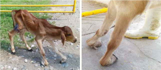
In a bovine production unit (BPU) in Martinez de la Torre, Veracruz, Mexico, a case was reported of a one-month-old male 3/4 Zebu x Holstein calf weighing 35 kg, with inability to extend the metacarpophalangeal joint (knuckle or fetlock) in both forelimbs. The calf did not walk normally, frequently presented a backward movement, falls and emaciation of one evolution week (Figure 1).
Clinical history Anamnesis
The calf was born with the joint alteration and until the third week of age was fed manually by bottle. After the fourth week, the animal stopped feeding properly and progressive emaciation was observed. The calf was joining less frequently and bleeding lesions were observed in the dorsal region of the pastern. It is mentioned that the site of the lesion has been bandaged since the onset of alopecia and inflammation of the area (from one week of age). Previous medical treatments included the placement of a temporary splint to stimulate joint extension, administration of non-steroidal anti-inflammatory drugs (diclofenac and tolfenamic acid), ADE and B-complex vitamins, beta-lactam antibiotics and local rubefacient/anti-inflammatory ointments based on methyl salicylate, camphor and carbolic acid for two weeks, with no positive evolution.
Physical examination
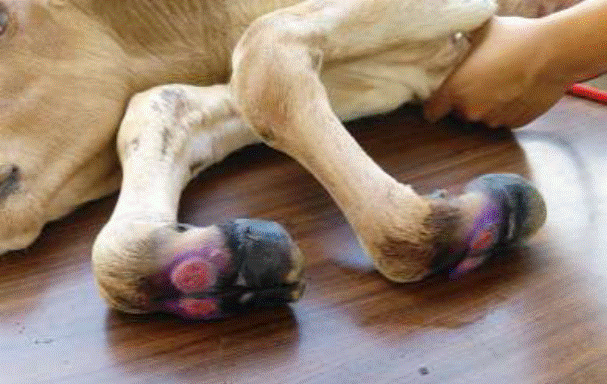
In the clinical examination, underweight, alopecia in legs and belly, as well as lesions in the dorsal region of the pastern were detected (Figure 2). The metacarpophalangeal joint could not be extended manually and pain was manifested on manipulation. During palpation, the superficial and deep digital flexor tendons of both limbs were felt to be contracted and under excessive tension. Physiological constants were within normal ranges for species and age (Piccione et al., 2010).
Diagnosis
It was determined that the calf presented bilateral contracture of superficial and deep digital flexor tendons in forelimbs. The alopecia in legs and belly was a consequence of prolonged contact with urine accumulated on the floor due to prolonged prostration time. It was considered that pharmacological treatment and physical therapy were no longer sufficient for the calf's recovery due to the severe degree of contraction and the previously mentioned lesions. Therefore, surgical intervention by tenotomy of the superficial and deep digital flexor tendons was decided.
Therapeutic approach Pre-surgical considerations
A blood sample was taken from the calf for a hemogram, the result of which was within normal ranges for the species. Prior to surgery, the patient was restricted from feed and water for 18 and 10 hours, respectively.
Surgical procedure
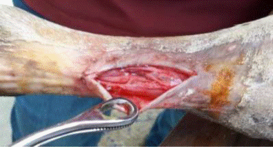
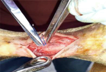
The surgery was performed, starting with deep sedation of the patient with Xylazine 2 % at a dose of 0.3 mg/kg of live weight intramuscularly (IM. This dose provides a decrease in muscle tone and good analgesia in cattle. The patient was placed in lateral decubitus, tricotomy of the metacarpal region was performed and the area was locally infiltrated with 20 ml of lidocaine 2 %. Antisepsis was performed with povidone iodine and a 6 cm longitudinal incision was made from the proximal metacarpal quarter to the middle of the metacarpal on the lateral aspect to avoid the medial palmar artery, just over the anatomical position of the superficial and deep flexor tendons (Figure 3). The superficial and deep fasciae were carefully approached to identify and avoid damaging the lateral palmar digital nerve and the palmar metacarpal arteries and veins. I was proceeded to separate these structures to protect them and locate the superficial and deep flexor tendons (Figure 4).
Subsequently, the superficial flexor tendon was elevated and extended with the aid of a blunt forceps (Figure 5a) and a surgical transection was performed with the aid of a scalpel (Figure 5b). Extensor movements of the metacarpophalangeal joint were performed, although it improved a little, the movement was still inadequate, therefore, it was proceeded to elevate, tighten and expose the deep flexor tendon and sectioned in the same way (Figure 5c). It is important that the incision of the deep tendon be made at a different site from the cut of the superficial tendon to avoid adhesion formation.

Figure 5 a) Presentation of superficial and deep flexor tendons, b) dissection of superficial flexor tendon and c) evaluation of deep flexor tendon function
The extension was corroborated and improvement was evident, therefore, there was no need for further intervention on another structure. The area was hydrated with saline and antibiotic and the fasciae were closed with 3.0-gauge synthetic absorbable suture in a continuous pattern. The skin was closed with single separate stitches with nylon #35. Micronized aluminum-based healing agent was applied, the phalangeal and metacarpal region was lightly bandaged, a layer of cotton was spread and a splint was placed in the distal-plantar region, from the crown of the hoof to the proximal portion of the metacarpal, to aid joint extension (Figure 6a). The same procedure was performed on both extremities.
Post-surgical treatment
An antibiotic based on G procaine penicillin was administered at a dose of 300,000 IU per 25 kg of weight (420,000 IU total) via IM for three days and meglumine flunixin as an anti- inflammatory and analgesic for 4 days at a dose of 1.5 mg/kg via IM. The splint was removed every third day for dressing change and aseptic treatment of the incision and the previously mentioned pre-surgical lesions. The nylon skin sutures were removed 10 days after surgery.
Case development and resolution
One week after surgery, the calf showed improved standing and began to lightly place the soles of hooves. Eighteen days later, the splinting was discontinued and the patient was walking on its own by planting soles of its hooves most of the time (Figure 6b).
Thirty days later, the calf was planting its hooves well on the ground, although it was walking slowly. The calf was discharged 35 days post-surgery, walking slowly, but normally, with no manifestation of pain or infection (Figure 6c).
DISCUSSION
Within the group of tendinopathies in cattle, congenital flexor tendon contracture is considered the most prevalent musculoskeletal abnormality in neonatal calves (Sato et al., 2020). It is important to consider that, if calves with severe cases are not treated, the skin and phalanges of the involved limbs can be severely injured and increase the likelihood of developing suppurating arthritis, as well as producing digital extensor tendon rupture as a sequela (Figure 7). Due to these consequences, and the difficulty to feed themselves, most cases end in the calf slaughter.
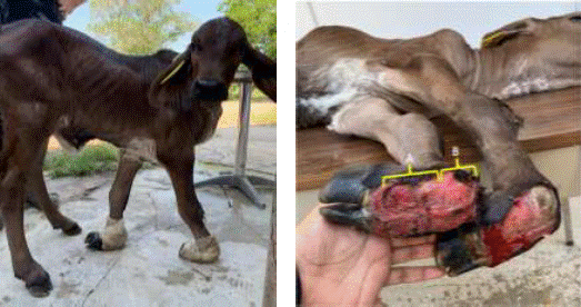
Figure 7 Dorsal lesion of the pastern joint (A) and fetlock (B) in an untreated severe case of flexor tendon contracture
In the present case, intervention was timely, since the secondary lesions were circumscribed in a small area of skin, superficial and easily treated (Figure 2). Avoiding larger lesions is key to the success or failure of surgery (Steiner et al., 2014). In many BPU, severe cases of this deformity are not adequately treated, due to reluctance or lack of knowledge to perform surgery and prolonged postoperative treatment, however, as can be seen, the surgical technique is quite feasible with the relevant anatomical knowledge of the area. One of the main objectives in the implementation of this type of surgical interventions is to shorten the postoperative recovery times of patients, to decrease the likelihood of splint and bandage injuries and for BPU owners to adopt these procedures. Recovery times vary from 8 to 10 weeks in some reports (Sohrt et al., 2013; Tamilmahan and Prabhakar, 2018), and in some cases, the injury does not fully recover (Sato et al., 2020). In this work, the recovery time was shortened by physiotherapy, postoperative pharmacological management of the incision and changing the splint and bandages every third day. In addition, the application of medications was only performed during the first week, and the calf was able to feed itself after one month, coinciding with the report of Fernandes et al. (2020), who reported one month of postoperative recovery. However, it is important to implement and evaluate various post-surgical protocols, with different breeds, drugs and degrees of injury, to assist in making decisions on appropriate interventions and to promote the welfare of the animals and their return to normal life.
The surgical technique used for tendon tenotomy in this report proved to be effective for this treatment and decreased invasion of tissues adjacent to the tendons. In some other species, apposition of the tendon ends post-transection using a far-near-near-near-far suture has been recommended in order to promote rapid healing (Fossum, 2019). Other authors, in bovines, have mentioned the "Z" incision of tendons to perform tendon elongation as a proposal to promote rapid recovery (Steiner et al., 2014), however, it is important to consider that tendons are a highly innervated structure and post-surgical pain could be greater. A study comparing the three techniques is recommended to determine the rapidity of fibroplasia re-union and recovery of the animals. To our knowledge, clinical case reports describing in detail the protocols for performing this surgical intervention in this type of tendinopathy are scarce (Tamilmahan and Prabhakar, 2018; Sato et al., 2020). There are some reports managed with conservative treatments in mild cases (Arieta and Fernández, 2011; Feito-Hermida et al., 2013), which in cases of severe contracture would not be sufficient. Other reports mention intervention as a complementary part of other conditions and in a single extremity (Sohrt et al., 2013).
One of the most important aspects to consider during postoperative treatment is the placement of an adequate splint, with rounded and protected edges, since one of the main postsurgical complications is the development of ulcers or ischemic necrosis due to pressure from the bandage and/or splint (Duncanson, 2010). In the present report, a splint was designed based on PVC tubing without corners, with filed edges and a thick pleated cotton cover fastened with industrial adhesive tape. This splint protected the skin and as soon as the calf was able to partially step on the hoof sole, the splint was removed so that the patient's own weight could promote joint extension.
Finally, before deciding to implement this surgery, it is important to perform an adequate diagnosis of the joint alteration, since it is necessary to differentiate between the different forms of contracture presentation and rule out other alterations such as arthrogryposis (Fernandes et al., 2020; Divers and Peek, 2017), which is an extreme form of tendon contraction that occurs in many joints and the contractions can be in extension and flexion (Blowey and Weaver, 2011). It should be considered that if flexion includes the carpal joint and calves support it to move, even if tenotomy is performed, the prognosis is usually poor.
Some recommendations: if necessary, trim the hooves to aid normal conformation and to promote proper placement on the ground. If the alteration is not corrected with tendon tenotomy, a demotomy of the suspensory ligament of the fetlock (interosseous muscle) may be considered.
CONCLUSION
A case of severe FTC in a calf with successful resolution is reported and the history, treatments and surgical process carried out are explained in detail. This case reinforces the veterinary medical literature that addresses these cases, since most of the existing reports have focused on cases of mild contracture that are resolved with drugs and physical therapy. A short post-surgical recovery time is reported due to the use of an adequate splint, pharmacological therapy and physical exercise stimulation as rehabilitation therapy.
LITERATURA CITADA
Alsaaod M, Huber S, Beer G, Kohler P, Schüpbach-Regula G, Steiner A. 2017. Locomotion characteristics of dairy cows walking on pasture and the effect of artificial flooring systems on locomotion comfort. Journal of Dairy Science. 100(10):8330-8337. https://doi.org/10.3168/jds.2017-12760 [ Links ]
Arieta RRJ, Fernandez FJA. 2011. Contractura a nivel de tendones en extremidades anteriores de un becerro: informe de un caso clínico. REDVET. 12(12):1-4. E-ISSN: 1695-7504. https://www.redalyc.org/pdf/636/63622039011.pdf [ Links ]
Blowey RW, Weaver AD. 2011. Diseases and disorders of cattle. 3th ed. China: Mosby- Elsevier. Pp. 99. ISBN: 978-0-7234-3602-7. https://books.google.com.mx/books?hl=es&lr=&id=roHrW4mc_8kC&oi=fnd&pg=PR1&dq=Diseases+and+disorders+of+cattle.+3th+ed+blowey&ots= [ Links ]
Bruijnis MR, Hogeveen H, Stassen EN. 2010. Assessing economic consequences of foot disorders in dairy cattle using a dynamic stochastic simulation model. Journal of Dairy Science. 93(6):2419-2432. https://doi.org/10.3168/jds.2009-2721 [ Links ]
Divers TJ, Peek SF. 2017. Rebhun's diseases of dairy cattle. 3rd ed. St. Louis, Missouri: Elsevier Health Sciences - Saunders. Pp. 704. https://doi.org/10.1016/C2013-0-12799-7 [ Links ]
Duncanson G. 2010. Veterinary treatment for working equines. 2nd ed. London: CABI. Pp. 104. ISBN-13: 978-1-84593-655-6. https://books.google.com.mx/books?hl=es&lr=&id=jBvAQriQJDMC&oi=fnd&pg=PR5&dq=Veterinary+treatment+for+working+equines+2+ed&ots= [ Links ]
Feito-Hermida J, Jareño-Moreno S, Mínguez-Rico A, Kawiecki-Peralta A. 2013. Retracción de tendones en terneros. REDUCA. 5:195-199. http://www.revistareduca.es/index.php/reduca/article/view/1632 [ Links ]
Fernandes MEDSL, de Albuquerque-Carvalho L, Chenard MG, Pitombo CA, dos Santos OJ, Caldas SA, Nogueira VA, Helayel MA. 2020. Management of a Congenital Flexural Deformity in a Calf-Surgical and Pathological Aspects. Acta Scientiae Veterinariae. 48(1):581. https://www.seer.ufrgs.br/ActaScientiaeVeterinariae/article/view/104033 [ Links ]
Fossum TW. 2019. Cirugía en pequeños animales. 5th ed. España: Elsevier. Pp. 1632. ISBN:978-84-9113-380-3. https://books.google.com.mx/books?hl=es&lr=&id=48nSDwAAQBAJ&oi=fnd&pg=PP1&d q= [ Links ]
Gaughan EM. 2017. Flexural limb deformities of the carpus and fetlock in foals. Veterinary Clinics of North America: Equine Practice. 33:331-42. http://dx.doi.org/10.1016/j.cveq.2017.03.004 [ Links ]
Kirkendall DT, Garrett WE. 1997. Function and biomechanics of tendons. Scandinavian Journal of Medicine and Science Sports. 7 (2):62-66. https://doi.org/10.1111/j.1600-0838.1997.tb00120.x [ Links ]
Piccione G, Casella S, Pennisi P, Giannetto C, Costa A, Caola G. 2010. Monitoring of physiological and blood parameters during perinatal and neonatal period in calves. Arquivo Brasileiro de Medicina Veterinária e Zootecnia. 62(1):1-12. https://doi.org/10.1590/S0102-09352010000100001 [ Links ]
Sato A, Kato T, Tajima M. 2020. Flexor tendon transection and post-surgical external fixation in calves affected by severe metacarpophalangeal flexural deformity. Journal of Veterinary Medical Science. 82(10):1480-1483. https://doi.org/10.1292/jvms.20-0057 [ Links ]
Sohrt JT, Heppelmann M, Rehage J, Staszyk C. 2013. Tenotomy of carpal and digital flexor tendons for correction of congenital neuromyodysplasia in a calf. Tierarztliche Praxis. 41(2):113-118. https://doi.org/10.1055/s-0038-1623160 [ Links ]
Steiner A, Anderson DE, Desrochers A. 2014. Diseases of the Tendons and Tendon Sheaths. Veterinary Clinics: North America-Food Animal Practice. 30(1):157-175. https://doi.org/10.1016/j.cvfa.2013.11.002 [ Links ]
Tamilmahan P, Prabhakar R. 2018. Surgical management of congenital flexor tendon deformity in calves: A review of three cases. Journal of Entomology and Zoology Studies. 6(3):544-546. http://krishikosh.egranth.ac.in/handle/1/5810139315 [ Links ]
Wavreille G, Fontaine C. 2009. Tendón normal: anatomía y fisiología. EMC-Aparato Locomotor. 42(1):1-12. https://doi.org/10.1016/S1286-935X(09)70909-8 [ Links ]
Weaver AD, Atkinson O, St. Jean G, Steiner A. 2018. Contracted flexor tendons. In: Weaver D, Atkinson O, Steiner A, St. Jean G. Eds. Bovine Surgery and Lameness. 3rd ed. Oxford: John Wiley and Sons Ltd. Pp. 267. ISBN: 9781119040514. https://books.google.com.mx/books?hl=es&lr=&id=UHFODwAAQBAJ&oi=fnd&pg=PP2&dq=Bovine+Surgery+and+Lameness+3rd&ots= [ Links ]
Received: January 28, 2021; Accepted: June 01, 2021


 text in


