SciELO - Scientific Electronic Library Online

 
vol.95 issue4Leaf anatomical and biochemical adaptations in Typha domingensis Pers. ecotypes for salinity toleranceThe phaeophyte Hizikia fusiformis extracts suppress rhizoid and blade formation in seaweeds author indexsubject indexsearch form
Home Pagealphabetic serial listing  

Services on Demand

Journal

Article

Indicators

Related links

  • Have no similar articlesSimilars in SciELO

Share


Botanical Sciences

On-line version ISSN 2007-4476Print version ISSN 2007-4298

Bot. sci vol.95 n.4 México Oct./Dec. 2017

https://doi.org/10.17129/botsci.894 

Structural Botany

Leaf morphoanatomy of the medicinal Maytenus imbricata (Celastraceae): an ecological approach

Silvia Ribeiro de Souza1 

Izabel Cristina Trindade2 

Maria Olívia Mercadante-Simões3  * 

Lucienir Pains Duarte4 

Grácia Divina de Fátima Silva4 

Maria Cristina Teixeira Braga Messias5 

Ariadna Conceição dos Santos3 

Sidney Augusto Vieira-Filho2 

1 Faculdade de Ciências da Saúde, Universidade de Brasília. Campus Darcy Ribeiro, Asa Norte, Brasília.

2 Departamento de Farmácia, Escola de Farmácia, Universidade Federal de Ouro Preto. Morro do Cruzeiro, Ouro Preto

3 Departamento de Biologia Geral, Centro de Ciências Biológicas e da Saúde, Universidade Estadual de Montes Claros. Campus Professor Darcy Ribeiro

4 NEPLAM, Departamento de Química, Instituto de Ciências Exatas, Universidade Federal de Minas Gerais

5 Departamento de Biodiversidade, Evolução e Meio Ambiente, ICEB, Universidade Federal de Ouro Preto.


Abstract:

Background:

The leaves of many species of Maytenus are used to prepare herbal drugs to treat stomach ulcers.

Questions:

In order to define key anatomical characters for the unambiguous species identification, the present study aimed to describe the leaf morphoanatomy of M. imbricata.

Species study:

Maytenus imbricata Mart. ex Reissek (Celastraceae)

Study site and dates:

Collected in an area of ‘campo rupestre’ - one of the Brazilian Savannah vegetation (20º 22’ 11.02” S, 43º 30’ 22.81” W) in August 2015.

Methods:

The plant material was processed according to commonly used techniques in plant anatomy studies.

Results:

The major anatomical characters for several species of Maytenus identification also present in M. imbricata are marginal teeth, conspicuous epicuticular wax, ciclocytic stomata, thick external periclinal wall with large pits on epidermal cells and vascular system with a closed loop shape in cross-section. Strips on the anticlinal walls on epidermal cells and conspicuous gelatinous fibers sheath are diagnostic for M. imbricata.

Conclusions:

Strips on the anticlinal wall of the epidermal cell are reported for the first time for the genus. The xeromorphic traits help the species to survive in arid environments and may contribute to quality control of the raw material used in the production of herbal medicines.

Keywords: cell wall strips; colleter; gelatinous fiber; xeromorphic

Resumen:

Antecedentes:

Las hojas de muchas especies de Maytenus se utilizan para la preparación de medicamentos para el tratamiento de las úlceras de estómago.

Preguntas:

Con la finalidad de contribuir a la identificación de la especie, este estudio tuvo como objetivo describir la morfología y anatomia de la hoja de M. imbricata.

Especie de estudio:

Maytenus imbricata Mart. ex Reissek (Celastraceae)

Sitio y años de estudio:

Recolecta de material en una zona de ‘campo rupestre’ - vegetación del cerrado brasileño (20º 22’ 11.02” S, 43º 30’ 22.81” W) en agosto del 2015.

Métodos:

El material vegetal se procesó mediante las técnicas que se utilizan en los estudios de anatomía vegetal.

Resultados:

Los principales caracteres anatómicos para la identificación de varias especies de Maytenus, que están presentes en M. imbricata, son los dientes marginales, la cera epicuticular gruesa, los estomas ciclocíticos, las células epidérmicas con la pared periclinal externa gruesa, con grandes punteaduras y la sección transversal del sistema vascular en un círculo cerrado. Las estrías en las paredes anticlinales y las fibras gelatinosas son caracteres diagnósticos para M. imbricata.

Conclusiones:

Las estrías que se forman en la pared de las células epidérmicas se reportan por primera vez en el género. Estos caracteres xeromórficos ayudan a la especie a sobrevivir en ambientes áridos y pueden contribuir al control de la calidad de la materia prima utilizada en la producción de medicamentos.

Palabras clave: caracteres xeromórficos; coléteres; estrías en la pared celular; fibras gelatinosas

Maytenus Molina is one of the largest genera of Celastraceae, found in tropical and subtropical regions, and distributed throughout the Brazilian territory (Lombardi et al. 2014). Many of the chemical components isolated from its species have pharmacological properties (Din et al. 2013, Duarte et al. 2013, Moo-Puc et al. 2014). A good example is M. ilicifolia Mart. ex Reissek, that is mentioned in the Brazilian Pharmacopoeia (ANVISA 2011) to treat gastric ulcer (Santos-Oliveira et al. 2009). Similarly, M. imbricata Mart. ex Reissek, (Silva et al. 2009, Rodrigues et al. 2012), M. aquifolium Mart., (Gonzalez et al. 2001), M. salicifolia Reissek, (Magalhães et al. 2011) and M. truncata Reissek, (Silva et al. 2005a) are used in folk medicine to treat the same stomach illness, and have the same popular name “espinheira-santa” (Niero et al. 2011). For these reasons it is necessary a careful quality control of raw material in the manufacture of drugs using Maytenus species and is required define anatomical key characters for the unambiguous species identification (Jacomassi & Machado 2004, Duarte & Debur 2005, Joffily & Vieira 2005, Nakamura et al. 2013). For five species of Maytenus, Den Hartog & Bass (1978) elected stomatal type and the presence of trichomes and crystals as safe parameters among others epidermal characteristics. Duarte & Debur (2005) proposed epidermal cells containing styloids, cuticular flanges and amphicrival vascular bundles as relevant characters for the diagnosis of M. ilicifolia.

M. imbricata is known for the presence of bioactive compounds (Silva et al. 2005b, 2007, Silva et al. 2009, Rodrigues et al. 2012) nevertheless there is no study to quality control. The goal of this work was to describe the morphological and anatomical features of the leaf and relate them to the environment where the species occurs.

Materials and methods

Fully expanded leaves of M. imbricata were obtained from three individuals in an area of ‘campo rupestre’ (one of the Brazilian Savannah vegetation, 20º 22’ 11.02”S, 43º 30’ 22.81”W) and they represent the morphological variation of the studied population. A voucher specimen (MCTB Messias, 2565) was deposited at the OUPR Herbarium (Universidade Federal de Ouro Preto, Minas Gerais, Brazil). Samples of the median region of the leaf blade and the petiole were fixed in Karnovsky solution (Karnovsky 1965) for 12 hours, dehydrated in a graded ethanol series (Jensen 1962), and cold-embedded (Paiva et al. 2011) in hydroxyethyl-methacrylate resin (Leica Microsystem Inc., Heidenbeg, Germany). Cross sections of the median rib, transverse and paradermal sections of the internerval region, and cross-sections of the margin and the petiole were obtained using a rotary microtome (Atago, Tokyo, Japan), stained with Toluidine blue pH 4.7, rutenium red (O’Brien et al. 1964, modified), basic Fuchsin and Astra blue and mounted in acrylic resin (Itacril, Itaquaquecetuba, Brazil). Phenolic compounds were identified by the red color obtained with basic Fuchsin and bright blue color obtained with Tolulidine blue, and acids polysaccharides with ruthenium red. Images were captured using an AxioCam MRc camera coupled to a AxioVision LE light microscope (Zeiss, Oberkochen, Germany).

Results

Morphology. The leaves are light green, congested, erect, imbricate, coriaceous and glabrous, and are coated on both sides by a conspicuous wax layer. Leaf shape varies from orbicular to oval, the base is slightly cordate and the apex is obtuse-emarginate. The margins are revolute, crenate, and have teeth. The petiole is 0.1 to 0.2 cm long, and the leaf blade is 1.5 to 3 cm long and 1 to 3 cm wide with a protruding central nerve on the adaxial side. Secondary nerves are evident at the leaf surface but are not protruding (Figure 1A). The venation is craspedodromous, and the marginal teeth contain multicellular secretory structures named colleters (Figure 1B-C).

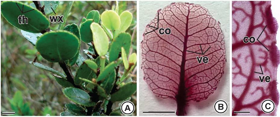
Figure 1 Leaf morphology of Maytenus imbricata Mart. ex Reiss. A) General view of the plant showing the rounded marginal teeth and epicuticular wax. B) Venation craspedodromous. C) Colleters on marginal teeth. co: colleters; th: teeth; ve: vein; wx: waxy. Bars: A-C = 1cm. 

Anatomy. At the midrib region the epidermal cells have elongated anticlinal wall and the vascular system has the shape of a closed loop in cross-section, and is surrounded by a conspicuous sheath of gelatinous sheath (Figure 2A). At the internerve region the cells of the adaxial face of the epidermis have thick external periclinal wall, and straight anticlinal wall with strips on its innermost region (Figure 2B-C). The mesophyll is dorsiventral with palisade parenchyma composed of two layers of transversely elongated cells, and spongy parenchyma composed of globoid cells compactly arranged facing the abaxial surface (Figure 2D-E). The presence of phenols is observed throughout the whole leaf tissues (Figure 2A-E).

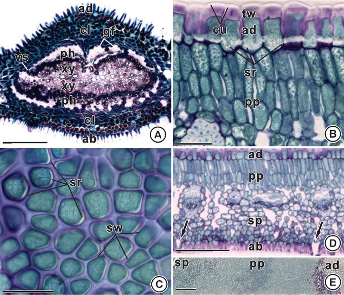
Figure 2 Leaf anatomy of Maytenus imbricata Mart. ex Reiss. A) Midrib region. Vascular system with a closed-loop shape (cross section). B-C) Cells of the adaxial epidermis with strips on the base of the straight anticlinal walls (cross section on B, and paradermic section - external view on C). D-E) Internerve region. Hypostomatic leaf (arrows). Dorsiventral mesophyll (cross section on D and paradermic-oblique section on E). ab: abaxial epidermis; ad: adaxial epidermis; cl: collenchyma; cu: cuticle; gf: gelatinous fibers; ph: phloem; pp: palisade parenchyma; sr: strips; sp: spongy parenchyma; sw: straigth anticlinal wall; tw: thick external periclinal wall; vs: vascular system; xy: xylem. Bars: A = 200 µm; B-E = 50 µm. 

The vascular bundles in secondary veins are collateral and gelatinous fibers are on both sides (Figure 3A). In cross section, subsidiary cells have pear-shaped and narrow diameter on the leaf surface, and are internally voluminous (Figure 3B). The leaf is hypostomatic displaying a higher number of ciclocytic stomata on the adaxial face (Figures 2D, 3B-C). The external walls of the ordinary cells of the abaxial epidermis are thick and show large pits such as pores (Figure 3B, D).

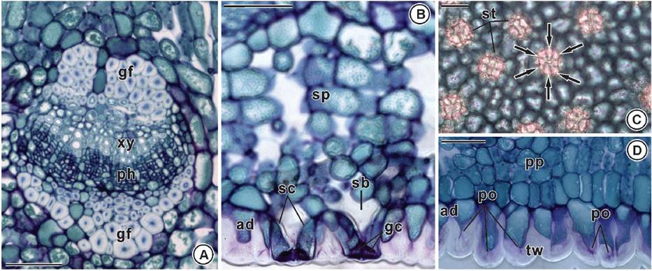
Figure 3 Leaf anatomy of Maytenus imbricata Mart. ex Reiss. A) Collateral vascular bundle with gelatinous fibers on both sides (cross section). B) Subsidiary pear-shaped cells with narrow diameter on the leaf surface, and voluminous internally (cross section). C) Ciclocytic stomata (paradermic section - internal view). D) Thick external wall of the ordinary cells of the abaxial epidermis with large pits as pores (cross section). gf: gelatinous fibers; ph: phloem; po: pore; sb: substomatal chamber; sc: subsidiary cell; sp: spongy parenchyma; st: stomata; tw: thick wall; xy: xylem. Bars = 50 µm. 

In leaf margin, the epidermal cells have thick cuticle and thick pectic walls. The mesophyll is homogeneous and involves a bicolateral vascular bundle with a prominent gelatinous fibers cap on one side (Figure 4A). The petiole outline is rounded with two lateral ribs on the adaxial side and is covered by epidermal cells with thick cuticle and thick pectic walls. The outer cortex is constituted by cells with periclinal divisions and the inner cortex has sclereids clusters. The vascular system is closed loop, and it is wrapped by a discontinuous sheath of gelatinous fibers and displays cambial activity. The secondary xylem is well developed and encircle a parenchymatous pith (Figure 4B).

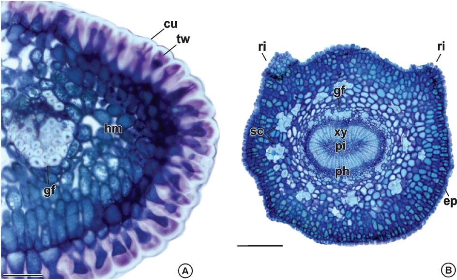
Figure 4 Leaf anatomy of Maytenus imbricata Mart. ex Reiss. A) Leaf margin: epidermal cells with thick cuticle and pectic walls, homogeneous mesophyll and a bicolateral bundle with gelatinous fibers on both sides. B) Petiole: ribs on the adaxial side, epidermal cells with thick cuticle and thick pectic walls, outer cortex cells with periclinal divisions, inner cortex with clusters of sclereids, vascular system closed loop, sheath of gelatinous fibers, cambial activity, secondary xylem and parenchymatous pith. cu: cuticle; ep: epidermis; gf: gelatinous fibers; hm: homogenous mesophyll; ph: phloem; pi: pith; ri: ribs; sc: sclereids; tw: thick wall; xy: xylem. Bars: A = 50 µm; B = 250 µm. 

Discussion

The structural features present in plant organs are useful tools for quality control in the manufacture of herbal medicines. The leaf margin teeth are an important feature in the identification of Maytenus species (Joffily & Vieira 2005, Lombardi & Groppo 2014). The thorns and scars present on the end of the teeth indicate the drop of the colleters observed at the primordial leaf margin. Some anatomical characters in leaves occur in response to environmental conditions (Voltolini & Santos 2011, Sack & Scoffoni 2013). Colleters secrete resin or mucilage to protect the stem apex against desiccation, which is favored by the low humidity of the air during the dry season in Brazilian Savannah (Mercadante-Simões & Paiva 2013).

Epidermal traits are particularly useful for quality control purpose, because they are well preserved even in pulverized material. In this work ciclocytic stomata were found in M. imbricata whereas anomoctic stomata were reported for M. aquifolia and M. ilicifolia (Jacomassi & Machado 2004). Acicular crystals on both sides of the epidermis were recorded for M. ardisiaefolia, M. brasiliensis, M. communis and M. obtusifolia (Joffily & Vieira 2005) but not observed in M. imbricata. The presence of epicuticular wax, thick cuticle and thick periclinal wall on the epidermis of M. imbricata has also been recorded in other species of Maytenus (Duarte & Debur 2005, Joffily & Vieira 2005). The thickening of the epidermal cell walls of M. imbricata seems to be related to the restriction of water loss by transpiration. Also, the reduced area of exposure of the subsidiary cell on the leaf surface minimizes water loss under high solar radiation conditions (Voltolini & Santos 2011).

The presence of pores in the wall of epidermal cells and the occurrence of guard cells arranged in rosette and subsidiary cells positioned below the surface in M. imbricata have been reported for other species of Celastraceae (Metcalfe & Chalk 1957). However, strips on the epidermal cell wall are reported for the first time for Maytenus and their role in the control of water traffic in apoplast is well recognized (Moreira & Isaias 2008, Yang et al. 2011). The ‘Phi thickening’ on the cell walls was also related to the water traffic control (Fernandez-Garcia et al. 2009, Idris & Collings 2015). On the other hand, the occurrence of large pits such as pore on the wall in M. imbricata may favor the deposition of deterrent compounds throughout the whole leaf surface protecting against herbivory and microorganisms infection (Yeats & Rose 2013).

Cells containing phenolic compounds are common in Maytenus species (Gupta & Sharma 2012) and have also been reported for other genera of Celastraceae (Mercadante-Simões et al. 2014). The secondary metabolites are responsible for the relation of the plant with the biotic and abiotic environment. Bioactive compounds accumulation can be induced by the severe water constraint conditions in the Brazilian Savannah (Di-Ferdinando et al. 2014).

The major branching of the craspedodromous venation in M. imbricata is related to the need of an adequate water supply. The vein length for area has a strong correlation with environmental aridity and higher values were found to sun plants when compared to shade plants (Sack & Scoffoni 2013). In our work a sheath of gelatinous fibers was found in M. imbricata. These characters were not observed in other species of Maytenus but a vascular bundle with a sclerenchymatic sheath at the margin were reported for M. aquifolia and M. ilicifolia (Jacomassi & Machado 2004) and perivascular fibers near the vascular system were observed in M. ardisiaefolia, M. brasiliensis, M. communis and M. obtusifolia (Joffily & Vieira 2005). The conspicuous sheath of gelatinous fibers surrounding the vascular bundles of M. imbricata contributes to the better use of water absorbed by the xylem. The gelatinous fibers have thickened walls rich in acid polysaccharides allowing water retention (Melo et al. 2011, Sonsin et al. 2012).

Conclusions

The structural features on medicinal plants are useful for their unequivocal identification, particularly when they are in small pieces to be used in herbal medicine production. The key characters to M. imbricata are strips on the epidermal cell anticlinal wall, reported here for the first time for the genus, and conspicuous gelatinous fibers sheath. These structural characters are related to the dry Brazilian Savanna conditions.

Acknowledgements

The authors wish to thank Dr. Rita Maria Carvalho-Okano (Botany Department of the Federal University of Viçosa) for providing botanical identifications and the Fundação de Amparo à Pesquisa de Minas Gerais (FAPEMIG) and Conselho Nacional de Pesquisa (CNPq) for providing financial support and scholarship.

Literature cited

Agência Nacional de Vigilância Sanitária (ANVISA). 2011. Formulário de fitoterápicos da Farmacopéia Brasileira. Brasília. 125p. [ Links ]

Di-Ferdinando M, Brunetti C, Agati G, Tattini M. 2014. Multiple functions of polyphenols in plants inhabiting unfavorable Mediterranean areas. Environmental and Experimental Botany 103: 107-116. DOI: 10.1016/j.envexpbot.2013.09.012 [ Links ]

Din AU, Uddin G, Hussain N, Choudary MI. 2013. Ficusonic acid: a new cytotoxic triterpene isolated from Maytenus royleanus (Wall. ex M.A. Lawson) cufodontis. Journal of the Brazilian Chemical Society 24: 663-668. DOI: 10.5935/0103-5053.20130084 [ Links ]

Duarte MC, Tavares JF, Madeiro SAL, Costa VCO, Barbosa-Filho M, Agra MF, Braz-Filho R, Silva MS. 2013. Maytensifolone, a new triterpene from Maytenus distichophylla Mart. ex Reissek. Journal of the Brazilian Chemical Society 24: 1697-1700. DOI: 10.5935/0103-5053.20130205 [ Links ]

Duarte MR, Debur MC. 2005. Steam and leaf morphoanatomy of Maytenus ilicifolia. Fitoterapia 76: 41-49. DOI: 10.1016/j.fitote.2004.10.003 [ Links ]

Fernandez-Garcia N, Lopez-Perez L, Hernandez M, Olmos E. 2009. Role of phi cells and the endodermis under salt stress in Brassica oleracea. New Phytologist 181: 347-360. DOI: 10.1111/j.1469-8137 [ Links ]

Gonzalez FG, Portela TY, Stipp EJ, Di-Stasi LC. 2001. Antiulcerogenic and analgesic effects of Maytenus aquifolium, Sorocea bomplandii and Zolernia ilicifolia. Journal of Ethnopharmacology 77: 41-47. DOI: 10.1016/S0378-8741(01)00268-9 [ Links ]

Gupta V, Sharma M. 2012. Phytochemical analysis and evaluation of antioxidant activities of methanolic extracts of Maytenus emarginata. Omics 16: 257-262. DOI: 10.1089/omi.2011.0051 [ Links ]

Den Hartog RM, Baas P. 1978. Epidermal characters of the Celastraceae sensu lato. Acta Botanica Neerlandica 27: 355-388. DOI: 10.1111/j.1438-8677.1978.tb00301.x [ Links ]

Idris NA, Collings DA. 2015. The life of phi: the development of phi thickenings in roots of the orchids of the genus Miltoniopsis. Planta 241: 489-506. DOI: 10.1007/s00425-014-2194-z [ Links ]

Jacomassi E, Machado SR. 2004. Características anatômicas da espinheira-santa (Maytenus ilicifolia Mart. ex Reissek e Maytenus aquifolia Mart.) e mata-olho (Sorocea bonplandii (Baill.) Burg. Lanj. & Boer.) para o controle de qualidade da matéria prima. Revista Brasileira de Plantas Medicinais 6: 84-96. [ Links ]

Jensen WA. 1962. Botanical histochemistry: principles and pratices. San Francisco, W. H. Freeman and Company. [ Links ]

Joffily A, Vieira RC. 2005. Anatomia foliar de Maytenus Mol. emend Mol. (Celastraceae), ocorrente no estado do Rio de Janeiro, Brasil. Acta Botanica Brasilica 19: 549-561. [ Links ]

Karnovsky MJ. 1965. A formaldehyde-glutaraldehyde fixative of high osmolality for use in eletron microscopy. Journal of Cell Biology 27: 137-138. [ Links ]

Lombardi JA, Groppo M, Biral L. 2014. Celastraceae in Lista de Espécies da Flora do Brasil. Jardim Botânico do Rio de Janeiro. < http://floradobrasil.jbrj.gov.br/jabot/floradobrasil/FB6763 > (Accessed August 27 2016) [ Links ]

Magalhães CG, Ferrari FC, Guimaraes DAS, Silva GDF, Duarte LP, Figueiredo RC, Vieira-Filho SA. 2011. Maytenus salicifolia Reissek, Celastraceae: triterpenes isolated from stems and antioxidant property of extracts from aerial parts. Brazilian Journal of Pharmacognosy 21: 415-419. DOI: 10.21577/1984-6835.20160107 [ Links ]

Melo JCF, Ceccantini G, Bona C. 2011. Wood ecological anatomy of Copaifera langsdorffii Desf. (Leguminosae) at different substrate conditions in the Cerrado of South Brazil. Iheringia 66: 189-200. [ Links ]

Mercadante-Simões MO, Paiva EAS. 2013. Leaf colleters in Tontelea micrantha (Celastraceae, Salacioideae): Ecological, morphological and structural aspects. Comptes Rendus Biologies 336: 400-406. DOI: 10.1016/j.crvi.2013.06.007 [ Links ]

Mercadante-Simões MO, Mazzottini-dos-Santos HC, Nery LA, Ferreira PRB, Ribeiro LM, Royo VA, Oliveira DA. 2014. Structure, histochemistry and phytochemical profile of the bark of the sobol and aerial stem of Tontelea micrantha (Celastraceae - Hippocrateoideae). Annals of the Brazilian Academy of Sciences 86: 1167-1179. DOI: 10.1590/0001-3765201420130105 [ Links ]

Metcalfe CR, Chalk L. 1957. Anatomy of the dicotyledons. New York: Oxford University Press. [ Links ]

Moo-Puc JA, Martín-Quintal Z, Mirón-López G, Moo-Puc RE, Quijano L, Mena-Rejón GJ. 2014. Isolation and antitrichomonal activity of the chemical constituents of the leaves of Maytenus phyllanthoides Benth. (Celastraceae). Quim Nova 37: 85-88. DOI: 10.1590/S0100-40422014000100016 [ Links ]

Moreira ASFP, Isaias RMS. 2008. Comparative anatomy of the absorption roots of terrestrial and epiphytic orchids. Brazilian Archives of Biology and Technology 51: 83-93. DOI: 10.1590/S1516-89132008000100011 [ Links ]

Nakamura SS, Valle JS, Jacomassi E, Linde GA, Colauto NB. 2013. Molecular authentication of Maytenus sp by PCR-RFLP. Semina: Ciências Agrárias 34: 627-634. DOI: 10.5433/1679-0359.2013v34n2p627 [ Links ]

Niero R, Andrade SF, Cechinel-Filho V. 2011. A review of the ethnopharmacology, phytochemistry and pharmacology of plants of the Maytenus Genus. Current Pharmaceutical Design 17: 1851-1871. DOI: 10.2174/138161211796391029 [ Links ]

O’Brien TP, Feder N, Mccully ME. 1964. Polychromatic staining of plant cell walls by toluidine blue O. Protoplasma 59: 368-373. [ Links ]

Paiva EAS, Pinho SZ, Oliveira DMT. 2011. Large plant samples: how to process for GMA embedding? In: Chiarini-Garcia H, Melo RCN. eds. Light microscopy: methods and protocols. New York: Springer Humana Press, 37-49. [ Links ]

Rodrigues VG, Duarte LP, Silva GDF, Silva FC, Góes JV, Takahashi JA, Pimenta LPS, Vieira-Filho SA. 2012. Evaluation of antimicrobial activity and toxic potential of extracts and triterpenes isolated from Maytenus imbricata. Quimica Nova 35: 1375-1380. DOI: 10.1590/S0100-40422012000700017 [ Links ]

Sack L, Scoffoni C. 2013. Leaf venation: structure, function, development, evolution, ecology and applications in the past, present and future. New Phytologist 198: 983-1000. DOI:10.1111/nph.12253 [ Links ]

Santos-Oliveira R, Coulaud-Cunha S, Colaço W. 2009. Review of Maytenus ilicifolia Mart. ex Reissek, Celastraceae. Contribution to the studies of pharmacological properties. Brazilian Journal of Pharmacognosy 19: 650-659. [ Links ]

Silva GDF, Silva SRS, Barbosa LCA, Duarte LP, Ribeiro SMR, Queiroz JH, Vieira-Filho AS, Oliveira MLR. 2009. Antioxidant activity of Maytenus imbricata Mart., Celastraceae. Brazilian Journal of Pharmacognosy 19: 530-536. DOI: 10.1590/S0102-695X2009000400004 [ Links ]

Silva JL, Silva RP, Jorge RM, Silva FGD, Vieira-Filho SA, Fonseca APND, Tagliati CA. 2005a. Evaluation of antiulcerogenic activity of Maytenus truncata Reiss (Celastraceae). Brazilian Journal of Pharmacognosy 15: 30-35. DOI: 10.1590/S0102-695X2005000100007 [ Links ]

Silva SRS, Silva GDF, Barbosa LC, Duarte LP, Vieira-Filho SA. 2005b. Lupane pentacyclic triterpenes isolated from stems and branches of Maytenus imbricata (Celastraceae). Helvetica Chimica Acta 88: 1102-1109. DOI: 10.1002/hlca.200590081 [ Links ]

Silva SRS, Silva GDF, Barbosa LCA, Duarte LP, King-Diaz B, Camacho FA, Hennsen BL. 2007. Uncoupling and inhibition properties of 34-seco-friedelan-3-oic acid isolated from Maytenus imbricata. Pesticide Biochemistry and Physiology 87: 109-114. DOI: 10.1016/j.pestbp.2006.06.008 [ Links ]

Sonsin JO, Gasson FLS, Barros CF, Marcati CR. 2012. A comparison of the wood anatomy of 11species from two cerrado habitats (Cerrado s.s. and adjacent gallery forest). Botanical Journal of the Linnean Society 170: 257-276. DOI: 10.1111/j.1095-8339.2012.01282.x [ Links ]

Voltolini CH, Santos M. 2011. Variações na morfoanatomia foliar de Aechmea lindenii (E. Morren) Baker var. lindenii (Bromeliaceae) sob distintas condições ambientais. Acta Botanica Brasilica 25: 2-10. DOI: 10.1590/S0102-33062011000100002 [ Links ]

Yang C, Zhang X, Zhou C, Seago-Jr JL. 2011. Root and stem anatomy and histochemistry of four grasses from the Jianghan Floodplain along the Yangtze River, China. Flora 206: 653-661. DOI: 10.1016/j.flora.2010.11.011 [ Links ]

Yeats TH, Rose JKC. 2013. The formation and function of plant cuticles. Plant Physiology 163: 5-20. DOI: 10.1104/pp.113.222737 [ Links ]

Associate Editor: Juan Núñez Farfán

Received: August 27, 2016; Accepted: May 12, 2017

* Corresponding author: Maria Olívia Mercadante-Simões, e-mail: omercadante@hotmail.com.

Author Contributions. Silvia Ribeiro de Souza, Izabel Cristina Trindade and Ariadna Conceição dos Santos performed the botanical histological study. Maria Olívia Mercadante-Simões, Lucienir Pains Duarte and Sidney Augusto Vieira-Filho conceived the work and guided its execution by the students. Grácia Divina de Fátima Silva and Maria Cristina Teixeira Braga Messias carried out the ethnobotanical study, the field collection and the botanical identification.

Creative Commons License This is an open-access article distributed under the terms of the Creative Commons Attribution License