


 text new page (beta)
 English (pdf)
 Article in xml format
 Article references
 Send this article by e-mail
 Cited by SciELO
  Similars in
SciELO
Similars in
SciELO 
 Permalink
Peridinium quinquecorne T.H. Abé 1927 is one of the few known benthic-planktonic dinoflagellates. It is worth mentioning that the author of this species noted that “no corresponding form was found in the genus” (Abé, 1927: 411), highlighting the superficial similarity between this species and Amylax triacantha (Jørgensen) Sournia 1984 (= Gonyaulax triacantha Jörgensen 1899). In Mexican waters, it is best known from the state of Veracruz, southwestern Gulf of Mexico, where it has caused recurrent blooms; the species has also been reported from other regions of the world (see references in Trigueros et al., 2000; Pertola et al., 2006; Aké-Castillo et al., 2014; Okolodkov et al., 2016; Rodríguez-Gómez et al., 2021; Hoppenrath et al., 2023).
The synonymy of Peridinium quinquecorne with Heterocapsa quadridentata F. Stein 1883 has almost always been avoided in the published literature, although the synonymy of the former with Protoperidinium quinquecorne (T.H. Abé) Balech 1974 is widely known. However, Horiguchi & Soto (1994) concluded that the transfer of Peridinium quinquecorne by Balech (1974) to the genus Protoperidinium Bergh 1881 is not appropriate: the former has five cingular plates, unlike Protoperidinium species that possess four cingulars, including a transitional plate.
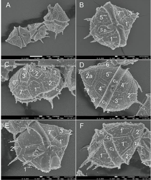
Figure 1 Scanning electron micrographs of Blixaea quinquecornis from a bloom in the coastal waters of Veracruz. A - three polygonal cells in ventral view, showing variability in size; B and E - cells in ventral view, showing variability in the cell size and the number of the antapical spines; C - cell in apical-ventral view, showing the apical pore complex and the apical spine (arrowhead) belonging to the 3’ plate; D - larger rounded cell in dorsal view; F - cell in ventral-left-side view with four antapical spines. Thecal plates: 1´-3´ - apicals; 1´´-7´´ - precingulars; 1´´´-5´´´ - postcingulars; 1´´´´ and 2´´´´ - antapicals; c1-c5 - cingulars; Po - the pore plate; S.p. - the sulcal posterior plate; x - the canal plate. Scale bar: 10 μm in A, 1 μm in B-F.
Iwataki (2008) noted that H. quadridentata had not been reported from any location since its original description and does not possess the characteristics of the genus Heterocapsa F. Stein emend. Iwataki et Fukuyo 2003. Although the line drawing of the taxon by Stein (1883: 13, pl. IV, fig. 3) is somewhat schematic, considering that the first apical plate (1´) is of ortho-type, the cell shape, its general proportions, the length, position and the number of the antapical spines, slightly attenuated apex and the displacement of the cavozone cingulum give it much in common with the original description and illustrations by Abé (1927). The morphologically similar Amylax triacantha has a different set of thecal features, and Stein’s taxon differs to a still greater extent from all known dinoflagellate species except for Peridinium quinquecorne.
Hansen (1995) states that H. quadridentata is undoubtedly conspecific with Peridinium quinquecorne and introduces a new combination, Peridinium quinquecorne (F. Stein) G. Hansen, noting that whether the name of P. quinquecorne is valid, its assignment to the genus Peridinium Ehrenberg is another question. We completely agree with Hansen’s opinion and that P. quinquecorne should be considered a junior synonym to H. quadridentata. A new variety, Peridinium quinquecorne T.H. Abé var. trispiniferumAké-Castillo et Vásquez 2011, was described, and it has only been reported from Sontecomapan Lagoon (la Laguna de Sontecomapan), southwestern Gulf of Mexico (Aké-Castillo & Vazquez, 2011).
The genus Blixaea (T.H. Abé) Gottschling in Gottschling et al. 2017 is currently monotypic. Cells of Blixaea quinquecornis possess a Chaetoceros-like diatom as endosymbiont (Horiguchi & Pienaar, 1991; Horiguchi & Takano, 2006), which is a unique feature among the so-called “dinotoms” (Imanian et al., 2011) that constitute the family Kryptoperidiniaceae, a well-supported monophyletic group (Gottschling et al., 2017). Cells have a stigma located in the sulcal area.
In the published literature, the number of the antapical spines emerging from the antapical plates in the dinoflagellate differs among the authors. Abé (1927) described the species from Mutsu Bay, northern Japan, based on one and half cells (the hypotheca) having four spines. Most authors also described and illustrated cells with this feature (Abé, 1981; Madariaga et al., 1989; Horiguchi & Pienaar, 1991; Horiguchi & Soto, 1994; Trigueros et al., 2000; Barón-Campis et al., 2005; Faust et al., 2005; Mohammad-Noor et al., 2007; Gárate-Lizárraga & Muñetón-Gómez, 2008; Okolodkov et al., 2016; Hoppenrath et al., 2023). Pertola et al. (2006) illustrated a cell germinated from the sediment from the eastern Baltic Sea with four spines, comparing it to the pictured cell from Belize with five spines. Furthermore, Satta et al. (2010: 116, fig. 2a, b) reported the species in high abundance in surface sediments at two sites in the Western Mediterranean Sea, mentioning five processes on the hypotheca; however, we could clearly distinguish only four spines. The authors could not confirm whether they found “resting cells” or cysts (resting or temporary). Horstmann (1980) described cells from Maribago Bay, Cebu, the Philippines, that usually had five spines and in some more cases, referring to the species as Peridinium cf. quinquecorne, distinguishing two cell shapes, roundish and angular, and noting that the latter is likely characteristic of older cells. According to Horiguchi & Soto (1994), the correct identification of Horstmann’s species has remained enigmatic; however, the morphological characteristics examined by these authors support the bloom-forming species from Maribago Bay being truly Peridinium quinquecorne. They also distinguished and illustrated the two morphotypes described by Horstmann. The same morphotypes were illustrated by Barón-Campis et al. (2005). Horiguchi (1990) reports three to five spines in the cells from Japan, although a cell illustrated in fig. C (p. 138) has six spines. Adachi (1972) describes a dinoflagellate as Peridinium sp. 1 with four to seven antapical spines (cited after Horstmann, 1980).
Abé (1981: 294-295) illustrated the cells with four antapical spines, mentioning that the cells with three spines are also common, and “their length appears to be reversely correlated to the size of the body”. He also distinguished an angular polygonal cell shape in younger specimens and a less angulated form often coupled with broader sutures (in older cells). In addition, the body of younger cells was strongly flattened dorsoventrally, unlike in older cells. It was concluded that the presence of “every intermediate form between these two” suggests their unity.
The question of why Peridinium quinquecorne var. trispiniferum cannot be a new species is reasonable; however, molecular studies should be performed to answer a range of questions about the taxonomic status of the cells that differ in the number of the antapical spines (3, 4, 5 or more) and if there are other features coupled with the number of spines. It is logical to suggest that slight genetic differences, being accumulated, may result in noticeable morphological differences such as the number of antapical spines, which can be indicative of interspecific differences. Despite numerous records of this bloom-forming species, its thecal morphology has not been examined in detail; the study by Horiguchi & Pienaar (1991) is a rare exception. Faust et al. (2005) showed the morphology of the apical pore complex consisting of the pore plate (Po) and the canal plate (x) in scanning electron micrographs. According to Aké-Castillo & Vázquez (2011), some features such as the cell shape, the number and arrangement of thecal plates as well as the presence of spines in general on the hypotheca of var. trispiniferum correspond to those of Peridinium quinquecorne, but the number of spines does not. Therefore, both scanning electron and light microscope images published in the cited literature do not allow us to describe the thecal plates in detail and their morphological variability or to conclude if this variability is infra- or interspecific.
Herein, we describe the morphological variability of the cell shape and some details of the theca of B. quinquecornis sampled during a bloom on 15 July 2008 in the coastal waters in the port of Veracruz, southwestern Gulf of Mexico (Fig. 1A-F). Fixed specimens were examined in a JEOL JSM-7600F (JEOL, Ltd., Tokyo, Japan) scanning electron microscope (SEM) at a working distance of 8 mm, a voltage of 5.0 kV after a preliminary wash in distilled water followed by dehydration in a series of ethanol solutions of increasing concentration (30, 50, 70, 90, and 100%). Specimens were then air dried on 0.5´´ aluminum mounts and sputter-coated with gold-palladium using a Polaron SC7640 High-Resolution Sputter Coater (Quorum Technologies, Newhaven, SXE, UK).
The trichocyst pores are crater-like, and rimmed, 0.14-0.20 μm in diameter, are irregularly distributed through all thecal plates, with a tendency to form rows along their margins (Fig. 1C). Some cells have a short spine belonging to the 3’ plate, inserted ventrally on the right side, near the apical pore complex (Fig. 1C). These morphological peculiarities have not been described in the literature.
Genetic variability and differentiation between populations of the same species with a wide geographical distribution is the principal tendency in speciation of the oceanic holoplanktonic taxa (Pierrot-Bults & Van der Spoel, 1976). It is considered that in the ocean the processes of speciation are slow and, in general, incomplete. Formation of many cryptic species inhabiting marine environments is common (Medlin, 1995). For example, recent studies on some common benthic dinoflagellate species, such as Prorocentrum lima (Ehrenberg) F. Stein and P. hoffmannianum M.A. Faust, proved the existence of species complexes (integrated by several or many cryptic species) rather than individual species under a given Latin species name (Zhang et al., 2015; Cembella et al., 2021). As an object of numerous genetic and biotechnological studies, the brackish-water-marine dinoflagellate Crypthecodinium cohnii (Seligo) Chatton, found associated with decaying macroalgae from the North Sea to Caribbean, by the mid-1990s had contained 52 reported sibling species (Steidinger & Tangen, 1996). Therefore, we consider that Blixaea quinquecornis, a neritic tropical-boreal dinoflagellate with a wide geographical distribution in both marine and brackish waters, including the water column, seagrasses, macroalgae, floating detritus, tropical tidal pools and sediments, tolerating temperatures up to 42oC (Faust et al., 2005), may represent a species complex that should be evaluated in detail through future molecular evidence.

