SciELO - Scientific Electronic Library Online

 
vol.33 issue3Acute myocarditis after administering the BNT162b2 (Pfizer-BioNTech) vaccine against COVID-19 in an adolescent patientReducing radiation exposure in an electrophysiology lab with the CARTO-UNIVU™ module author indexsubject indexsearch form
Home Pagealphabetic serial listing  

Services on Demand

Journal

Article

Indicators

Related links

  • Have no similar articlesSimilars in SciELO

Share


Cardiovascular and metabolic science

On-line version ISSN 2954-3835Print version ISSN 2683-2828

Cardiovasc. metab. sci vol.33 n.3 Ciudad de México Jul./Sep. 2022  Epub May 29, 2023

https://doi.org/10.35366/107626 

Clinical cases

Persistent angina without persistent ST-segment elevation: do not forget a myocardial infarction with acute occlusion of a coronary artery

Angina persistente sin elevación persistente del segmento ST: que no se te olvide el infarto de miocardio con oclusión aguda de una arteria coronaria

Albina Aldomà-Balasch1  * 

Pedro Kristian Rivera-Aguilar1 

Marta Zofia Zielonka1 

1 Servicio de Cardiología. Hospital Universitari Arnau de Vilanova, IRB Lleida. Lleida. Spain.


Abstract:

Case report:

A 57-year-old man consulted for chest pain suggestive of angina. The electrocardiogram (ECG) showed a right bundle branch block (RBBB), not present in previous ECGs, with secondary abnormalities of repolarization without meeting the criteria for ischemia. Due to persistent angina despite treatment, an emergent coronary angiography was performed, which showed an acute thrombotic occlusion of the right coronary artery (RCA) with good collateral circulation (CC) from the left coronary tree, which did not present significant stenosis. After reperfusion, angina progressively disappeared, and RBBB resolved. The infarct size was smaller than expected, as well as the poor electrocardiographic expressiveness, due to the good heterocoronary CC that was observed in the infarcted territory.

Conclusion:

Myocardial infarction with acute occlusion of a coronary artery must be kept in mind in patients with persistent angina despite treatment, and the ECG does not show the typical abnormalities.

Keywords: transmural acute myocardial infarction; right bundle branch block; repolarization abnormalities; coronary collateral circulation

Resumen:

Caso clínico:

Paciente de 57 años que consultó por dolor torácico sugestivo de angina, con un electrocardiograma (ECG) que mostraba un bloqueo de rama derecha (BRD) no presente previamente, con las correspondientes alteraciones de la repolarización en contexto de bloqueo de rama sin cumplir criterios de isquemia. Por persistencia de angina a pesar del tratamiento se decidió realizar coronariografía emergente objetivando una oclusión trombótica aguda de la arteria coronaria derecha con buena circulación colateral (CC) heterocoronaria desde el árbol coronario izquierdo, el cual no presentaba lesiones significativas. Después de la revascularización, la angina desapareció progresivamente y se resolvió el BRD. Dada la buena CC heterocoronaria del territorio infartado, el tamaño del infarto fue menor de lo esperado y esto justificaría la escasa expresividad del ECG.

Conclusión:

El infarto de miocardio con oclusión aguda de una arteria coronaria debe tenerse en cuenta en pacientes con angina persistente a pesar del tratamiento y que el ECG no muestre las anomalías típicas.

Palabras clave: infarto agudo de miocardio transmural; bloqueo de rama derecha; alteraciones de la repolarización; circulación colateral coronaria

Introduction

When a patient consults for chest pain with anginal characteristics, a 12-lead electrocardiogram (ECG) should be performed immediately to rule out an acute coronary syndrome (ACS) with persistent ST-segment elevation that requires urgent reperfusion. We present a case of a patient who came to the emergency room for progressive angina despite treatment, with pathological ECG but without persistent ST-segment elevation.

Case presentation

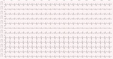
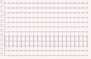
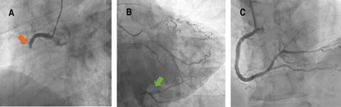
A 57-year-old man with obesity, hypertension and former smoker as cardiovascular risk factors, and previous history of Sjögren’s syndrome and gastroesophageal reflux, consulted his general practitioner for chest pain compatible with angina on moderate exertion. The ECG showed a right bundle branch block (RBBB) not present in previous ECGs (Figures 1 and 2). Treatment with ASA 100 mg od, statin and beta-blocker were started, and the patient was referred preferentially to cardiology for further evaluation. A conventional stress test was performed two weeks later. However it was stopped in the middle of the second stage of the Bruce protocol (6.8 METs and 88% of maximum heart rate) due to angina, without presenting electrocardiographic repolarization changes suggestive of ischemia. Progressive angina persisted despite treatment, so the patient consulted the emergency room four days later due to persistent chest pain at rest. The ECG did not show changes compared to the previous one, however biomarkers of myocardial necrosis were elevated (peak Tn I-hs 5140 pg/mL and CK 411 U/L), so the patient was admitted to the coronary care unit with the diagnosis of non-ST-segment elevation myocardial infarction (NSTEMI). On admission, an echocardiogram showed akinesia of the inferior basal and inferolateral basal segments with preserved parietal thickness, and normal biventricular ejection fraction, without significant valve disease or other notable findings. Antithrombotic therapy was started with ASA 100 mg, clopidogrel 300 mg, fondaparinux 2.5 mg sc, and intravenous nitroglycerin, with slight improvement, but without complete resolution of angina, so an emergent coronary angiography was decided to perform. Acute thrombotic occlusion of the right coronary artery (RCA) was observed, with collateral circulation (CC) from the left coronary tree, which did not present significant stenosis (Figure 3). Angioplasty was performed with implantation of a drug-eluting stent, with subsequent resolution of angina and disappearance of the RBBB. The patient presented a favorable clinical evolution, being discharged on the fourth day of admission.

Figure 1: ECG at initial evaluation shows an RBBB with secondary repolarization abnormalities. 

Figure 2: Previous ECG with narrow QRS complex without repolarization abnormalities. 

Figure 3: Coronary angiography shows A) total occlusion of RCA (orange arrow) with B) good heterocoronary collateral circulation from the left coronary tree (green arrow). C) RCA after stent implantation. 

Discussion

When a patient presents with symptoms suggestive of angina, a 12-lead resting ECG should be performed immediately to assess repolarization changes suggestive of ischemia, especially to identify early persistent ST-segment elevation, which requires emergent reperfusion.1 Characteristic ECG abnormalities in ACS without persistent ST-segment elevation include ST-segment depression, transient ST-segment elevation, and T-wave changes. Even though the ECG in this setting may be normal in more than 30% of patients,2 If the patient has signs or symptoms suggestive of ongoing myocardial ischemia and the standard leads are inconclusive, additional leads should be recorded. The left circumflex artery occlusion may be detected only in posterior leads (V7-V9) or right ventricular MI only in V3R and V4R.3 In case of persistent or recurrent symptoms or diagnostic uncertainty, it is also recommended to obtain additional 12-lead ECGs. In patients with a preexisting left bundle branch block (LBBB), specific ECG criteria (Sgarbossa’s criteria) help detect candidates for immediate coronary angiography.4 Patients with high clinical suspicion of ongoing myocardial ischemia and LBBB, regardless of whether it was previously known, should be treated similar to those with STEMI.1 In patients with RBBB, ST-elevation indicated of STEMI, while ST-segment depression in the leads I, aVL, and V5-6 is indicative of NSTE-ACS.5 Our patient did not strictly meet ECG criteria for persistent ST-segment elevation. However the patient persisted with symptoms of ongoing angina despite treatment and presented with a recent RBBB, so it was decided to perform emergent coronary angiography. Given the good heterocoronary CC observed in the infarcted territory, the infarct size was smaller than expected, as well as the poor electrocardiographic expressiveness. Therefore, it is important to always keep in mind the possibility of a MI with acute occlusion of a coronary artery in the presence of persistent angina despite adequate treatment and few repolarization abnormalities (even with a normal ECG) or new bundle branch block.

Conclusions

Myocardial infarction with acute occlusion of a coronary artery must be kept in mind in patients with persistent angina despite treatment, and the ECG does not show the typical abnormalities.

References

1. Collet JP, Thiele H, Barbato E, Barthélémy O, Bauersachs J, Bhatt DL et al. 2020 ESC Guidelines for the management of acute coronary syndromes in patients presenting without persistent ST-segment elevation. Eur Heart J. 2021; 42 (14): 1289-1367. [ Links ]

2. Mandelzweig L, Battler A, Boyko V, Bueno H, Danchin N, Filippatos G et al. The second Euro Heart Survey on acute coronary syndromes: Characteristics, treatment, and outcome of patients with ACS in Europe and the Mediterranean Basin in 2004. Eur Heart J. 2006; 27 (19): 2285-2293. [ Links ]

3. Thygesen K, Alpert JS, Jaffe AS, Chaitman BR, Bax JJ, Morrow DA et al. Fourth universal definition of myocardial infarction (2018). Circulation. 2018; 138 (20): e618-e651. [ Links ]

4. Smith SW, Dodd KW, Henry TD, Dvorak DM, Pearce LA. Diagnosis of ST-elevation myocardial infarction in the presence of left bundle branch block with the ST-elevation to S-wave ratio in a modified Sgarbossa rule. Ann Emerg Med. 2012; 60 (6): 766-776. [ Links ]

5. Neumann JT, Sörensen NA, Rübsamen N, Ojeda F, Schäfer S, Keller T et al. Right bundle branch block in patients with suspected myocardial infarction. Eur Heart J Acute Cardiovasc Care. 2019; 8 (2): 161-166. [ Links ]

1Funding/support: no financial support was received for this study.

2Conflict of interest: the authors declare no conflict of interest.

Received: February 28, 2022; Accepted: July 26, 2022

*Corresponding author: Albina Aldomà-Balasch. E-mail: albinaab81@hotmail.es

Creative Commons License This is an open-access article distributed under the terms of the Creative Commons Attribution License