


Services on Demand
Journal
Article
 text in
  English (pdf)
 Article in xml format
 Article references
 Send this article by e-mail
Indicators
-
 Cited by SciELO
 -
 Access statistics
Related links
-
 Similars in
SciELO
Similars in
SciELO
Share

 Permalink
Abanico veterinario
On-line version ISSN 2448-6132Print version ISSN 2007-428X
Abanico vet vol.13 Tepic Jan./Dec. 2023 Epub Oct 27, 2023
https://doi.org/10.21929/abavet2023.12
Clinical case
An unusual intravesical blood clot in a dog
*
1
 http://orcid.org/0000-0002-5332-3896
**
1
http://orcid.org/0000-0002-1348-0336
1Facultad de Medicina Veterinaria y Zootecnia, Universidad Veracruzana. Veracruz, México.
In canines, when a mass in the bladder is observed by ultrasonography, a malignant tumor is a presumptive diagnosis. In humans, it is common in the presence of clots due to uncontrolled hematuria. We describe the case of a 7-year-old male mixed breed dog, with a clinical history of hematuria 6 months and presence of ticks. Ultrasound studies of the bladder were performed for one month. The evaluations showed a mass in the bladder trigone, covering 80% of the bladder lumen without acoustic shadow, and then a cystectomy was performed. An oval mass was found with irregular borders and clotted blood appearance, with not adherent to the bladder tissue. The histological diagnosis of the mass showed erythrocytes and polymorphonuclear inflammatory cells: neutrophils and mononuclear cells such as macrophages, mixed with moderate fibrin threads compatible with a bladder clot. Therefore, the diagnosis was a benign mass and not a transitional cell carcinoma as usual. Masses found in the bladder are not necessarily a neoplastic proliferation.
Keywords: cystectomy; hematuria; neoplasic proliferation
En perros, cuando se observa una masa en la vejiga a través del uso de ultrasonografía, el diagnóstico presuntivo es de un tumor maligno. En humanos, es común la presencia de coágulos por causas de hematuria no controlada. El caso clínico se trata de un perro macho de raza mixta de 7 años de edad, con antecedentes clínicos de hematuria 6 meses y presencia de garrapatas. Estudios ecográficos de vejiga se realizaron durante un mes. Los estudios mostraron una masa en el trígono vesical, que cubría el 80 % del lumen vesical sin sombra acústica. Se realizó una cistotomía y se encontró una masa ovalada, con bordes irregulares y aspecto de sangre coagulada, que no estaba adherida al tejido vesical. El diagnóstico histológico mostró que la masa mostraba eritrocitos y células inflamatorias polimorfonucleares: neutrófilos y células mononucleares como macrófagos, mezclados con moderados hilos de fibrina compatibles con un coágulo vesical. Por lo tanto, el diagnóstico fue una masa benigna y no un carcinoma de células transicionales como suele ser habitual. No necesariamente las masas encontradas en vejiga son una proliferación neoplásica.
Palabras clave: cistectomía; hematuria; proliferación neoplásica
INTRODUCTION
Dogs with lower urinary tract disease are often associated predisposing factors like anatomic anomalies, neoplasia, polyps, uroliths, pyelonephritis, prostatitis, ureterocele and urachus remnant, endocrine diseases, and immunosuppression (Visser et al., 2020). Bladder tumours represent 2% of cancer in dogs, and 90% of those tumours are malignant tumours and epithelial tumours. Among primary epithelial neoplasms of the urinary bladder, transitional cell carcinoma represents 75 to 90 percent in dogs (Rasteiro et al., 2022). They can lead to partial or total obstruction of the ureters and urethra, and 50 to 90 percent of them may metastasize into lungs, lymph nodes, kidneys, liver, and prostate, although sometimes also in bones or eyes (Martins-Leal et al., 2012). In human, clot formation in the bladder is common (Xu et al., 2020). Haematuria is the presence of three or more red blood cells in a urinalysis field. It occurs as a result of other abnormalities of the lower urinary tract such as tract infections, urolithiasis, trauma, alterations of the renal parenchyma, neoplasmss urethral lesions, prostatic hyperplasia treatments, bladder tumors, nephroliths and cystitis secondary to radiation treatment (Moloney et al., 2014). Dogs present clinical findings as dysuria, haematuria, stranguries, urinary frequency and incontinence. In case of obstruction of the urinary tract, acute kidney injury can develop, and the patient may suffer anorexia and recurrent vomit, with high serum levels of creatinine, urea, phosphorus and CK activity (Dunaevich et al., 2020). The diagnosis is based on physical examinations findings, clinical signs, and diagnostics tests such as urinalysis and bacteriological urine culture. To reduce the risk of neoplastic seeding urine may be obtained via voiding or catheterization rather than cystocentesis (Fulkerson & Knapp, 2015). An ultrasound scanning, a cytology performed by cystoscopy and/or urine sample, cystotomy for excisional biopsy or the histopathologic analysis of the sample, may help to rule out the presence of a bladder neoplasia (Harriman et al., 2016). There is a wide discussion about diagnosis when bladder masses are observed by ultrasound. Commonly, the presence of a malignant neoplasia is an unfavourable prognosis to the patient. In veterinary medicine, histopathologic analysis before deciding on the next stage of treatment is essential. In this study, we report a clinical case of a dog with the presence of recurrent haematuria with the formation of a bladder mass that does not necessarily have a neoplastic origin.
Case Report
A 7-year-old male mixed breed dog was clinical examination. The owner reported haematuria, oliguria, and dysuria and ticks a few months ago. A clinical examination and abdominal ultrasound were developed. Findings revealed a hyperechogenic structure, surrounding inside the bladder, with not presence of acoustic shadow in the bladder trigone. The size of the mass occupied 80% of the bladder lumen. There was no mobility of the mass at the time of fanning with ultrasound (Figure 1). Complete blood count, serum biochemistry and urine specific gravity were performed (Table 1).

Table. 1 Complete blood count, serum biochemistry and urine specific gravity
| Value | Reference Interval1 | |
|---|---|---|
| Red blood cells, x106/µL | 3.6 | 3.3 - 7.8 |
| Haemoglobin, g/dL | 70.2 | 120 - 180 |
| Haematocrit, % | 21.4 | 37.0 - 55.0 |
| White Blood Cells, x103/µL | 6.1 | 6.0 - 17.0 |
| Platelets, x103/µL | 162.0 | 200 - 500 |
| Blood urea nitrogen, mg/dL | 40.77 | 10 - 24 |
| Creatinine, mg/dL | 2.03 | 0.7 - 1.2 |
| Urine specific gravity | 1.050 | 1.001 - 1.060 |
The hematologic profile of the patient revealed hypochromic normocytic anaemia, with thrombocytopenia. Significant serum biochemistry findings and urine specific gravity were post-renal azotemia, matching with acute kidney injury. Peripherial blood smear showed morulae in the lymphocytes and confirmed Ehrlichia infection. Prior to surgery, dog was stabilized with doxycycline (10 mg/kg BID twice a daily for 4 weeks). A cystectomy was developed to explore and extract the mass located in the bladder lumen. A pre-anaesthetic protocol was used, that consisted of buprenorphine 0.02 mg/kg IV, meloxicam 0.2 mg/kg IV, ketamine 3 mg/kg IV, xylazine 2 mg/kg IV, propofol 3 mg/kg IV for induction.
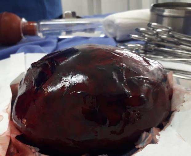
Anaesthesia was maintained with isoflurane at 2% through surgery. The bladder was incised in the medial part and an oval mass of 25 cm of diameter was observed, with irregular edges, not adhered to the bladder tissue and with coagulated blood appearance (Figure 2).
The mass was removed uneventfully, kept in formalin at 10% and subsequently send to histopathologic examination. The microscopic report revealed erythrocytes and polymorphonuclear inflammatory cells: neutrophils and mononuclear cells as macrophages, mixed with fibrin strands to a moderate degree compatible with a blood clot in the bladder lumen (Figure 3).
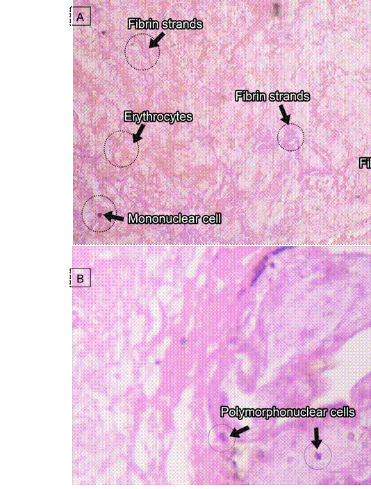
Figure 3 Histopathologic findings of mass removed from the bladder. A: erythrocytes and mononuclear cells as macrophages, mixed with fibrin strands to a moderate degree. Hematoxylin and Eosin Stain, 10x. B: erythrocytes and polymorphonuclear inflammatory cells: neutrophils mononuclear cells as macrophages. Hematoxylin and Eosin Stain, 20x.
The patient returned for removal of stitches from the surgical wound 10 d later. There were no findings of hematuria. Doxycycline treatment continued for 30 d, as well as pain management with tramadol 3 mg/kg for 8 d and meloxicam 0.1 mg/kg for 4 d. Dog came back for check-up six months later with no pathological findings by ultrasound evaluation. The urinalysis reported no apparent pathologic changes. Blood chemistry analysis showed normal concentrations of creatinine, BUN, and urea. There were no evidences of post-renal azotemia. Doctors in Veterinary Medicine specialists in animal surgery performed the surgery and procedures in compliance with the animal regulations enacted by Mexican laws. Clinical case took place in The Small Animal Hospital - Facultad de Medicina Veterinaria y Zootecnia - Universidad Veracruzana, localized in eastern Mexico at 19°11’ N and 96°08’ W.
DISCUSSION
The diagnosis of a bladder lumen blood clot was established with the histopathologic examination report. The foregoing describes that it may be related to sterile haemorrhagic cystitis that occurs in animals treated for neoplastic/immunological diseases such as the use of antineoplastic drugs like cyclophosphamide for example, that causes ulcers of the mucosa, haemorrhage and edema. The clot also can be secondary to the presence of urolithiasis or transitional cell carcinoma (Nunura & Navarro, 2004). Our patient did not had previous neoplastic treatment or findings of urolithiasis, crystals and neoplastic cells, but there was a suspicion of rickettsia disease. Upon surgical inspection, the tissues were homogeneous and without the presence of masses inside the bladder. Childress et al. (2011) indicated that the use of cystography is useful for the detection of a bladder mass process. The abdominal ultrasound developed in our patient was useful to decide for an exploratory cystectomy. Tumours found in the bladder are generally reported to be 90% malignant (Rasteiro et al., 2022), however the patient was within the other 10%, improving the patient's prognosis.
Meloxicam was used as premedication despite acute kidney injury in the patient. De Santis et al. (2022) indicated that non-steroidal analgesics could be used in patients with not chronic renal disease. Our patient had a urinary density of 1.050 suggesting a postrenal azotemia because of a clot obstructing the urine outflow. Zhao et al. (2022), in mice with induced acute kidney injury, found that meloxicam inhibited inflammatory cytokines and cell apoptosis in the kidney.
The clot in the bladder lumen of the dog could induced by the rickettsial disease because of anaemia, thrombocytopenia and coagulation disorders (Chochlios et al., 2019; Rodríguez-Alarcón et al., 2020). Rickettsial infection in dog-associated tucks in tropical México is common (Boria-Gamboa et al., 2019).
It is concluded that not all the masses observed in the bladder lumen of dogs are uroliths or neoplasms of malignant origin. Patients with haematuria, dysuria, and oliguria, it is necessary to develop cabinet studies for a successful medical and surgical treatment, and a good outcome for the patient's well-being.
REFERENCES
Boria-Gamboa BP, Villagómez-Cortes JA, Martínez-Herrera D. 2019. Clinical Evidence of lyme disease in dogs and disease awareness among students and veterinarians in Veracruz, Mexico. Veterinary Science Research. 1(2):10-17. ISSN: 2661-3867. https://doi.org/10.30564/vsr.v1i2.1280 [ Links ]
Childress MO, Adams LG, Ramos-Vara JA, Freeman LJ, He S, Constable PD, Knapp DW. 2011. Results of biopsy via transurethral cystoscopy and cystotomy for diagnosis of transitional cell carcinoma of the urinary bladder and urethra in dogs: 92 cases (20032008). Journal of the American Veterinary Medical Association. 239(3):350-356. ISSN:0003-1488. https://doi.org/10.2460/javma.239.3.350 [ Links ]
Chochlios TA, Angelidou E, Kritsepi-Konstantinou M, Koutinas CK, Mylonakis ME. 2019. Seroprevalence and risk factors associated with Ehrlichia canis in a hospital canine population. Veterinary Clinical Pathology. 48(2):305-309. ISSN:1939-165X. https://doi.org/10.1111/vcp.12736 [ Links ]
De Santis F, Boari A, Dondi F, Crisi PE. 2022. Drug-dosing adjustment in dogs and cats with chronic kidney disease. Animals. 12(3):262. ISSN: 2076-2615. https://doi.org/10.3390/ani12030262 [ Links ]
Dunaevich A, Chen H, Musseri D, Kuzi S, Mazaki-Tovi M, Aroch I, Segev G. 2020. Acute on chronic kidney disease in dogs: Etiology, clinical and clinicopathologic findings, prognostic markers, and survival. Journal of Veterinary Internal Medicine. 34(6):2507-2515. ISSN:1939-1676. https://doi.org/10.1111/jvim.15931 [ Links ]
Fulkerson CM, Knapp DW. 2015. Management of transitional cell carcinoma of the urinary bladder in dogs: a review. The Veterinary Journal. 205(2):217-225. ISSN:15322971. http://doi.org/10.1016/j.tvjl.2015.01.017 [ Links ]
Harriman GM, Popovitch C, MacLeod A. 2016. Ultrasound evaluation of the urinary bladder in dogs following cystotomy with full thickness suture placement. Journal of American Animal Hospital Association. 52(4):212-219. ISSN:1547-3317. http://doi.org/10.5326/JAAHA-MS-6381 [ Links ]
Martins-Leal L, Fernandes-Machado MR, Bignotto-Ferreira F, Didoné EM, Bruno de Souza I. 2012. Transitional cell carcinoma in dogs: a report of two cases, mother and daughter. Revista de Investigaciones Veterinarias del Perú. 23(4):516-522. ISSN:16823419. http://doi.org/10.15381/rivep.v23i4.973 [ Links ]
Moloney F , Murphy KP, Twomey M, O'Connor OJ, Maher MM. 2014. Haematuria: An Imaging Guide. Advances in Urology. ISSN: 16876377, 16876369. https://doi.org/10.1155/2014/414125 [ Links ]
Nunura J, Navarro J. 2004. Hemorrhagic cystitis in bone marrow transplant recipients: First report of cases in our country. Anales de la Facultad de Medicina. 65(2):138-146. ISSN:1025-5583 http://dev.scielo.org.pe/pdf/afm/v65n2/a09v65n2.pdf [ Links ]
Nuñez-Ochoa L, Bouda J. 2007. Patología Clínica Veterinaria. Universidad Nacional Autonoma de México, Ciudad de México. ISBN:9703245501. Pp. 233. [ Links ]
Rasteiro AM, Sá E Lemos E, Oliveira PA, Gil da Costa RM. 2022. Molecular markers in urinary bladder cancer: applications for diagnosis, prognosis and therapy. Veterinary Science. 28(9):107. ISSN:2306-7381. http://doi.org/10.3390/vetsci9030107 [ Links ]
Rodríguez-Alarcón CA, Beristain-Ruiz DM, Olivares-Muñoz A, Quezada-Casasola A, Pérez-Casio F, Álvarez-Martínez JA, Tapia-Alanía J, Lira-Amaya JJ, Rivera-Barreno R, Cera-Hurtado OS, Ibancovichi-Camarillo JA, SoonGómez L, Adame-Gallegos JR, Figueroa-Millan JV. 2020. Demonstrating the presence of Ehrlichia canis DNA from different tissues of dogs with suspected subclinical ehrlichiosis. Parasites & Vectors. 13(1):518. ISSN:1756-3305. http://doi.org/10.1186/s13071-020-04363-0 [ Links ]
Visser J, Kummeling A, van Nugteren MA, Grinwis GCM, Brocks BAW. 2020. Resection of urachal anomalies in dogs with recurrent lower urinary tract disease. Veterinary Surgery. 49(1):214-221. ISSN:1532-950X. https://doi.org/10.1111/vsu.13311 [ Links ]
Xu M, Jin L, Shan Y, Zhu J, Xue B. 2020. A simple and effective method for bladder blood clot evacuation using hydrogen peroxide. The Journal of International Medical Research. 48(5). ISSN: 13412051. https://doi.org/10.1177/0300060520924546 [ Links ]
Zhao S, Cui L, Zheng X, Ji Y, Yu C. 2022. Meloxicam alleviates sepsis-induced kidney injury by suppression of inflammation and apoptosis via upregulating GPNMB. Applied Bionics and Biomechanics. e1790104. ISSN: 11762322. https://doi.org/10.1155/2022/1790104 [ Links ]
Received: January 09, 2023; Accepted: May 18, 2023
 Este es un artículo publicado en acceso abierto bajo una licencia Creative Commons
