


Services on Demand
Journal
Article
 text in
  English (pdf)
 Article in xml format
 Article references
 Send this article by e-mail
Indicators
-
 Cited by SciELO
 -
 Access statistics
Related links
-
 Similars in
SciELO
Similars in
SciELO
Share

 Permalink
Abanico veterinario
On-line version ISSN 2448-6132Print version ISSN 2007-428X
Abanico vet vol.11 Tepic Jan./Dec. 2021 Epub Apr 04, 2022
https://doi.org/10.21929/abavet2021.44
Clinical case
Surgical management and immunotherapy of Pythium insidiosum in a horse: case report
1
*
 http://orcid.org/0000-0002-8143-0269
1
http://orcid.org/0000-0002-2767-7527
1
http://orcid.org/0000-0001-8762-3988
1Facultad de Medicina Veterinaria y Zootecnia, Universidad Veracruzana. Veracruz, Ver., México.
2 Instituto de Investigaciones Agropecuarias y Forestales-Universidad Michoacana de San Nicolás de Hidalgo. Hidalgo, México.
Equine cutaneous pythiosis is caused by the oomycete Pythium insidiosum, which generally produces rapidly progressive granulomatous ulcerative lesions on the distal parts of limbs and abdomen. It is a globally distributed disease with higher prevalence in tropical and subtropical regions. The objective of this case report was to describe granulomatous lesions with necrotic tissue and fibrin bloody exudate in the palpebral region of the left eye caused by Pythium insidiosum in a quarter horse mare in Veracruz State, Mexico. The diagnosis was based on the clinical characteristics of the lesion and histopathology. Due to the extent of lesions, surgical resection was performed in combination with immunotherapy. Treatment with immunotherapy proved to be an effective alternative to achieve complete resolution of skin lesions.
Keywords: Pythium insidiosum; pythiosis; equine; immunotherapy
La pitiosis cutánea equina es causada por el oomiceto Pythium insidiosum, el cual generalmente produce lesiones ulcerativas granulomatosas de progresión rápida en las partes distales de las extremidades y del abdomen. Es una enfermedad distribuida globalmente y con mayor prevalencia en regiones tropicales y subtropicales. El objetivo de este reporte de caso fue describir las lesiones granulomatosas, con tejido necrótico y exudado fibrinosanguinolento en la región palpebral del ojo izquierdo ocasionadas por Pythium insidiosum en una yegua cuarto de milla en el estado de Veracruz, México. El diagnóstico se fundamentó en las características clínicas de la lesión y la histopatología. Debido a la extensión de las lesiones, se realizó la resección quirúrgica en combinación con la inmunoterapia. El tratamiento con la inmunoterapia demostró ser una alternativa eficaz para lograr la resolución completa de las lesiones cutáneas.
Palabras clave: Pythium insidiosum; pitiosis; equino; inmunoterapia
INTRODUCTION
The etiological agent Pythium insidiosum (P. insidiosum) belongs to the class Oomycetes, whose zoospores lack chitin and ergosterol in their cellular composition, and causes a medical condition known as equine cutaneous pythiosis, which occurs in tropical and subtropical regions (Gaastra et al., 2010, Santos et al., 2011a, Cardona et al., 2013).
Equine is the species with the most reported cases, although there are also reports in canids (intestinal form), felines, wildlife, cattle, sheep (Loreto et al., 2014 and humans (He et al., 2016, Chitasombat et al., 2020). Skin lesions are more frequent on the distal parts of the limbs, ventral and lateral abdomen, pectoral region and mouth, perhaps because of the greater contact of these areas with potentially zoospore-contaminated water. Lesions are granulomatous ulcerative, with irregular borders, fistulous tracts or cavitations and with the presence of hard necrotic masses that detach easily (Atiba et al., 2020, Souto et al., 2021).
Diagnosis is based primarily on observation of the clinical features of the skin lesions and histopathology (Schanzembach et al., 2019). Surgical resection of all affected tissue, in combination with an immunotherapeutic containing P. insidiosum antigens, is one of the most effective treatments currently available (Mendoza et al., 2003; Mendoza et al., 2004).
This report describes the clinical characteristics of a case of pythiosis in an equine, with lesions focused in the palpebral region, its histopathological diagnosis and its treatment by immunotherapy.
CASE DESCRIPTION
An 11-year-old Quarter Horse mare was referred to the outpatient service of the Veterinary Hospital for Large Species of the Faculty of Veterinary Medicine and Zootechnics of the Veracruzan University, for evaluation of a fast-growing granulomatous lesion that did not respond to treatment.
During the medical evaluation, it was observed that the mare presented a granulomatous ulcerative lesion with necrotic tissue and fibrin bloody exudate in the palpebral region of the left eye, measuring approximately 20 cm x 16 cm (Figure 1). During palpation of the affected area, the presence of fistulous tracts and yellowish-white caseified exudate was evident. The mare presented intense pruritus, pain in the affected region, pale mucous membranes and progressive weight loss. In the blood cytometry the relevant findings were anemia and leukocytosis due to neutrophilia.

Figure 1 Granulomatous ulcerative lesion with necrotic tissue and bloody fibrin exudate in the palpebral region of the left eye
Tissue samples were taken and preserved in 10% formalin for subsequent histopathological analysis. Histopathological evaluation showed an intense infiltrate of abundant eosinophils, reactive mast cells, presence of areas of multifocal necrosis and Splendore-Hoeppli phenomenon (Figure 2); likewise, the presence of occasionally septate, branched and smooth-walled hyphae coinciding with P. insidiousum was identified (Figure 3).
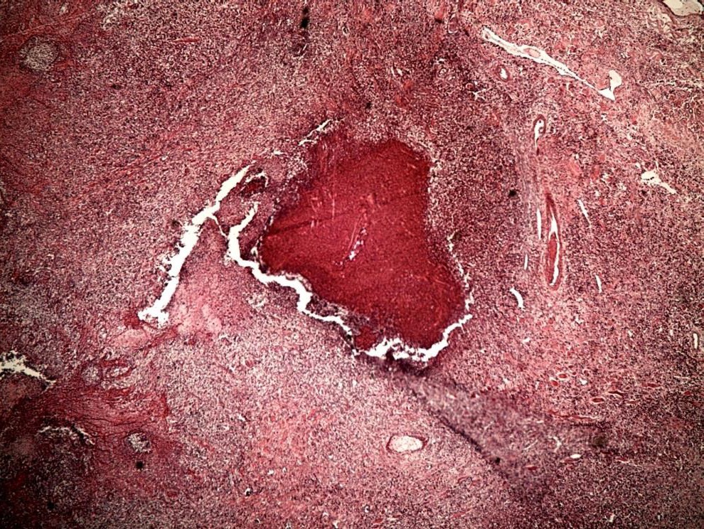

Figure 3 Branched and moderately septate hyphae of Pythium insidiosum (hematoxylin and eosin stain, 100X)
After clinical evaluation and histopathological findings, the affected tissue was removed by surgical resection with the mare on station. The mare was fasted for 12 hours prior to the surgical procedure, a 14 G catheter was placed in the jugular vein, and gentamicin (6.6 mg/kg IV), meglumine flunixin (1.1 mg/kg IV) and 22 000 IU/kg procaine penicillin were administered intramuscularly.
The mare was kept sedated with an initial bolus of 0.01 mg/kg detomidine hydrochloride and maintenance with a constant infusion of the same product, at a total dose of 10 mg in 250 mL of saline solution (0.04 mg/mL), and regional blocks were performed with 2% lidocaine hydrochloride. The affected area was prepared with aseptic technique with 8% povidone iodine and sterile water. During the surgical procedure it was possible to remove all the granulomatous tissue and concretions ("kunkers") and enucleation of the eyeball was avoided.
Due to the extent of the lesions and the risk of hyphae in deep tissues, it was decided to use a commercial immunotherapeutic developed from hyphal proteins and metabolites of P. insidiosum. The treatment consisted of applying 1 mL subcutaneously in the distal third of the neck on days 0 (day of surgery), 7 and 21 after the surgical procedure, according to the manufacturer's specifications (Pan American Veterinary Laboratories, Lexington, Texas®). After inoculation of the immunotherapeutic, no side effects were observed in the area of application.
Post-surgical treatment consisted of a compressive bandage, with a cross pattern, so as not to interfere with the field of vision of the contralateral eye and wound cleaning was carried out using isotonic solution every 24 hours. At the same time, she was maintained on gentamicin (6.6 mg/kg IV), procaine penicillin (22,000 IU/kg IM) and meglumine flunixin (1.1 mg/kg IV) for five days. During the postoperative period, the wound proliferation phase was monitored to ensure adequate granulation tissue formation and appropriate epithelialization and tissue remodeling.
DISCUSSION
Most of the cases reported in equines are in South America (Salas et al., 2012, Cardona et al., 2014, Dória et al., 2015, Schanzembach et al., 2019, Paz et al., 2021) which is related to its tropical and subtropical regions. Veracruz state presents similar environmental conditions, for example, abundant rainfall that favors water stagnation in pastures and high temperatures, which benefit the etiological agent and its contact with equines (Gaastra et al., 2010).
The skin lesions found in the periorbital region of the mare agree with the description of granulomatous skin lesions described in other case reports in different countries (Bezerra- Junior et al., 2010, Mosbah et al., 2012, Cardona et al., 2014, Tartor et al., 2020), where cutaneous lesions are reported in anatomical regions in direct contact with stagnant water contaminated with zoospores of P. insidiosum, so lesions in high regions of the head are not common. However, the first case with pythiosis lesions within the nasal cavity of a pregnant mare and antibodies for P. insidisum in three horses was reported in Thailand (Tonpitak et al., 2018, Mar Htun et al., 2021).
The diagnosis of pythiosis is primarily based on the observation of the clinical features of skin lesions. Similarly, it is important to establish the differential diagnoses of these lesions, as they are often misdiagnosed as cutaneous habronemiasis, exuberant granulation tissue, or sarcoids, so it is important to perform histopathological studies for definitive diagnosis.
The histopathological findings of this case agree with those described by Márquez et al. (2010), where the Splendore-Hoeppli phenomenon and the identification of septate hyphae could be observed. This phenomenon is considered an adaptive strategy of the etiological agent with the aim of ensuring its proliferation and survival in the host tissue, which can be confirmed by the presence of viable hyphae within the eosinophilic reaction (Martins et al., 2012).
The treatment of choice for P. insidiosum remains surgical resection of all affected tissue. However, some studies have shown that the combination of surgical resection and systemic antibiotics such as amphotericin B and miconazole, administered via regional perfusion in the affected extremities, obtained a resolution of 92% (Worster at al., 2000, Dória et al., 2012). In contrast, Pires et al. (2013) only obtained 60% resolution of pythiosis skin lesions with amphotericin B. This type of combination therapy is performed when the lesion has invaded other types of anatomical structures, such as muscles, tendons and bones, which cannot be surgically removed.
In case surgical resection is partial due to the depth of the lesion, immunotherapy based on proteins from P. insidiosum hyphae can be used, which has had an efficacy of 90% in cases of acute lesions of less than 60 days and 20% in chronic cases that exceed two months (Mendoza et al., 2003, Mendoza et al., 2005). The immunological response was similar to that reported by Pereira et al. (2011), where seven days after inoculation of the first dose of the immunotherapeutic, the mare no longer showed evidence of P. insidiosum lesions or pruritus and began the wound remodeling phase (Figure 4).

Figure 4 Initial phase, proliferation and remodeling. (a) Initial post-surgical lesion; (b-f) images show the proliferation and remodeling phases. At the end of the treatment only a scar lesion is seen with complete resolution of the case.
The success of immunotherapy is based on the fact that it promotes a shift from an antibody-mediated to a cell-mediated response. Normally, a type 2 (Th2) helper lymphocyte-regulated response predominates, with release of interleukins (IL) 4 and 5 and activation of eosinophils and mast cells, resulting in eosinophilic inflammation. In contrast, immunotherapy favors a response regulated by type 1 helper lymphocytes (Th1), with release of IL-2 and IFN-y and activation of T lymphocytes and macrophages, which destroy P. insidiosum (Loreto et al., 2014). Despite the curative properties of immunotherapy, Santos et al. (2011b) reported that the antibodies produced are probably not sufficient to prevent reinfection in the short and long term.
REFERENCES
Atiba A, Ghazy A, Hamad MH. 2020. Evaluating the efficacy of surgical excision and topical dimethyl sulphoxide (DMSO) in the treatment of equine cutaneous pythiosis. Iranian Journal of Veterinary Research. 21(4):301-307. ISSN: 2252-0589 PMC7871735 https://www.ncbi.nlm.nih.gov/pmc/articles/PMC7871735/ [ Links ]
Bezerra-Junior P, Pedroso P, Pavarini S, Dalto A, Santurio J, Driemeier D. 2010. Equine intestinal pythiosis in Southern Brazil. Arquivo Brasileiro de Medicina Veterinária e Zootecnia. 62(2):481-483. ISSN: 1678-4162. https://doi.org/10.1590/S0102-09352010000200031 [ Links ]
Cardona JA, Vargas-Viloria M, Perdomo S. 2013. Pitiose cutânea em equinos: uma revisão. Revista CES Medicina Veterinaria y Zootecnia. 8(1):58-67. ISSN: 1900-9607. http://www.redalyc.org/articulo.oa?id=321428109005 [ Links ]
Cardona JA, Vargas-Viloria M, Perdomo S. 2014. Frecuencia de pythiosis cutánea en caballos de producción en explotaciones ganaderas de córdoba, Colombia. Revista de la Facultad de Medicina Veterinaria y de Zootecnia. 61(1):31-43. ISSN: 0120-2952. http://dx.doi.org/10.15446/rfmvz.v61n1.43882 [ Links ]
Chitasombat MN, Jongkhajornpong P, Lekhanont K, Krajaejun T. 2020. Recent update in diagnosis and treatment of human pythiosis. Peer J. 8:e8555. ISSN: 2167-8359. https://doi.org/10.7717/peerj.8555 [ Links ]
Dória RG, Carvalho MB, Freitas SH, Laskoski LM, Colodel EM, Mendonça FS, Silva M A, Grigoletto R, Fantinato NP. 2015. Evaluation of intravenous regional perfusion with amphotericin B and dimethylsulfoxide to treat horses for pythiosis of a limb. BMC Veterinary Research. 11:152. ISSN: 1746:6148. https://doi.org/10.1186/s12917-015-0472-z [ Links ]
Gaastra W, Lipman LJ, De Cock AW, Exel TK, Pegge RB, Scheurwater J, Vilela R, Mendoza L. 2010. Pythium insidiosum: an overview. Veterinary Microbiology. 146(1-2):1-16. ISSN: 1873-2542. https://doi.org/10.1016/j.vetmic.2010.07.019 [ Links ]
He H, Liu H, Chen X, Wu J, He M, Zhong X. 2016. Diagnosis and Treatment of Pythium Insidiosum Corneal Ulcer in a Chinese Child: A Case Report and Literature Review. The American Journal of Case Reports. 17:982-988. ISSN: 1941-5923. https://doi.org/10.12659/ajcr.901158 [ Links ]
Loreto ÉS, Tondolo JSM, Zanette RA, Alves SH, Santurio JM. 2014. Update on pythiosis immunobiology and immunotherapy. World Journal of Immunology. 4(2):88-97. ISSN: 2219-2824. http://dx.doi.org/10.5411/wji.v4.i2.88 [ Links ]
Mar Htun Z, Laikul A, Pathomsakulwong W, Yurayart C, Lohnoo T, Yingyong W, Kumsang Y, Payattikul P, Sae-Chew P, Rujirawat T, Jaturapaktrarak C, Chongtrakool P, Krajaejun T. 2021. An initial survey of 150 horses from Thailand for anti-Pythium insidiosum antibodies. Journal de Mycologie Medicale. 31(1): 101085. ISSN: 1773-0449. https://doi.org/10.1016/j.mycmed.2020.101085 [ Links ]
Márquez A, Salas Y, Canelón J, Perazzo Y, Colme-Nárez V. 2010. Descripción anatomopatológica de pitiosis cutánea en equinos. Revista de la Facultad de Ciencias Veterinarias UCV. 51(1):7-42. ISSN: 0258-6576. https://www.redalyc.org/articulo.oa?id=373139075005 [ Links ]
Martins TB, Kommers, GD, Trost ME, Inkelmann MA, Fighera RA, Schild AL. 2012. A comparative study of the histopathology and immunohistochemistry of pythiosis in horses, dogs and cattle. Journal of Comparative Pathology. 146(2-3):122-131. ISSN: 1532-3129.https://doi.org/10.1016/j.jcpa.2011.06.006 [ Links ]
Mendoza L, Newton JC. 2005. Immunology and immunotherapy of the infections caused by Pythium insidiosum. Medical Mycology. 43(6):477-486. ISSN: 1460-2709. https://doi.org/10.1080/13693780500279882 [ Links ]
Mendoza L , Mandy W, Glass R. 2003. An improved Pythium insidiosum-vaccine formulation with enhanced immunotherapeutic properties in horses and dogs with pythiosis. Vaccine. 21(21-22):2797-2804. ISSN:1873-2518.https://doi.org/10.1016/s0264-410x(03)00225-1 [ Links ]
Mendoza L , Prasla SH, Ajello L. 2004. Orbital pythiosis: a non-fungal disease mimicking orbital mycotic infections, with a retrospective review of the literature. Mycoses. 47(1-2):14-23. ISSN: 1439-0507 https://doi.org/10.1046/j.1439-0507.2003.00950.x [ Links ]
Mosbah E, Karrouf G, Younis E, Saad H, Ahdy A, Zaghloul A. 2012. Diagnosis and surgical management of pythiosis in draft horses: Re-port of 33 cases in Egypt. Journal of Equine Veterinary Science. 32(3):164-169. ISSN: 0737-0806. https://doi.org/10.1016/j.jevs.2011.08.014 [ Links ]
Paz G, Camargo GG, Cury JE, Apolonio E, Garces HG, Prado A, Chechi JL, Oliveira A L, Watanabe MJ, Bagagli E, Bosco S. 2021. Outbreak of equine pythiosis in a southeastern region of Brazil: Environmental isolation and phylogeny. Transboundary and Emerging Diseases. 10.1111/tbed.14135. ISSN: 1865-1682. https://doi.org/10.1111/tbed.14135 [ Links ]
Pereira C, Soares R, Santurio J, Marques L. 2011 Eficácia da imunoterapia no tratamento de pitiose facial em equino. Acta Scientiae Veterinariae. 39(1):955. ISSN: 1679-9216. http://www.ufrgs.br/actavet/39-1/PUB%20955.pdf [ Links ]
Pires L, Bosco S, da Silva NF, Jr , Kurachi C. 2013. Photodynamic therapy for pythiosis. Veterinary Dermatology. 24(1): 130-6.e30. ISSN: 1365-3164. https://doi.org/10.1111/j.1365-3164.2012.01112.x [ Links ]
Salas, Y, Márquez A, Canelón J, Perazzo Y, Colmenárez V, López JA. 2012. Equine pythiosis: report in crossed bred (Criole Venezuelan) horses. Mycopathologia. 174(5-6):511-517. ISSN: 1573-0832. https://doi.org/10.1007/s11046-012-9562-7 [ Links ]
Santos C, Santurio J, Marques C. 2011a. Pitiose em animais de produção no Pantanal Mato-grossense. Pesquisa Veterinária Brasileira. 31(12):1083-89. ISSN:1678-5150. https://doi.org/10.1590/S0100-736X2011001200008 [ Links ]
Santos CE, Marques LC, Zanette RA, Jesus FP, Santurio JM. 2011b. Does immunotherapy protect equines from reinfection by the oomycete Pythium insidiosum? Clinical and Vaccine Immunology. 18(8):1397-1399. ISSN: 1556-679X. https://doi.org/10.1128/CVI.05150-11 [ Links ]
Schanzembach M, Brayer D, Sallis S,César D, Matto C, Almeida R, Nan F, Rodríguez V, Parodi P, Pereira M, Gianneechini R, Rivero R. 2019. Descripción de un caso de pitiosis cutánea equina y su diagnóstico mediante diversas técnicas. Veterinaria (Montevideo). 55(212):96-101. ISSN: 0376-4362. http://dx.doi.org/10.29155/vet.55.212.8. [ Links ]
Souto EPF, Maia LA, Neto EMN, Kommers GD, Junior FG, Riet-Correa F, Galiza GJN, Dantas AFM. 2021. Pythiosis in equidae in Northeastern Brazil: 1985-2020. Journal of Equine Veterinary Science. 105. 103726. ISSN: 0737-0806. https://doi.org/10.1016/j.jevs.2021.103726 [ Links ]
Tartor YH, Hamad MH, Abouzeid NZ, El-Belkemy FA. 2020. Equine pythiosis in Egypt: clinicopathological findings, detection, identification and genotyping of Pythium insidiosum. Veterinary Dermatology. 31(4):298-e73. ISSN:1365-3164. https://doi.org/10.1111/vde.12845 [ Links ]
Tonpitak W, Pathomsakulwong W, Sornklien C, Krajaejun T, Wutthiwithayaphong S. 2018. First confirmed case of nasal pythiosis in a horse in Thailand. JMM Case Reports. 5(1) e005136. ISSN: 2053-3721. https://doi.org/10.1099/jmmcr.0.005136 [ Links ]
Worster AA, Lillich JD, Cox JH, Rush BR. 2000. Pythiosis with bone lesions in a pregnant mare. Journal of the American Veterinary Medical Association. 216(11):1795-1760. ISSN: 0003-1488. https://doi.org/10.2460/javma.2000.216.1795 [ Links ]
Received: September 11, 2021; Accepted: December 07, 2021
 Este es un artículo publicado en acceso abierto bajo una licencia Creative Commons
