


Services on Demand
Journal
Article
 text in
  English (pdf)
 Article in xml format
 Article references
 Send this article by e-mail
Indicators
-
 Cited by SciELO
 -
 Access statistics
Related links
-
 Similars in
SciELO
Similars in
SciELO
Share

 Permalink
Abanico veterinario
On-line version ISSN 2448-6132Print version ISSN 2007-428X
Abanico vet vol.8 n.1 Tepic Jan./Apr. 2018
https://doi.org/10.21929/abavet2018.81.5
Original Article
Experimental infection of Gnathostoma binucleatum in Canis familiaris from the municipality of Tepic in Nayarit, Mexico
1Secretaría de Investigación y Posgrado. Universidad Autónoma de Nayarit. México.
2Unidad Académica de Medicina Veterinaria y Zootecnia, Universidad Autónoma de Nayarit. Posgrado en Ciencias Biológicas Agropecuarias y Pesqueras, universidad Autónoma de Nayarit.
Gnathostoma binucleatum is the etiological agent of animal and human gnathostomiasis in the Nayarit state. In four infected female dogs, parasite phases were found in the stomach. Only one female dog eliminated eggs and adult parasite phases in feces. In this dog, the prepatent period lasted 22 weeks and the patency period 14 weeks. Necropsy results showed a copiously vascularized 10-cm diameter fibrous nodule lodged in the greater curvature of the stomach. Two female dogs that did not eliminate any eggs showed 1-2 cm diameter nodules on the gastric wall with 5 juvenile phases in each. One female dog that did not eliminate any eggs and exhibited no gastric nodules showed juvenile parasites on the gastric wall. New data on the pathological and parasitological aspects of canine gnathostomiasis are presented.
Key words: Gnathostoma binucleatum; nodules; infected dogs
Gnathostoma binucleatum es el agente etiológico de la gnathostomiasis animal y humana en el estado de Nayarit. En cuatro perras infectadas se encontraron huevos y parásitos adultos en heces. El período de prepatencia fue de 22 semanas y el de patencia 14 semanas. Los resultados de la necropsia demostraron un nódulo vascularizado de 18 cm de diámetro localizado en la curvatura mayor del estómago. Dos perras que no eliminaron huevos presentaron nódulos de 1-2 cm de diámetro sobre la pared gástrica, recuperándose cinco parásitos juveniles en cada uno. En una perra que no eliminó huevos ni presentó nódulos gástricos se observaron cinco parásitos juveniles. Los resultados confirman a los perros como hospederos definitivos de este parásito. Se presentan datos nuevos sobre los aspectos patológicos y parasitológicos de la gnathostomiasis canina.
Palabras clave: Gnathostoma binucleatum; nódulos; perros infectados
Introduction
Gnathostoma binucleatum is the only confirmed species in the American Continent, which causes animal and human gnathostomiasis. This disease represents a serious public health problem in the state of Nayarit, Mexico. Between 1995 and 2005, 6328 cases were reported in Nayarit (SUAVE, 2005).
The earliest intermediate hosts of all identified Gnathostoma species are usually copepods; the stuaring fishes (Cathorops fuerthi, Dormitator latifrons, Pomadasys macrachanthus, Mugil curema) , and a few other species of turtles (Kinosternon integrum and Trachemys scripta), which act as second intermediate and paratenic hosts respectively (Álvarez Guerrero y Lamothe Argumedo 2000; León-Regagnon et. al. 2002; Álvarez Guerrero y Alba Hurtado 2007). The role of carnivorous mammals (ichthyophages) as definitive hosts in the biological cycle of Gnathostoma sp species is widely documented. Adult parasites of G. binucleatum were found in gastric nodules of naturally infected cats and ocelots (Almeyda Artigas et al., 1991), as well as in dogs and cats experimentally by Koga et al. (1999) and Álvarez-Guerrero et al, (2014). However, the effect caused by the parasites on their hosts has not been well studied.
The present study aims to experimentally infect dogs with G. binucleatum larvae to describe the pathological alterations caused by parasites in organs and tissues.
Material and Methods
For the experimental study, four clinically healthy female dogs from 2 to 4 months of age of undefined race were selected. At the arrival of the animals, coproparasitoscopic (cps) analyzes of Faust were performed to identify helminth eggs that could alter the study. Positive animals were treated with a formula of praziquantel, pyrantel pamoate and febantel (Drontal plus de Bayer). The ectoparasites were removed with sprays of 2-methylethoxyphenyl carbamate (Bolfo Bayer) and they were immunized against distemper and parvovirus. The animals were kept in individual cages; cleaning was performed daily and animals consumed water and balanced feed ad libitum.
The G. binucleatum larvae were recovered from the fish muscle of Cathorops fuerthi, collected in the Agua Brava lagoon, located in the northern part of Nayarit state. The specimens were individually dissected and the meat was ground in a homemade mill (Pica-lica, Moulinex). One volume of ground meat (50 g) was placed between two crystals 15 cm wide by 18 cm long and 4 mm thick. The tissue was compressed to form a transparent cloth and was observed with a manual magnifying glass against a light of 100 watts, to recover larvae of advanced third stage (L3A) for its inoculation. The L3A were isolated with entomological needles for their experimental inoculation Álvarez-Guerrero y Alba-Hurtado (2007).
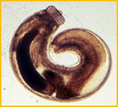
The animals were inoculated orally with 50 larvae (L3A) of G. binucleatum; Faust coproparasitoscopic analyses were performed daily to detect eggs, (see Figure 1) denoting the presence of adult parasites. The animals were kept in individual cages within the laboratory of parasitology belonging to Secretariat for Research and Graduate Studies at the Autonomous University of Nayarit. The cleaning was done daily and the maintenance consisted of water and balanced foods of the Purina mark ad limitum.

Dog necropsy was performed at 9, 9, 7 and 13 months postinoculation (PI); this included a comprehensive review of abdominal and thoracic cavity organs; the stomach was removed to detect adult nodules and parasites. In animals that did not develop nodules, the stomach was processed by artificial digestion in the search for evolutionary phases of the parasite. One cm3 of nodular tissue was taken and fixed in 10 % of formaldehyde for 48 hours and embedded in paraffin to make histological sections of 4μ thickness, stained with hematoxylin and eosin under conventional techniques. The morphology and morphometry of adult larvae and parasites were determined according to the criteria established by Miyazaki (1991).
The variables considered by the author are: total length, maximum width, number of rings in the cephalic bulb, bulb length, width of the head bulb, and number of hooks per cephalic ring, difference between the averages of the first and last row of the bulb cephalic, localization of the cervical papilla and distance from the sewer to the posterior end. In adult parasites (male) the pattern of cuticular spines, spicule shape and size, and location of the vulva in females are considered. In the juvenile parasites intermediate forms are observed depending on their evolution in the organism. The variables described were performed with optical microscopy and scanning electron Bozzola and Russell (1992). The morphometry of 30 larvae (measured) was performed with a calibrated composite microscope. The study was approved by the internal committee for the care of experimental animals, under the postgraduate program in health sciences and animal production of the Autonomous University of Mexico.
Results
The morphometry of the larvae (see Figure 2), showed average values of 3.988 mm of total length, 0.308 of maximum width, 4 concentric rings in the cephalic bulb, which presented 0.147mm long and 0.235mm wide. The number of hooks in the four concentric rows of the cephalic bulb was in the 1: 38.4, 2: 41.6, 3: 43.8 and 4: 46.2 ring; the difference between the averages of the fourth to the first row of the cephalic bulb was 7.3. The cervical papilla was located in the transverse row 14.6, and the distance from the cloaca to the posterior end was 0.065mm. The mouth presented a pair of lips with two papillae each and between the two papillae the amphidia were located. The cuticle of the body has more than 200 transversal striae with single-pointed spines. The esophagus occupies 30 % of the body of the parasite, parallel to this organ were four cervical sacs that dilate and contract the cephalic bulb. The intestine presented blood-red granules.
The four inoculated dogs developed stages of the parasite in the stomach; in dog one the removal of eggs in feces was presented at 22 weeks PI (period of prepatency) and eggs were maintained for a period of 14 weeks, plus (patency period). Necropsy was performed at 9 months PI, with a fibrous nodule 18 cm in diameter, located in the major curvature of the stomach, from which 11 adult parasites (5 females, 6 males) were recovered. The nodule had perforations communicating with the abdominal cavity and a small opening approximately 2 mm in diameter communicating with the gastric lumen. The perforations contained exudate of mucosanguinolento aspect, probably provoked by the stay and internal migration of the parasites.
Dissecting the nodule only recovered male worms that were embedded in the tissue; while females were removed in feces after ovoposition. In dog 2 no eggs were detected; necropsy was performed at 9, 9, 7 and 13 months PI, with five gastric nodules 1 to 2 cm in diameter and by artificial digestion; 3 juvenile parasites and 2 moulting larvae were recovered. In dog 3 eggs removal was not observed, necropsy was performed at seven-month PI and no gastric nodules were detected. During the artificial digestion of the stomach, five juvenile phase parasites were recovered. In dog 4, no eggs were detected in feces, necropsy was performed at 13 months PI, with 3 nodules of 1 to 2 cm in diameter, located in the gastric mucosa and five juvenile parasites were recovered by artificial digestion.
The histopathological examination of the nodule of 18 cm in diameter, showed abundant vascularization; internally, caves with mucosanguinolent exudate and adult worms included in the tissue were observed. One of the caverns had communication with the abdominal cavity and an adult (female) parasite was observed outside the nodule and in contact with the viscera. In two more dogs nodules 1 to 2 cm in diameter were found, with juvenile parasites located in the wall of the stomach. Another infected dog had no nodules, but by artificial digestion juvenile parasites were recovered in the stomach wall.
Necropsy performed on the four infected dogs revealed muscle atrophy, hepatomegaly, splenomegaly, mesenteric lymphangitis, pancreatitis, gastric hypertrophy, chronic gastritis and small ulcers in the gastric mucosa. Histologically, the gastric nodules presented zones of fibrosis, small areas of necrosis, large numbers of eggs captured within the nodular tissue and infiltration of plasma cells, macrophages and eosinophils surrounding the eggs.
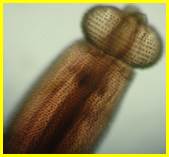
In scanning electron microscopy, the cephalic bulb of the adult parasites (Figure 3) showed 8 to 9 rows of hooks, and the mouth mouths with a pair of papillae each, and between the two papillae the amphidia were located. The pattern of cuticular spines in the anterior region was 2 to 3 points; in the middle and posterior region decreased to 2 and 1 point. The male's tail presented four pairs of large lateral papillae and four more pairs in the middle position and a pair of uneven spikes, which project through the cloaca. The vulva in the female was located in the middle and ventral region of the body.
Discussion and Conclusion
Three species of the genus Gnathostoma sp., have been reported in Mexico, G. binucleatum (Almeyda-Artigas, 1991), Gnathostoma turgidum (Lamothe-Argumedo et al., 1998) and Gnathostoma lamothei, Bertoni-Ruiz et al., 2005; Hernández-Gómez et al., 2010). The four dogs inoculated with L3A larvae of G. binucleatum, developed adult and juvenile forms in their stomach; only one of them eliminated adult eggs and parasites in feces, in the rest only juvenile phases were found. The lesions developed in the infected dogs were nodules of different size in the gastric wall. These lesions have been reported in the majority of the definitive hosts of the fourteen species of Gnathostoma spp, identified in the world, except G. nipponicum, which is located in esophagus, G. miyazakii and G. viet-namicum in kidney, G. didelphis and G. brasiliense in liver (Miyazaki, 1991).
It is important to mention that in the dog that eliminated eggs, the elimination of females in fecal matter was observed and at necropsy only males were found embedded in the wall of the nodule; this different behavior between males and females has not been clarified, but probably because females that have finished producing eggs, deplete their metabolic reserves and die. The observation of the completely empty uterus in the eliminated females and the loss of coloration of the pseudocelloma seem to confirm their biological exhaustion. Microscopically the nodule which had adult worms, presented large numbers of eggs trapped within the tissue and a strong reaction of macrophages and eosinophils around. The presence of large numbers of inflammatory cells close to or attached to the egg is probably a result of its antigenicity and contributes to the growth of the nodule. The results confirm the dogs as definitive hosts of this parasite. In this paper we present new data on the pathological and parasitological aspects of canine gnathostomiasis.
Literatura Citada
Almeyda-Artigas RJ. 1991. Hallazgo de Gnathostoma binucleatum en felinos silvestres y el papel de peces dulceacuícolas y oligohalinos como vectores de la gnatostomiasis humana en la cuenca baja del río Papaloapan, México. An. Inst. Cienc. del Mar y Limnol. 18 (2): 137-155. http://biblioweb.tic.unam.mx/cienciasdelmar/instituto/1991-2/articulo386.html [ Links ]
Alvarez-Guerrero C, Lamothe-Argumedo R. 2000a. Larvas de Gnathostoma sp. en peces estuarinos de Nayarit, México. An. Inst. Biol. Ser. Zool. 71 (2): 179-184. http://www.ejournal.unam.mx/zoo/071-02/ZOO71205.pdf [ Links ]
Alvarez-Guerrero C, Alba-Hurtado F. 2007. Estuarine fish and turtles as intermediate and paratenic hosts of Gnathostoma binucleatum in Nayarit, México. Parasitol. Res. 102: 117-122. https://doi.org/10.1007/s00436-007-0738-x [ Links ]
Alvarez-Guerrero C, Muñoz Guzmán MA, Alba-Hurtado F. 2014. Pathological and Parasitological traits in experimentally infected cats Gnathostoma binucleatum Spirurida: Gnathostomatidae. Vet. Parasitol. (204), 279-284. https://doi.org/10.1016/j.vetpar.2014.04.027 [ Links ]
Bertoni-Ruiz F, García-Prieto L, Osorio-Zarabia D, León-Régagnon V. 2005. A new species of Gnathostoma (Nematoda: Gnathostomidae) in Procyon lotor hernandezii from México. J. Parasitol. 91: 1143-1149. https://doi.org/10.1645/GE-516R.1 [ Links ]
Bozzola JJ, Russell LD. (1992). Electrón microscopy. Principles and Techniques for biologists. Jones and Bartlett publishers. 542 p. [ Links ]
Koga M, Akahane H, Ogata K, Lamothe AR, Osorio SD, García-Prieto L, Martínez-Cruz JM. 1999. Adult Gnathostoma cf. binucleatum obtained from dogs experimentally infected with larvae as an etiological agent in Mexican Gnathostomiasis: External morphology. J. Helminthol. 66 (1): 41-46. http://bionames.org/bionames-archive/issn/1049-233X/66/41.pdf [ Links ]
León-Régagnon V, Osorio-Zarabia D, García-Prieto L, Akahane H, Lamothe-Argumedo R, Koga M, Messina RM, Álvarez-Guerrero C. 2002. Study of the ethiological agent of gnathostomosis in Nayarit, México. Inter. Parasitol. 51: 201-204 https://doi.org/10.1016/S1383-5769(02)00014-4 [ Links ]
Miyazaki I. 1991. An Illustrated Book of Helminthic Zoonoses. SEAMIC Publ. 62: 368-408 [ Links ]
SUAVE. Sistema único automatizado para la vigilancia epidemiológica. 2005. Secretaría de Salud de Nayarit (1995-2005) [ Links ]
Received: February 27, 2017; Accepted: August 08, 2017
 Este es un artículo publicado en acceso abierto bajo una licencia Creative Commons
