


 text new page (beta)
 English (pdf)
 Article in xml format
 Article references
 Send this article by e-mail
 Cited by SciELO
  Similars in
SciELO
Similars in
SciELO 
 Permalink
Background
Endoscopic ultrasound-guided fine-needle aspiration biopsy (EUS-FNAB) and EUS-fine-needle biopsy (FNB) are widely known techniques for obtaining tissue in multiple diagnostic conditions. EUS-FNAB is routinely performed to obtain samples of various tissues such as pancreas, liver, gastric wall, adrenal glands, kidney, and lymph nodes adjacent to the gastrointestinal tract1,2.
Vilmann et al. reported the first case of EUS-FNAB in 1992; since then, its use has become more popular in clinical diagnosis. According to the literature, the diagnostic accuracy of this method has ranged from 70 to 100% for mediastinal tumors or intraabdominal tumors and from 38 to 100% for gastrointestinal wall lesions such as submucosal tumors3.
The EUS-FNAB is performed using a linear transducer to pass a needle through the working channel of an endoscope4. Afterwards, the needle is inserted into the lesion of interest under direct vision, and several degrees of suction are applied to obtain a sample by aspiration, unlike the EUS-FNB, in which practically no suction or aspiration is involved5.
Several types of needles and different degrees of aspiration are commonly used; nevertheless, there is no clear consensus on the optimal aspiration or nonaspiration technique. There are two key aspects regarding the samples sent for cytopathological evaluation: quality (qualitative analysis of structural cell morphology) and cellularity (quantitative analysis of cells).
The aspiration biopsy technique (FNAB-EUS) has undergone some modifications; two variations are in current use: the wet technique and the capillary technique. Both were developed with the aim of improving the quality of the aspirate for cytopathological diagnosis.
The wet EUS-FNAB technique consists in filling the needle shaft with 1.8 mL of saline solution before aspiration to replace the air contained in it6.
A review article by Wani et al. reported a diagnostic accuracy of 96.5% with the capillary technique compared to 88.5% with a standard suction technique. In a prospective randomized study, Attam et al. reported a diagnostic accuracy of 87.5%7.
Iwashita et al. studied the capillary EUS-FNB technique and demonstrated a diagnostic accuracy of 96%. The biopsy is performed by removing the stylet from the needle at the same time as the needle is inserted into the tissue to be biopsied. No aspiration is used8.
Previous studies Dabizzi et al.9, have compared the capillary technique and the non-wet or conventional aspiration technique, but no studies have compared the capillary technique and the wet technique.
The authors of the present study believe that comparing both techniques is necessary to determine which is the best option for routine use in cases such as digestive neoplasm.
Materials and methods
The study protocol and procedures were approved by the ethics committee of the Centro Medico Nacional Siglo XXI, which belongs to a public institution known as the Mexican Social Security Institute (IMSS, according to its Spanish initials). The protocol number is NCT03460197 and can be found in the website ClinicalTrials.gov, where it was accepted on March, 2018. All patients signed a written informed consent after the study protocol was fully explained to them. Since there are no studies comparing the wet and capillary techniques, the authors of the present study set out to obtain a non-probabilistic sample of the first thirty consecutive cases that arrived at the hospital, which were then randomized by means of the lottery method. This sample was used to perform a pilot study in order to make a more accurate calculation of the required sample size for a second phase study in which we will explore diagnostic accuracy test reporting sensibility and specificity of each technique (capillarity versus wet).
The general objective was to compare two different methods for obtaining tissue (capillary technique versus wet technique) in patients with a suspected malignancy. In order to compare both techniques, the cellularity and quality of the tissue samples were recorded according to each graded category: cellularity, graded 0 = no cells, 1 = sparsely cellular, 2 = moderately cellular, and 3 = highly cellular. Tissue smear and cell block were assessed of blood and insertion tissue contamination with this scale: graded 0 = free of contamination, 1 = contaminated, and 2 = highly contaminated, with or without blood clots. We also evaluate biopsy diagnosis characteristics with this scale: 0 = not adequate, 1 = suspicious for particular etiology, 2 = diagnostic for etiology, e.g. cancer, and 3= negative for neoplastic cells.
The Papanicolaou classification was used for evaluating pancreatobiliary cells in biopsy samples.
I. Non-diagnostic: the specimen does not provide information about whether the lesion is cystic or solid.
II. Negative (for malignancy): the specimen has adequate cellularity and/or extracellular material that can be used to define a lesion identified on an image.
III. Atypical: there are cells with architectural, nuclear, or cytoplasmic atypia that are not consistent with reactive changes. However, these findings are not enough to conclusively diagnose a malignancy or a suspected malignancy.
IV. Neoplasia: benign and others. Benign neoplasm: a sample has elements characteristic of a benign neoplasm. Other neoplasias: may be a pre-malignant lesion such as lowlevel dysplasia, intermediate or high-grade dysplasia, or a low-grade neoplasm with malignant behavior.
V. Suspected of malignancy: cytological characteristics support the diagnosis of malignancy but, quantitatively or qualitatively, are not enough to confirm it.
VI. Positive/malignant: cytological changes correspond unequivocally to malignancy.
The patients were sent to the endoscopy department by their corresponding basic medical units. The sample included patients over 18 years of age, of both genders, with suspected tumor in the pancreas or liver or lymph node metastasis. Patients who did not require evaluation by EUS were excluded.
Equipment and procedures
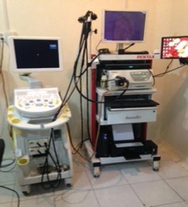
The biopsies were taken (with both techniques) by an endosonographer using an EUS system (Hitachi, EZU-MT30-S1 Hi Vision Avius); a radial echoendoscope (Pentax, EG-3670URK) was used for diagnosis and characterization of lesions; a linear echoendoscope (Pentax, EG-3870UTK) was used for the sampling and characterization of tissue by FNAB. All the equipment is part of the biomedical services provided by the Mexican Institute of Social Security (Fig. 1).
The procedures required general anesthesia and a pre-operative assessment. The lesion was localized and explored through EUS (Figs 2-4). Subsequently, the two biopsy techniques under study were used to extract tissue, taking care not to damage blood vessels.


In the capillary technique, the stylet is usually not removed from the needle until the puncture is performed to obtain a biopsy. The removal of the stylet is carried out in a synchronized manner with the to-and-fro movements for inserting the needle. The removal of the stylet is stopped at the moment the puncture is finished, and the device is removed from the echoendoscope. In the wet suction technique, the needle is filled with 1.5 mL of saline solution after removing the stylet and before inserting the needle. A 10-mL suction syringe attached to the needle is used to generate maximum suction from a locked position on the proximal end of the needle just when the punctures are made to obtain a biopsy. The syringe is removed or blocked to stop the suction at the moment when the puncture is finished and the device is removed from the echoendoscope needle. Both techniques require delivering the samples onto slides by passing the stylet through the lumen of the needle until all the biopsy materials are pushed out. Air is then passed through the lumen of the needle using a 10 mL syringe to extract the remaining biopsy material. Finally, the biopsy material is smeared and placed in a bottle containing 98% alcohol. Differently labeled bottles were used to store the biopsy samples obtained with the capillary (bottle 1) and wet (bottle 2) techniques.
After finishing the preparation of the samples obtained by both techniques, they were sent to the pathology department for analysis (in a blinded manner) by two independent pathologists. First, they were analyzed by the pathologist on duty that day; 1 week later, they were analyzed by another pathologist recruited into the study (Pathologist A and Pathologist B). None of them knew the diagnosis made by the other. The pathology reports documented the quality and the cellularity of the samples, as well as the diagnosis made from the smears regardless of the biopsy technique used.
The statistical analysis was carried out using the statistical software SPSS (version 21) for macOS by Apple Inc. Descriptive statistics were used to summarize demographic and clinical characteristics. Data were analyzed with Chi-square test, assuming a significant p = 0.05. A concordance analysis between the two participating pathologists to measure the degree of agreement between their diagnoses was performed using the weighted kappa coefficient.
Results
A total of 26 patients were included; 69.23% (n = 18) were female and 30.76% (n = 8) male; the average age of the patients was 61 ± 12 with a range of 32-78 years. Four patients were excluded because they were not characterized as probable cases of malignant tumors by the clinical researchers based on EUS evaluation. An initial diagnosis was made of all the patients under study based on an evaluation by EUS. The EUS diagnoses were distributed as follows: pancreatic tumor: 61.5% (n = 16); cholangiocarcinoma: 7.7% (n = 2); pancreatic head tumor versus pseudotumoral lesions of the pancreas: 3.8% (n = 1); pancreatic head-and-neck tumor with infiltration of the distal bile duct and hepatic hilum T3N × M: 3.8% (n = 1); neuroendocrine tumor versus mesenchymal tumor: 3.8% (n = 1); gallbladder cancer: 3.8% (n = 1); systemic ganglion disease: 3.8% (n = 1): mediastinal lymph node disease: 3.8% (n = 1); liver metastasis: 3.8% (n = 1); and metastasis of retropancreatic lymph nodes: 3.8% (n = 1). Table 1 shows the histological reports of each pathologist, the diagnostic category and the tissue sampling technique used.
Table 1 Percentage distribution of histological diagnoses reported by pathology by both techniques
| Capillarity % (n) | Wet % (n) | |||
|---|---|---|---|---|
| Pathologist A | Pathologist B | Pathologist A | Pathologist B | |
| Pancreatic Adenocarcinoma | 46.2 (12) | 46.2 (12) | 46.2 (12) | 50 (13) |
| Choledochal Adenocarcinoma | 7.7 (2) | 7.7 (2) | 3.8 (1) | 3.8 (1) |
| Muco-producer Adenocarcinoma with cystic degeneration | 3.8 (1) | 3.8 (1) | 3.8 (1) | 3.8 (1) |
| Well-differentiated Pancreas Adenocarcinoma | 3.8 (1) | 3.8 (1) | 3.8 (1) | 3.8 (1) |
| Pancreas Adenocarcinoma with muco-producer zones | 3.8 (1) | 3.8 (1) | 3.8 (1) | 3.8 (1) |
| Bile duct Adenocarcinoma | 3.8 (1) | 3.8 (1) | 3.8 (1) | 3.8 (1) |
| Poorly differentiated Pancreas Adenocarcinoma | 3.8 (1) | 3.8 (1) | 3.8 (1) | 3.8 (1) |
| Stomach Adenocarcinoma | 3.8 (1) | 3.8 (1) | 3.8 (1) | 3.8 (1) |
| Moderately differentiated liver Adenocarcinoma | 3.8 (1) | 3.8 (1) | 3.8 (1) | 3.8 (1) |
| Adenocarcinoma metastases to regional ganglia | 3.8 (1) | 3.8 (1) | 3.8 (1) | 3.8 (1) |
| Insufficient sample | - | - | 7.7 (2) | 7.7 (2) |
| Inadequate sample | - | - | 3.8 (1) | - |
| Positive cases for malignancy | 84.6 (22) | 84.6 (22) | 80.7 (21) | 84.6 (22) |
| Negative for neoplastic cells | 15.4 (4) | 15.4 (4) | 7.7 (2) | 7.7 (2) |
| Blood contamination Free | 15.4 (4) | 15.4 (4) | 23.07 (6) | 19.2 (5) |
| Contaminated | 73.0 (19) | 84.6 (22) | 57.6 (15) | 57.6 (15) |
| Highly | 11.53 (3) | - | 19.2 (5) | 23.07 (6) |
Pathologist A was able to make a diagnosis from the biopsy material obtained by the capillary technique in 96.15% (n = 24) of the cases and in 88.46% (n = 22) of the cases on biopsy material obtained by the wet technique. Pathologist B was able to make a diagnosis from the biopsy material obtained by the capillary technique in 96.15% (n = 24) of the cases and in 92.30% (n = 23) of the cases on biopsy material obtained by the wet technique. Combining the cytopathological results of both techniques allowed to make a diagnosis in 100% of the cases, either to confirm malignancy or to discard it.
Table 2 shows the degree of agreement (kappa coefficient) between both pathologists regarding their histopathological diagnoses with the two tissue sampling techniques. Without revealing to them the tissue sampling technique used, the pathologists were asked to report their preference between pairs of smears with material obtained using each of the techniques under study. The smears corresponding to the capillary technique were preferred 80.8% (n = 21) of the time; the pathologists considered that they showed better cellularity and quality. The smears corresponding to the wet technique were preferred only 19.2% (n = 5) of the time (p = 0.004).
Table 2 Measure of agreement between observers for histopathological diagnosis according to the technique used
| Kappa* | p | |
|---|---|---|
| Capillarity | 1 | < 0.001 |
| Wet | 0.948 | < 0.001 |
| Preference for Capillarity technique | 0.708 | < 0.001 |
*Weighted u Ordinary Kappa for two proportions
The histological diagnoses reported by both pathologists were analyzed using the Chi-square test and Fisher's exact test. In the comparison between the diagnoses corresponding to the capillary technique and the wet technique by pathologist A and pathologist B, the values of p were p ≤ 0.001 (gl = 120) and p ≤ 0.001, (gl = 100), respectively.
Discussion
It is difficult to obtain tissue samples from some tumors located in the vicinity of the gastrointestinal tract using conventional methods guided by computed tomography or ultrasound.
FNB guided by EUS has become the procedure of choice for the diagnosis of various digestive neoplasms10. This technique is currently used in numerous endoscopic centers, and it is evident that it has a beneficial effect on the therapeutic management of patients, as it allows to make a definitive diagnosis of their lesions. The results of the capillary and the wet techniques are superior to the results obtained by means of conventional percutaneous methods guided by tomography and ultrasound10,11. Other studies have also shown that the capillary and wet techniques yield better results in terms of cytopathological diagnosis than those obtained with conventional techniques12.
Various biopsy techniques are currently in use around the world13,14, such as the wet suction technique, capillarity, and the use of core needles, but none seems to clearly improve the collection of optimal tissue samples for cytopathological analysis15.
Sensitivity and specificity with EUS-FNAB of solid lesions of pancreas are documented higher than 85%16. Nowadays, there is no work that compares both techniques (capillarity and wet). The purpose of this work was to assess the quality of cytological samples, comparing two different FNA techniques, in order to optimize tissue acquisition and present our outcomes as a preliminary report and subsequently (with the inclusion of more patients), to compare sensibility and specificity between the techniques to obtain the diagnostic accuracy.
In our first findings, we observed that the pathologists found blood contamination in both techniques, but this situation was showed with a greater "high pollution" in the wet technique. The explanation could be in relation to the use of physiological solution which has to be placed in the light of the needle instead of the stylus, it favors the aspiration of histological material by "slipping" better the sample and by this pre-humidity-lubrication as well. Nevertheless, the authors consider that placing fluid in the needle also brings on more blood cells. This is an advantage in obtaining a higher sample quality in the capillarity technique. From our point of view, this is only an inference which would have to be confirmed with more extensive studies.
We had some weaknesses throughout the study, between them, the number of patients included, this fact does not make our results robust and convincing. It is also important to mention the order in which the procedures were realized. In all cases, we use the same needle for both procedures starting with capillarity, followed by the wet technique. Some authors refer that the edge of stylet is lost with the passage and the number of "passes" that are made, and this could influence in the greatest amount of tissue damage when the wet technique is executed. However, there was a tendency in obtaining superior quality samples with the capillary technique according the data distribution we found. However, we consider doing more extensive work in order to confirm and ratify these initial results.
The use of both techniques in the same patient possibly ensures 100% of the histological diagnosis, therefore that is a strong recommendation from the authors.
Conclusion
Given the way in which the resulting data were distributed, we can state that in this preliminary study, the tissue sampling technique by capillarity was superior to the wet technique in terms of the number of adequate samples, cellularity, quality, and diagnostic results. The pathologists were able to make a diagnosis from all the samples when combining both techniques.
However, further studies should be carried out on this subject with to draw a definitive conclusion.

