SciELO - Scientific Electronic Library Online

 
vol.10 issue2Evaluation of free flow constructed wetlands with Sagittaria latifolia and Sagittaria lancifolia in domestic wastewater treatmentGenotype-environment interaction on tuber and chip quality of potato genotypes grown in Central Mexico author indexsubject indexsearch form
Home Pagealphabetic serial listing  

Services on Demand

Journal

Article

Indicators

Related links

  • Have no similar articlesSimilars in SciELO

Share


Ingeniería agrícola y biosistemas

On-line version ISSN 2007-4026Print version ISSN 2007-3925

Ing. agric. biosist. vol.10 n.2 Chapingo Jul./Dec. 2018  Epub May 23, 2022

https://doi.org/10.5154/r.inagbi.2017.12.018 

Scientific article

Synthesis of silver nanoparticles using aqueous tejocote extracts as reducing and passivating agent

Elba Ronquillo-de Jesús1  * 

Miguel Angel Aguilar-Méndez2 

Patricia López-Perea1 

José Guzmán-Mendoza2 

Victoriano Hernández-Martínez1 

Nathaly Quiroz-Reyes2 

Miguel Angel Cruz-Hernández3 

Marineth Ortiz-Balderas1 

1Universidad Politécnica de Francisco I. Madero, Dirección de Ingeniería Agroindustrial, Domicilio conocido, Tepatepec, Hidalgo, C. P. 42660, MÉXICO.

2Instituto Politécnico Nacional, Centro de Investigación en Ciencia Aplicada y Tecnología Avanzada. Legaria núm. 694, Col. Irrigación, Ciudad de México, C. P. 11500, MÉXICO.

3Colegio de Posgraduados. Carretera México-Texcoco km 36.5. Montecillo, Texcoco, México, C. P. 56230, MÉXICO.


Abstract

Introduction:

Biosynthesis has emerged as an option for obtaining nanometric materials due to the need to use more environmentally-friendly synthesis methods.

Objective:

To synthesize silver nanoparticles (Ag NPs) with aqueous Crataegus gracilior Phipps (tejocote) bark extract as precursor.

Methodology:

Ag NPs were synthesized with AgNO3 and aqueous Crataegus gracilior bark extracts, and later characterized by ultraviolet-visible spectroscopy (UV-Vis), Fourier-transform infrared spectroscopy (FTIR), transmission electron microscopy (TEM) and X-ray diffraction (XRD). In addition, their size distribution and zeta potential were obtained.

Results:

The presence of Ag NPs reached maximum values at concentrations of 10 % (w/v). Mostly spherical nanoparticles were found in the range of 20 to 50 nm in size. FTIR confirmed the stabilization of the nanoparticles through their interactions with functional groups of carbohydrates and proteins. XRD and TEM results were explained by their face-centered cubic (FCC) structure with a size of 26 nm, a mean hydrodynamic diameter of 108 nm and polydispersity index of 0.24. The zeta potential values in the dispersions were -21.9 ± 5.11 mV, denoting colloidal stability.

Limitations of the study:

The characteristics of the nanoparticles obtained are only valid under the following synthesis conditions: 10 % (w/v) solids and pH 10.

Originality:

A non-reported plant material was used, capable of acting as a reducing and passivating agent of silver nanoparticles.

Conclusions:

Biosynthesis of Ag NPs with tejocote extract is an efficient, low-cost and environmentally-friendly method.

Keywords metal nanoparticles; biosynthesis; Crataegus gracilior; characterization

Resumen

Introducción:

La biosíntesis ha surgido como una opción para obtener materiales nanométricos debido a la necesidad de utilizar métodos de síntesis más amigables con el ambiente.

Objetivo:

Sintetizar nanopartículas de plata (NPs Ag) con extracto acuoso de corteza de Crataegus gracilior (tejocote) como precursor.

Metodología:

Se sintetizaron NPs Ag con AgNO3 y extractos acuosos de corteza de Crataegus gracilior, posteriormente se caracterizaron mediante espectroscopia ultravioleta-visible (UV-vis), espectroscopia infrarroja por transformada de Fourier (FTIR), microscopía electrónica de transmisión (MET) y difracción de rayos X (XRD), además se obtuvo su distribución de tamaño y potencial zeta.

Resultados:

La presencia de NPs Ag alcanzó los valores máximos a concentraciones de 10 % (m/v). Se encontraron nanopartículas mayormente esferoidales en el rango de 20 a 50 nm de tamaño. La FTIR confirmó la estabilidad de las nanopartículas mediante su interacción con grupos funcionales de carbohidratos y proteínas. Los resultados de XRD y MET se explicaron por su fase cúbica centrada en las caras (FCC) con tamaño de 26 nm, diámetro hidrodinámico medio de 108 nm e índice de polidispersidad de 0.24. Los valores del potencial zeta en las dispersiones fueron -21.9 ± 5.11 mV, denotando estabilidad coloidal.

Limitaciones del estudio:

Las características de las nanopartículas obtenidas solo son válidas bajo las siguientes condiciones de síntesis: 10 % (m/v) de sólidos y pH de 10.

Originalidad:

Se utilizó un material vegetal no reportado capaz de actuar como agente reductor y pasivante de nanopartículas de plata.

Conclusiones:

La biosíntesis de NPs Ag con extracto de tejocote es un método eficiente, de bajo costo y amigable con el ambiente.

Palabras clave: nanopartículas metálicas; biosíntesis; Crataegus gracilior; caracterización

Introduction

Silver nanoparticles (Ag NPs) have been extensively studied because of their unique optical, magnetic, electrical, and catalytic properties, with applications in pharmacology, medicine, food processing, and agriculture (Luo, Zhang, Zeng, Zeng, & Wang, 2005; Petica, Gavriliu, Lungu, Buruntea, & Panzaru, 2008; Sondi & Salopek-Sondi, 2004). The synthesis of Ag NPs can be achieved by several methods, including chemical reduction, electrochemical reduction, UV-radiation, photochemical reduction, and ultrasonic assisted, microwave, and laser ablation (Kumar, Smita, Cumbal, & Debut, 2017; Mahendran & Ranjitha-Kumari, 2016). These processes usually have a high cost and involve use of organic solvents and toxic reducing agents such as sodium borohydride and N,N-dimethyl formamide (Raja, Ramesh, & Thivaharan, 2017).

On the other hand, biological synthesis has emerged as a new option for obtaining nanoscale materials, primarily as a result of the increasing need to use environmentally-friendly synthetic methods. Biosynthesis involves the use of microorganisms (bacteria, yeasts, fungi) or plant extracts to achieve the reduction of metal ions, including silver, gold and copper (Basavaraja, Balaji, Lagashetty, Rajasab, & Venkataraman, 2008; Bradley, Schmid, Shevchenko, & Weller, 2004; He et al., 2007; Sastry, Ahmad, Khan, & Kumar, 2003; Vigneshwaran et al., 2007).

Of particular interest herein is that the use of plant extracts acting as reducing agents may provide a quick way of preparing nanoparticles. Examples include the synthesis of metal nanoparticles using as reducing or passivating agents extracts of coriander (Narayanan & Sakthivel, 2008), lemon verbena (Cruz et al., 2010), orange peel (Kaviya, Santhanalakshmi, Viswanathan, Muthumary, & Srinivasan, 2011), or banana peel (Bankar, Joshi, Kumar, & Zinjarde, 2010).

Crataegus spp., a Mexican plant commonly known as tejocote, has medicinal, industrial and ornamental uses (Nieto-Ángel, Pérez-Ortega, Núñez-Colín, Martínez-Solís, & González-Andrés, 2009). The Crataegus gracilior plant extract contains a high percentage of pectin, which is composed mainly of D-galacturonic acid and, in less proportion, D-galactose, L-arabinose, D-xylose and L-rhamnose. In Ag NP synthesis, pectin forms polymer networks in which silver nanoparticles are embedded, also acting as a reducing agent and silver ion protector (Krivorotova et al., 2016; Zahran, Ahmed, & El-Rafie, 2014; Zainudin, Wong, & Hamdan, 2018). To our knowledge, however, this has never been addressed in the literature. Therefore, the objective of the present study was to synthesize Ag NPs using aqueous Crataegus gracilior bark extract as precursor.

Materials and methods

Ag NPs were synthesized using silver nitrate (AgNO3, > 99.8 %, Sigma-Aldrich®, USA) as precursor and aqueous tejocote bark extracts as reducing and passivating agent. Deionized water (18 MΩ·cm-1; Easypure II, Thermo ScientificTM, Spain) was used throughout the experiments and sodium hydroxide (NaOH) was used to adjust the pH.

For the synthesis, 100 mL of aqueous tejocote bark extract were placed in a round-bottomed flask, where the pH was adjusted with 0.1M HCl and 0.1 M NaOH. Then, the solution was heated to 75 °C under constant stirring. When the desired temperature was reached, 10 mL of 0.1 M AgNO3 were added. The final mixture was stirred for 30 min at the same temperature. Preliminary analyses were conducted at solid concentrations of 1, 5, and 10 % (w/v) and pH0 6, 8, and 10. Samples with a 10 % solid concentration and pH 10 contained the smallest and most stable nanoparticles.

Characterization of nanoparticles

Ultraviolet-visible spectroscopy (UV-Vis). Optical absorption spectra for Ag NP suspensions were obtained using a Cary 50 spectrophotometer (Varian®, USA). Ag NP suspensions were diluted at a 5:1 ratio with deionized water (18 μΩ·cm-1) prior to analysis. Deionized water was used as a reference.

Fourier-transform infrared spectroscopy (FTIR). The Ag NP suspension was centrifuged at 13 000 rpm (Sigma 2-16, Sigma Laborzentrifugen GmbH®, Germany) and subsequently dried at room temperature. Dried powdered plant material and Ag NPs were separately mixed with KBr at a 1:10 ratio to form pellets. The spectra were recorded using a spectrometer (Spectrum One, Perkin Elmer®, USA), with a 4 000 to 400 cm-1 frequency range.

Transmission Electron Microscopy (TEM). Ag NPs were examined in a JEM-2010 microscope (JEOL®, Japan) operating at 200 kV and 115 μA, for which a single drop of the colloidal solution was placed on a 200-mesh carbon-coated copper grid (Electron Microscopy Sciences, USA). Additionally, the selected area electron diffraction (SAED) was obtained at a wavelength of 0.0025 nm and a camera length of 20 cm. Average size and size distribution values were obtained by analyzing 150 particles, using PhotoImpact 11 software (Ulead Systems®, USA).

X-ray diffraction (XRD). It was performed in a D8 Advance diffractometer (Bruker®, Germany). The patterns were recorded by CuKα (1.54 A°) with a cupper monochromator. The scanning of Ag NPs was done in a region of 2θ from 5 to 90° at 0.05°/10 s.

Determination of zeta potential and size distribution. The hydrodynamic diameter and polydispersity index of the nanoparticles were determined by dynamic light scattering (DLS) and zeta potential (ZS ZEN3600, Malvern Instruments®, UK). Measurements were performed in triplicate, with each measurement conducted using 1 mL of suspension at room temperature (25 °C). The calculations of electrophoretic mobility were automatically converted into zeta potential values, based on the Smoluchowski model; ten readings were obtained to calculate the average electrical charge.

Results and discussion

UV-Vis. After the addition of AgNO3 to the extract solution, the color of the solution changed from colorless to brown indicating the formation of silver nanoparticles. It is highly possible that pectin, hydrolysable tannins, polyphenols and flavonoids present in the tejocote extract have acted as reducing agents to produce Ag0. Maximum absorbance values for Ag NP with tecojote extracts suspensions were obtained (10 % w/v) at 500 nm.

The position and shape of the absorption band for a surface plasmon depend on particle size and shape: increases in size cause the absorption band to shift towards higher wavelengths (Slistan-Grijalva et al., 2008). Based on the spectral signals obtained (Mitra & Bhaumik, 2007), nanoparticles obtained with the tejocote extracts presented an average size ≤ 30 nm (Figure 1).

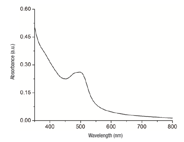
Figure 1 UV-Vis absorption spectrum of a colloidal system of silver nanoparticles with aqueous tejocote (Crataegus gracilior) bark extract. 

FTIR analysis. Shown in Figure 2 are the FTIR spectra of the precursors (AgNO3 and tejocote extract) and Ag NPs. The spectrum of tejocote extract denotes absorption bands at 1 045 cm-1 and 1 115 cm-1 (corresponding to CO vibrations), 3 417 cm-1 (amide A, NH stretching and OH bending and stretching), 1 619 cm-1 (amide I, C=O stretching), 1 520 cm-1 (amide II, bending vibrations of NH and CN stretching), and 1 254 cm-1 (amide III, bending vibrations of NH (Hayashi & Mukamel, 2008). FTIR spectra for AgNO3 revealed an intense absorption band at 1 376 cm-1, characteristic of the Ag+NO3 - ion pair.

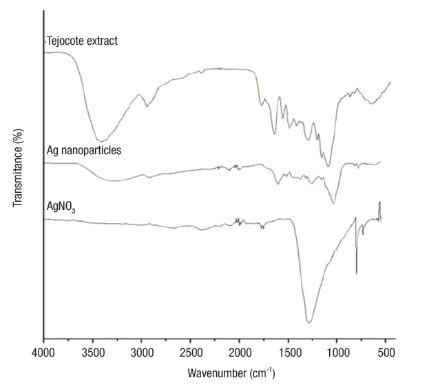
Figure 2 FTIR spectra of AgNO3, tejocote extract and silver nanoparticles, the last of which interact with different functional groups of carbohydrates and proteins. 

On the other hand, in the spectrum of the silver nanoparticles, the absorption bands corresponding to the amide groups shifted to lower wavenumber values. These changes on the position bands may be due to the interactions of proteins that are possibly bound to Ag NPs through the amine groups. Additionally, the band intensity of the ion pair Ag+NO3 - decreased and shifted to a higher wavenumber value. This peak, centered at 1 385 cm-1, is characteristic of the NO3 - ion in free form, and the absorption band displacement is caused by a change in the electronic environment of the anion, as a result of the separation of its counterpart Ag+ (Cho & So, 2006).

TEM. Figure 3a shows the Ag NPs embedded in a polymer network at tejocote concentrations of 10 %. Dark areas corresponded to Ag NPs and light areas to different compounds present in the plant extract. Ag NPs showed an average diameter of 30 nm and a spherical morphology, although the magnification showed an ovoid shape. Figure 3b exemplifies an electron diffraction pattern of the Ag NPs. High magnification evidenced a face-centered cubic (FCC) structure, a spatial group of Fm-3m (225), and a lattice parameter a = 4.09 Å.

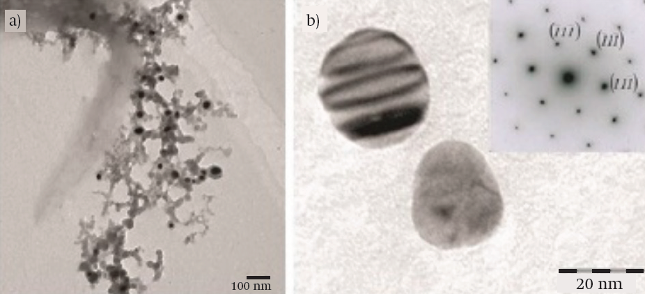
Figure 3 Transmission electron microscopy images of silver nanoparticles (Ag NPs): (a) Ag NPs embedded in a polymer network (10 Kv) and (b) magnified image and electron diffraction pattern of Ag NPs (120 kV). Extinction contour lines shown in the top section of the particle confirmed a variable thickness.  

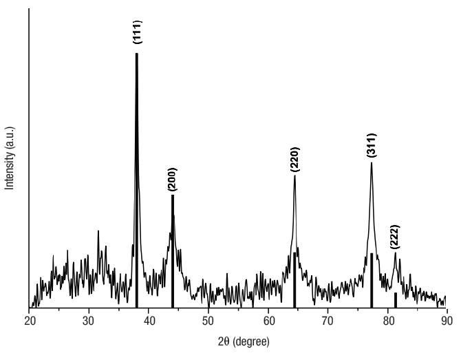
XRD. Results obtained by TEM and calculations using the Scherrer equation were best explained by an average size of 26 nm. Figure 4 shows the X-ray diffraction pattern obtained for Ag NPs. Five Bragg reflections are observed in the diffractogram at 2θ angles centered at 38.0°, 44.0° 64.4°, 77.32°, and 81.34° corresponding to planes (111), (200), (220), (311), and (222) respectively, from the FCC of the Ag with spatial group Fm-3m (225), according to the diffraction analysis PDF-04-0783. The diffractogram indicates the presence of nanometric-size crystals without preferential direction for growth.

Figure 4 Diffractogram of silver nanoparticles embedded in a polymer network. 

Nanoparticle size distribution and zeta potential. According to the DLS analysis, the particles obtained are a polydisperse mixture with a mean hydrodynamic diameter of 108 nm and a polydispersity index of 0.24. On the other hand, the colloidal solution of Ag NPs had a zeta potential value of -21.9 ± 5.11 mV, indicating good NP stability. This stability mainly depends on the surface charge; hence particles with a similar surface charge repel each other due to electrostatic repulsion forces (Khan, Mukherjee, & Chandrasekaran, 2011). According to Sun et al. (2014), a suspension with an absolute zeta potential lower than 20 mV is considered unstable, and its constituent particles will tend to precipitate, whereas an absolute zeta potential greater than 20 mV is indicative of a stable solution.

Conclusions

Biosynthesis of Ag NPs was performed using AgNO3 and aqueous tejocote (Crataegus gracilior) bark extracts as precursors, achieving an efficient, low-cost and environmentally-friendly synthesis. FTIR results confirmed the stabilization of Ag NPs by interacting with organic components in the extract, primarily carbohydrates and proteins. TEM showed that Ag NPs were mostly spherical, with an average size of 30 nm. Additionally, electron diffraction patterns confirmed an FCC structure.

Acknowledgments

The authors thank the Consejo Nacional de Ciencia y Tecnología (CONACyT) and the Secretaría de Investigación y Posgrado del Instituto Politécnico Nacional (SIP-IPN) for the funding provided.

References

Bankar, A., Joshi, B., Kumar, A. R., & Zinjarde, S. (2010). Banana peel extract mediated novel route for the synthesis of silver nanoparticles. Colloids and Surfaces A: Physicochemical and Engineering Aspects, 368(1-3), 58-63. doi: 10.1016/j.colsurfa.2010.07.024 [ Links ]

Basavaraja, S., Balaji, S. D., Lagashetty, A., Rajasab, A. H., & Venkataraman, A. (2008). Extracellular biosynthesis of silver nanoparticles using the fungus Fusarium semitectum. Materials Research Bulletin, 43(5), 1164-1170. doi: 10.1016/j.materresbull.2007.06.020 [ Links ]

Bradley, J. S., Schmid, G., Shevchenko, E. V., & Weller, H. (2004). Synthesis of metal nanoparticles. In: Schmid, G. (Ed), Nanoparticles: From theory to application (pp. 185-238). Germany: Wiley-VCH. doi: 10.1002/3527602399 [ Links ]

Cho, J. W., & So, J. H. (2006). Polyurethane-silver fibers prepared by infiltration and reduction of silver nitrate. Materials Letters, 60(21-22), 2653-2656. doi: 10.1016/j.matlet.2006.01.072 [ Links ]

Cruz, D., Falé, P. L., Mourato, A., Vaz, P. D., Serralheiro, M. L., & Lino, A. R. (2010). Preparation and physicochemical characterization of Ag nanoparticles biosynthesized by Lippia citriodora (Lemon Verbena). Colloids and Surfaces B: Biointerfaces, 81(1), 67-73. doi: 10.1016/j.colsurfb.2010.06.025 [ Links ]

Hayashi, T., & Mukamel, S. (2008). Two-dimensional vibrational lineshapes of amide III, II, I and A bands in a helical peptide. Journal of Molecular Liquids, 141(3), 149-154. doi: 10.1016/j.molliq.2008.02.013 [ Links ]

He, S., Guo, Z., Zhang, Y., Zhang, S., Wang, V., & Gu, N. (2007). Biosynthesis of gold nanoparticles using the bacteria Rhodopseudomonas capsulata. Materials Letters, 61(18), 3984-3987. doi: 10.1016/j.matlet.2007.01.018 [ Links ]

Kaviya, S., Santhanalakshmi, J., Viswanathan, B., Muthumary, J., & Srinivasan, K. (2011). Biosynthesis of silver nanoparticles using citrus sinensis peel extract and its antibacterial activity. Spectrochimica Acta Part A: Molecular and Biomolecular Spectroscopy, 79(3), 594-598. doi: 10.1016/j.saa.2011.03.040 [ Links ]

Khan, S. S., Mukherjee, A., & Chandrasekaran, N. (2011). Impact of exopolysaccharides on the stability of silver nanoparticles in water. Water Research, 45(16), 5184-5190. doi: 10.1016/j.watres.2011.07.024 [ Links ]

Krivorotova, T., Cirkovas, A., Maciulyte, S., Staneviciene, R., Budriene, S., Serviene, E., & Sereikaite, J. (2016). Nisin-loaded pectin nanoparticles for food preservation. Food Hydrocolloids, 54(49-56). doi: 10.1016/j.foodhyd.2015.09.015 [ Links ]

Kumar, B., Smita, K., Cumbal, L., & Debut, A. (2017). Green synthesis of silver nanoparticles using Andean blackberry fruit extract. Saudi Journal of Biological Sciences, 24(1), 45-50. doi: 10.1016/j.sjbs.2015.09.006 [ Links ]

Luo, C., Zhang, Y., Zeng, X., Zeng, Y., & Wang, Y. (2005). The role of poly(ethylene glycol) in the formation of silver nanoparticles. Journal of Colloid and Interface Science, 288(2), 444-448. doi: 10.1016/j.jcis.2005.03.005 [ Links ]

Mahendran, G., & Ranjitha-Kumari, B. D. (2016). Biological activities of silver nanoparticles from Nothapodytes nimmoniana (Graham) Mabb. fruit extracts. Food Science and Human Wellness, 5(4), 207-218. doi: 10.1016/j.fshw.2016.10.001 [ Links ]

Mitra, A., & Bhaumik, A. (2007). Nanoscale silver cluster embedded in artificial heterogeneous matrix consisting of protein and sodium polyacrylate. Materials Letters, 61(3), 659-662. doi: 10.1016/j.matlet.2006.05.039 [ Links ]

Narayanan, K. B., & Sakthivel, N. (2008). Coriander leaf mediated biosynthesis of gold nanoparticles. Materials Letters, 62(30), 4588-4590. doi: 10.1016/j.matlet.2008.08.044 [ Links ]

Nieto-Ángel, R., Pérez-Ortega, S. A., Núñez-Colín, C. A., Martínez-Solís, J., & González-Andrés, F. (2009). Seed and endocarp traits as markers of the biodiversity of regional sources of germplasm of tejocote (Crataegus spp.) from Central and Southern Mexico. Scientia Horticulturae, 121(2), 166-170. doi: 10.1016/j.scienta.2009.01.034 [ Links ]

Petica, A., Gavriliu, S., Lungu, M., Buruntea, N., & Panzaru, C. (2008). Colloidal silver solutions with antimicrobial properties. Materials Science and Engineering: B, 152(1-3), 22-28. doi: 10.1016/j.mseb.2008.06.021 [ Links ]

Raja, S., Ramesh, V., & Thivaharan, V. (2017). Green biosynthesis of silver nanoparticles using Calliandra haematocephala leaf extract, their antibacterial activity and hydrogen peroxide sensing capability. Arabian Journal of Chemistry, 10(2), 253-261. doi: 10.1016/j.arabjc.2015.06.023 [ Links ]

Sastry, M., Ahmad, A., Khan, M. I., & Kumar, R. (2003). Biosynthesis of metal nanoparticles using fungi and actinomycete. Current science, 85(2), 162-170. Retrieved from https://pdfs.semanticscholar.org/238b/7f05453ab4e0bcbdca2865e74b4078fb8b17.pdfLinks ]

Slistan-Grijalva, A., Herrera-Urbina, R., Rivas-Silva, J. F., Ávalos-Borja, M., Castillón-Barraza, F. F., & Posada-Amarillas, A. (2008). Synthesis of silver nanoparticles in a polyvinylpyrrolidone (PVP) paste, and their optical properties in a film and in ethylene glycol. Materials Research Bulletin, 43(1), 90-96. doi: 10.1016/j.materresbull.2007.02.013 [ Links ]

Sondi, I., & Salopek-Sondi, B. (2004). Silver nanoparticles as antimicrobial agent: a case study on E. Coli as a model for Gram-negative bacteria. Journal of Colloid and Interface Science, 275(1), 177-182. doi: 10.1016/j.jcis.2004.02.012 [ Links ]

Sun, Q., Cai, X., Li, J., Zheng, M., Chen, Z., & Yu, C. (2014). Green synthesis of silver nanoparticles using tea leaf extract and evaluation of their stability and antibacterial activity. Colloids and Surfaces A: Physicochemical and Engineering Aspects, 444(5), 226-231. doi: 10.1016/j.colsurfa.2013.12.065 [ Links ]

Vigneshwaran, N., Ashtaputre, N. M., Varadarajan, P. V., Nachane, R. P., Paralikar, K. M., & Balasubramanya, R. H. (2007). Biological synthesis of silver nanoparticles using the fungus Aspergillus flavus. Materials Letters, 61(6), 1413-1418. doi: 10.1016/j.matlet.2006.07.042 [ Links ]

Zahran, M. K., Ahmed, H. B., & El-Rafie, M. H. (2014). Facile size-regulated synthesis of silver nanoparticles using pectin. Carbohydrate polymers, 111, 971-978. doi: 10.1016/j.carbpol.2014.05.028 [ Links ]

Zainudin, B. H., Wong, T. W., & Hamdan, H. (2018). Design of low molecular weight pectin and its nanoparticles through combination treatment of pectin by microwave and inorganic salts. Polymer Degradation and Stability, 147, 35-40. doi: 10.1016/j.polymdegradstab.2017.11.011 [ Links ]

Received: December 19, 2017; Accepted: August 02, 2018

*Corresponding author: eronquillo@upfim.edu.mx, tel. 01 (738) 724 1174 ext. 165.

Creative Commons License This is an open-access article distributed under the terms of the Creative Commons Attribution License