


 text in
  English (pdf)
 Article in xml format
 Article references
 Send this article by e-mail
 Cited by SciELO
  Similars in
SciELO
Similars in
SciELO 
 Permalink
Highlights:
The growth regulators for the induction of somatic embryos of Echinocactus parryi were evaluated.
2, 4-D was involved in inducing globular embryo in mature seeds and seedlings explants.
Kinetin (0.5 mg∙L-1 ) induced all the somatic embryo phases in green compact callus explant.
The compact callus explants were the more efficient to induce 19.2 somatic embryos per explant.
Other endogenous phytohormones likely contributed to the torpedo phase that did not germinate.
Introduction
Cacti are one of the groups of plants more endangered in México due to illegal extraction and habitat destruction (Villanueva, 2016). Echinocactus parryi Engelm. is an endemic species from the municipality of Juárez, Chihuahua, México. Its ecosystem is characterized by rocky and gravely slopes in a desert environment, mainly in xerophilous and crassicaule communities (Quiñónez et al., 2018). However, the illegal collection, mining, and extraction of construction materials have reduced the native populations (Secretaría del Medio Ambiente y Recursos Naturales [SEMARNAT], 2010). Ecological studies on the population conditions of E. parryi are scarce. This species is propagated for seed only so that natural propagation has limitations that imply problems with their seed viability. The growth of plants is too slow, limiting the restoration of wild populations.
An option to improve the populations of threatened species is the in vitro plant tissue culture. Somatic embryogenesis is the most efficient in vitro technique for mass propagation. It has an excellent application for genetic diversity compared to organogenesis (Salma, Kundu, Ali, & Mandal, 2019). Embryogenesis can be carried out in a short time without seasonal dependence (Santos-Díaz, Pérez-Molphe, Ramírez-Malagon, Núñez-Palenius, & Ochoa-Alejo, 2011). The induction of somatic embryos has many advantages over conventional direct organogenesis; for example, producing valuable plants, artificial seeds, transgenic plants, conservation of genetic resources, and somaclonal variation (Brand, Quimbaya, Tohme, & Chavarriaga-Aguirre, 2019; Quiroz-Figueroa, Rojas-Herrera, Galaz-Avalos, & Loyola-Vargas, 2006). Somatic embryogenesis has been induced for a broad type of species, but for cactus species, a few of them reported, such as Opuntia ficus-indica (L.) Mill. (Bouamama et al., 2011; Linhares et al., 2006; Kaaniche-Elloumi, Jeddi, Mahmoud, Chakroun, & Jemmali, 2015) and Capiapoa tenuissima Ritt. (Lema-Ruminska, 2011; Lema-Ruminska, Goncerzewicz, & Gabriel, 2013). The problem of maturity for somatic embryo and their germination is not common however the accumulation of 2, 4-D (dichlorophenoxy acetic acid) inside globular embryos is reported, affecting the internal levels of indol-3-acetic acid (IAA) and altering the polarization of the cells (Garcia et al., 2019). It could affect the further embryo development and absence of apical meristem due to morphological abnormalities (Baskaran, & Van Staden, 2017; Nugent, Chandler, Whiteman, & Stevenson, 2001). Our recent studies in the in vitro clonal propagation of E. parryi showed the lower number of shoots (2.9 per explant) and the highest mass of callus formation, which limited the efficient clonal propagation (García-González, Santos-Díaz, Flores-Margez, & Osuna-Ávila, 2020). This study proposes the continuous exploration of this species and to study if the somatic embryo is more efficient than the clonal propagation route.
The use of auxins in the culture media is necessary to promote cell polarity and asymmetric cell division in the initiation of somatic embryogenesis (Yang, Wang, Le, & Dong, 2020). Typically, 2, 4-D synthetic auxin is used to induce or multiplicate somatic embryos that develop like zygotic embryos (Vondráková, Eliásová, Fischerová, & Vágner, 2011). The 6-furfurylaminopurine (kinetin) and 6-benzylaminopurine (BAP) promotes somatic embryo germination or shoot differentiation (Manokari, Latha, Priyadharshini, Jogam, & Shekhawat, 2020; Wu, Wei, Wang, & Wei, 2020). These cytokinins also function as a synergistic since combined with 2, 4-D to induce the somatic embryos (Anzidei, Benicci, Schiff, & Mori, 2000). The somatic embryos could be formed directly from the cells of the explant or indirectly from previous callus formation. First, the pro-embryogenic mass is promoted, and the subsequent somatic embryos are expressed on the peripheral of the callus (Alvez & Oropeza, 2015; Jhong, Pintado, & Jiménez, 2019). During the callus induction ploidy changes and mutations can occur (Bednarek & Orlowoska, 2020). The regenerated plants coming from somatic embryos may exhibit the somaclonal variation (Torres-Silva et al., 2018). In the case of E. parryi, there are no reports of somatic embryogenesis, so it is necessary to elucidate whether this species has the embryogenic capacity, which would be a more efficient alternative for its propagation. Thus, the main objective of this work was to evaluate the effects of the growth regulators for the induction of somatic embryos from mature seeds, shoots, and compact green callus. Also, was established a histological analysis of the induced embryogenic structures.
Materials and methods
Indirect somatic embryo structures induction from different explants
Mature seed explants
Seeds of E. parryi were collected randomly from mature fruits of native plants in August 2018, located in the municipality of Juárez, Chihuahua, México. The fruits were placed in paper bags and transported to the laboratory of plant biotechnology in the Universidad Autonoma de Ciudad Juarez. Fruits longitudinally were dissected with scissors, and the seeds were placed in a paper bag in dark conditions with a temperature of 25 ± 1 °C until use. The seeds were sterilized with 50 % commercial bleach (Cloralex® 5 % sodium hypochlorite solution) for 15 minutes, rinsed with sterile distilled water, and soaked in sterile water for 24 h (García-González et al., 2020). Half of the seed coat was removed using tweezers and a scalpel in sterile conditions. Ten scarified seeds per jar and each treatment were germinated in basal culture media MS (Murashige & Skoog, 1962) supplemented with 2, 4-D at 0, 2, 4, and 6 mg∙L-1 for callus induction. The embryogenic callus was estimated as the percentage of globular structures in the callus after 60 days. The callus maintained for 60 days were subcultured every 30 days in MS medium with the same concentrations of 2, 4-D. The embryogenic and non-embryogenic callus formation, color, and number of regenerated shoots were registered after 120 days.
Seedling explants
Sterile seedlings from seed germination, with 40 days of in vitro culture, were used as explants. The seedlings were dissected in three sections: apical, middle, and basal (the root was removed). Thirty-six explants per section were placed in Petri dishes containing 15 mL of basal MS culture media supplemented with 0, 2, 4, and 6 mg∙L-1 2, 4-D to induce embryogenic callus formation. The induced calli were divided randomly into 27 pieces per treatment and subcultured three times every 30 days in the same culture medium. After 90 days, the callus bearing globular structures (27 pieces of 1-2 cm) were transferred to MS with 0, 0.5, 1.0, 1.5, and 2.0 mg∙L-1 of 2, 4-D, BAP and kinetin (K) (Caisson Labs). The morphogenetic response and the color of the callus were evaluated after 120 days.
Compact green callus explants
Green compact calli from shoot apex and cultivated in different BAP concentrations for 30 weeks came from previous micropropagation in vitro experiments used for this study. Nine calli (1-2 cm size) explants per treatment were placed on Petri dishes (100 x 15 mm) containing 0, 0.5, 1.0, 1.5, and 2.0 mg·L-1 2,4-D, BAP or K, to compare their effect on somatic embryogenesis induction. The experiments were evaluated after 30 days with the imaging software NIS- Elements (Nikon) to count somatic embryos.
The three types of explants were incubated in a bioclimatic chamber light with photoperiod conditions (16 hours light). The fluorescent light bulbs gave the intensity of light (5 000 lux) at 25 ± 2 °C. The calli were observed under the stereoscope Nikon SMZ 800 to identify shoots or somatic embryos. The images were taken with a stereomicroscope attached to a Nikon Digital Sight DS-Fi2 camera.
Histology analysis
Histological analyses were performed on calli containing shoots, globular, and torpedo stages to verify the typical bipolar structure of somatic embryos. The sections were dissected using tweezers and a scalpel observed with a stereoscope. The samples were fixed in FAA (formalin 4 %, acetic acid, alcohol using 1:1:1 proportion) during 48 hours at room temperature. After fixation, the samples were dehydrated in ethanol solutions (80, 90, and 100 %) and xylol, and finally were embedded in paraffin (Tissue-Tek). Later the 5 µm sections were cut using a rotatory microtome (Microm HM 325) and stained with blue toluidine 1 % for 10 minutes. The samples were observed in an optical microscope Nikon Eclipse Ni.
Statistical analysis
Because of wide scatter in the data, including zero values, the log transformation ln (x+1) was used to include zero during transformation to approximate the data to the normal distribution. For mature seeds, the effect of 2,4-D treatments on embryogenic callus, percent of callus, and number of shoots was evaluated by analysis of variance to identify significant differences. The experiment was a completely randomized design using the univariate GLM procedure; subsequently, a Tukey test was performed to compare means. Embryogenic callus data in seedling explants were analyzed using a factorial arrangement where the factors were 2, 4-D at four levels (0, 2, 4 and 6 mg∙L-1) and explant section at three levels (apical, middle and basal); in addition, a Tukey's comparison of means was used since main effects and interaction were significant. The number of somatic embryos per green callus explant under the 2,4-D, BAP and kinetin treatments at four levels (0.5, 1.0, 1.5 and 2.0 mg∙L-1), with no interaction in this case, was analyzed in a completely randomized design. All significance was at the 95 % confidence limit (P < 0.05) using SPSS software version 25 (IBM Corp., 2017).
Results and discussion
Indirect somatic embryo structures induction from different explants
Mature seed explants
The incipient callus was white and derived directly from the cotyledon after 20 days of culture. Later, the callus presented intense pink, light green, and white areas. The ANOVA transformed data displayed that 2, 4-D had a significant difference in the embryogenic callus percentage than control (Table 1). After 60 days of culture, globular embryogenic structures were observed in the peripheral of the calli in all the 2, 4-D concentrations tested (Table 2). The subculture of embryogenic callus in media with or without 2, 4-D at 120 days did not promote their maturation.
Table 1 Analysis of variance for embryogenic callus and callus formation in mature seed explants of Echinocactus parryi under 2, 4-D treatments.
| Source | df | Callus | Embryogenic callus | ||||
|---|---|---|---|---|---|---|---|
| MS | F | P | MS | F | P | ||
| 2, 4-D | 3 | 0.295 | 2.25 | 0.099 | 50.31 | 37834 | 0 |
| Error | 36 | 0.131 | 0.001 | ||||
| Total | 39 | ||||||
df = degrees of freedom; MS = mean square.
Table 2 Effect of treatment with 2, 4-D in callus and embryogenic callus formation from mature seeds explants of Echinocactus parryi at 60 days of in vitro culture.
| 2, 4-D (mg∙L-1) | No. seeds | Callus (%) | Transformed means ln (x+1) | Embryogenic callus (%) | Transformed means ln (x+1) |
|---|---|---|---|---|---|
| 0 | 10 | 80 a | 2.375 | 0 b | 0 |
| 2 | 10 | 100 a | 2.718 | 89 ± 1.2 a | 4.487 |
| 4 | 10 | 100 a | 2.718 | 88.5 ± 1.0 a | 4.487 |
| 6 | 10 | 100 a | 2.718 | 89 ± 1.2 a | 4.487 |
Average values ± standard error. Means with different letters are statistically different between concentrations of 2, 4-D based on Tukey's test (P ≤ 0.05).
The globular structures in E. parryi, probably did not evolve to the more advanced stages because of 2, 4-D accumulation, occurring inside the globular embryos. The auxin 2, 4-D in the culture medium induces an embryogenic response associated with the increase of the endogenous levels of IAA and the polar transport, which is needed for the formation of the apical-basal axis (Garcia et al., 2019). Consequently, morphological abnormalities can occur, like the absence of apical meristem that could affect further embryo development (Baskaran & Van Staden, 2017; Nugent, Chandler, Whiteman, & Stevenson, 2001). In this study, the 2, 4-D treatments promoted embryogenic callus being the globular phase the dominant structure without reaching the next heart stage of maturity. Similar results were reported by Pinheiro, Soares, and Arnholdt-Schmitt (2001), which obtained globular structures from O. ficus-indica, incapable of regenerating plants. It suggested that probably this response is due to a deficiency in the supplemented carbohydrates in the culture media. However, Vondrákova et al. (2011) found that the developed somatic embryos of Abies alba Mill. cultivated with 2, 4-D failed to complete their maturation. However, the embryogenic callus from Mediocactus coccineus (DC.) Britton et Rose induced globular structures that turn onto a more defined cotyledonal stage (Infante, 1992). Stuppy and Nagl (1992) also induced embryogenic green callus from Ariocarpus retusus Scheidw. and after four months, the first somatic embryo appeared in the cotyledonary phase.
E. parryi non-embryogenic calli were also promoted and generated the shoot formation with trichomes (Figure 1). The ANOVA transformed data showed that 2, 4-D had a significant difference (P = 0.007) in the shoot number compared to the control. The Tukey mean comparison (P ≤ 0.05) for shoot number was superior in medium with 2.0 mg∙L-1 2, 4-D after 120 days (Table 3). Thus E. parryi calli were able to form organogenic and embryogenic calli. The number of indirect shoot regeneration depends on the plant genotype and the addition of plant growth regulators (Asad et al., 2019; Baskaran, Kumari, Naidoo, & Van Staden, 2016). For example, Pedda et al. (2019) used BAP 3.0 mg∙L-1 + ANA (1-naphthaleneacetic acid) 0.5 mg∙L-1 in Hylocereus costaricensis (F. A. C. Weber) Britton et Rose. and produced 13.2 shoots. In contrast, Wakhlu and Bhau (2000) reported 23.6 shots from callus using K 1.3 mg∙L-1 in Coryphantha elephantidens Lem. The presence of trichomes in the shoots is considered a morphological marker which excludes the possibility that they are somatic embryos (Saptari & Susila, 2019).
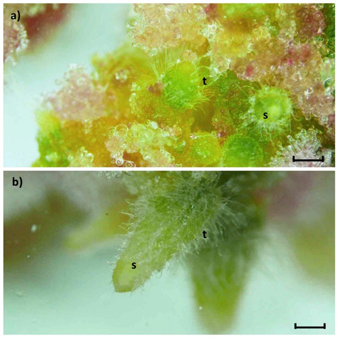
Figure 1 Regenerated shoots of Echinocactus parryi on subcultivated callus at 60 days of in vitro culture. a) massive rounded shoots (s) with trichomes (t) in the periphery of the callus, with 2,4-D 2.0 mg∙L-1. b) elongated shoots (s) showing trichomes (t) with kinetin 2.0 mg∙L-1. The bars represent 1 000 µm (1X).
Table 3 Effect of treatment with 2, 4-D in subcultivated callus from mature seed explants of Echinocactus parryi at 120 days of in vitro culture.
| 2, 4-D (mg∙L-1) | No. callus | Shoot number | Shoot mean | Transformed means ln (x+1) |
|---|---|---|---|---|
| 0 | 27 | 0 | 0 b | 0 |
| 2 | 27 | 48 | 0.791 ± 0.15 a | 0.23 |
| 4 | 27 | 17 | 0.259 ± 0.18 ab | 0.438 |
| 6 | 27 | 32 | 0.550 ± 0.12 ab | 0.583 |
Average values ± standard error. Means with different letters are statistically different according to Tukey's test (P ≤ 0.05).
Seedling explants
The ANOVA transformed data showed that 2, 4-D at four levels, the type of explant at three levels as main effects and their interactions were significant for embryogenic callus formation (Table 4). The Tukey's comparison of means (P ≤ 0.05) indicated that embryogenic callus induction was higher in the middle explant section (30.86 ± 0.40) and in the 2, 4-D treatment at 6 mg∙L-1, after 30 days of in vitro culture (Table 5). The apical, middle and basal sections of seedlings were competent to produce embryogenic callus at all evaluated concentrations of 2, 4-D compared to the control. The best interaction response was observed at all concentrations evaluated with the middle section explant (100 % embryogenic callus) and basal section explant with 2 and 6 mg∙L-1 of 2, 4-D (100 % and 92.9 ± 0.04 embryogenic callus, respectively). These explants showed globular structures after 30 days of in vitro culture (Table 6).
Table 4 Analysis of variance for embryogenic callus data of Echinocactus parryi in different seedling explant sections and 2, 4-D treatments.
| Source | df | MS | F | P |
|---|---|---|---|---|
| Explant | 2 | 11.57 | 643.3 | 0 |
| 2, 4-D | 3 | 116.1 | 6 451.2 | 0 |
| Explant*2, 4-D | 6 | 2.1 | 116.8 | 0 |
| Error | 96 | 0.01 | ||
| Total | 107 |
df = degrees of freedom; MS = mean square.
Table 5 Main effect of seedling explant section and 2, 4-D on the percentage of embryogenic callus formation of Echinocactus parryi at 30 days of in vitro culture.
| Factor | Embryogenic callus (%) | Data transformed ln (x+1) |
|---|---|---|
| Explant | ||
| Apical | 10.51 ± 0.28 c | 2.4436 |
| Medium | 30.86 ± 0.40 a | 3.4613 |
| Basal | 28.55 ± 0.39 b | 3.386 |
| 2, 4-D (mg∙L-1) | ||
| 0 | 0 ± 0 d | 0 |
| 2 | 58.83 ± 0.16 c | 4.0916 |
| 4 | 49.08 ± 0.19 b | 3.9136 |
| 6 | 79.05 ± 0.06 a | 4.3827 |
± standard error (n = 36). Means with different letters are statistically different between explant sections and between doses of 2, 4-D based on Tukey's test (P ≤ 0.05).
Table 6 Effect of the interaction of 2, 4-D treatment and seedling explant section on the percentage of embryogenic callus formation of Echinocactus parryi at 30 days of in vitro culture.
| 2, 4-D (mg∙L-1) | Explant | Embryogenic callus (%) | Transformed means In (x+1) |
|---|---|---|---|
| 0 | Apical | 0 ± 0 e | 0 |
| 2 | Apical | 20.0 ± 0 d | 3.044 |
| 4 | Apical | 14.5 ± 0.10 d | 2.738 |
| 6 | Apical | 53.1 ± 0.03 c | 3.991 |
| 0 | Medium | 0 ± 0 e | 0 |
| 2 | Medium | 100 ± 0 a | 4.615 |
| 4 | Medium | 100 ± 0 a | 4.615 |
| 6 | Medium | 100 ± 0 a | 4.615 |
| 0 | Basal | 0 ± 0 e | 0 |
| 2 | Basal | 100 ± 0 a | 4.615 |
| 4 | Basal | 79.4 ± 0.12 b | 4.387 |
| 6 | Basal | 92.9 ± 0.04 ab | 4.542 |
± standard error of the mean (n = 9). Means with different letters are statistically different according to Tukey's test (P ≤ 0.05).
The apical section still preserved a high percentage of intact tissue (Figure 2). The globular structures were white, green, and pink color varying from light to intense. After 120 days of culture, the globular stage did not evolve towards mature embryo development. The auxin 2, 4-D is hard to eliminate and stays in this tissue for an extended period. The more exposed the somatic embryos with this potent auxin can promote many abnormal embryos which cannot reach the following stages in some plant species (Garcia et al., 2019). Contrary, Osuna and Barrow (2004) observed that the shoot apexes of Bouteloua eriopoda (Torr.) Torr., cultivated with 2, 4-D or Dicamba (6-dichloro-o-anisic acid) for six weeks, the somatic embryos progressed from the globular to mature embryos resembled those naturally developed in seeds.

Figure 2 Response of apical, middle, and basal section of seedling explants of Echinocactus parryi after 30 days of culture. a) high cell division and embryogenic callus (ec) and shoot apex with intact tissue: apical meristem (am), cotyledons (c), and embryogenic callus (ec). b) middle section shows the embryogenic callus (ec) covering 100 % of the original tissue. c) basal section with embryogenic callus (ec) just in the cut part of the explant. The bars represent 1 000 µm (1X).
Compact green callus explants
In the case of E. parryi compact green callus (30 weeks old) cultivated in 2, 4-D, BAP and K, the data showed that only callus grown in medium with 0.5 mg∙L-1 K, promoted the highest number of somatic embryos after 30 days of culture. This treatment induced a mean of 19.2 embryos per explant. In contrast, the calli cultured on BAP or 2, 4-D treatments did not generate somatic embryos or shoots. The results indicated that compact green calli in E. parryi can produce somatic embryos without synthetic auxins. Also, Al-Dein, Al-Ramamneh, Sriskandarajah, and Serek (2006) used 1.5 mg∙L-1 K to achieve somatic embryos in globular, torpedo, and cotyledonal stage from Schlumbergera truncata (Haw.) Moran.
The somatic embryos of E. parryi were easily distinguished due to the typical white color, highly contrasting from the green color of the callus. The white color indicates the presence of carbohydrates and protein, essential for embryo development and germination in Ledebouria ovatifolia (Bak.) Jess. (Baskaran et al., 2016). The same characteristics were reported in Santalum album L. (Rugkhla & Jones, 1998), O. ficus-indica (Linhares et al., 2006), Zingiber officinale Rosc. (Lincy, Remashree, & Sasikumar, 2009) and Musa sp. (Debbrama, Sudhakar, Kumar, & Soorianathasundaram, 2019). The somatic embryos of E. parryi were transferred to the MS culture media without plant growth regulators to induce their germination. After 30 days, the somatic embryos did not germinate, and the number of embryos decreased drastically by about 50 %. The no germination of the somatic embryos could be due to an ineffective maturation due to malformations of the meristems (Correia, Cunha, Slagueiro, & Canhoto, 2012). Garcia et al. (2019) claim that abnormalities generated by genetic changes in the DNA of somatic embryos are hardly anomalies reversible; however, abnormalities caused by epigenetic changes can be reversible and abnormal embryos can produce plants in most cases. In contrast, other cactus species had successful germination of somatic embryos in a basal MS culture media. Some examples are the M. coccineus (Infante, 1992), Ariocarpus kostchoubeyanus (Lem.) K. Schum. (Moebius-Goldamer, Mata-Rosas, & Chávez-Avila, 2003), S. truncata (Al-Dein et al., 2006), and O. ficus-indica (Bouamama et al., 2011).
The induction of somatic embryos is possible only in genetically reprogramed totipotent plant cells, and their morphology relates to zygotic embryos (Osuna & Barrow, 2004; Silveira et al., 2013; Wu et al., 2020). Each embryogenesis comprises four stages: globular, heart, torpedo, and cotyledonal (Quiroz-Figueroa et al., 2006; Saptari & Susila, 2019). The somatic embryos of E. parryi, passed through all the stages at the same time (Figure 3). The achievement depends on the species and the supplementation of plant growth regulators. Kinetin had a more significant influence to induce all the somatic embryo phases than the rest of the treatments. Unfortunately, the latest stages did not germinate even though the compact calli were not cultured under 2, 4-D conditions. This lack of germination could be due to the plant genotype, which sometimes has ineffective maturation during the morphogenetic process. For example, the stored orchid somatic embryos (synthetic seeds) at different stages were germinated (91 %) on MS containing BAP and Kinetin at 0.5 mg∙L-1 each (Manokari et al., 2020). Regardless of somatic embryos have been induced in many plant species, there are still problems to be resolved in E. parryi.

Histology
The histological examination of the embryogenic callus samples showed the globular structures from seed explants (Figure 4). Figure 4a shows meristematic cells organized to form a globular embryo. The cells show intensely staining nuclei and thick cell walls. Meristematic cells are pluripotent and are the origin of the somatic embryos (Hui-Ju, Jen-Tsung, Hsiao-Hang, & Wei-Chin, 2018). In Figure 4b there are meristematic cells with a solid stained nucleus giving place to the formation of globular embryos. The protoderm were observed in an isolated globular embryo. The meristematic cells show a stained nucleus, dense cytoplasm, and starch grains (Figure 4c). The histological analysis revealed the tracheid cells in the vascular tissue surrounded by vacuolated cells (Figure 4d). Pinheiro et al. (2001) reported in O. ficus-indica, an unorganized tracheid structure observed in E. parryi, which probably means a disorder in endogenous metabolism and distribution auxin inside the cells that affect the maturation of the somatic embryos.

Figure 4 Histological analysis of the globular structures of embryogenic callus from seeds of Echinocactus parryi in 2.0 mg∙L-1 2, 4-D treatments. a) subepidermal globular embryo (arrow). b) meristematic cells (mc) with an intensely stained nucleus (arrow). c) isolated globular embryo with protoderm cells (p), meristematic cells with nuclei (arrow), and starch grains (sg) inside the cells. d) tracheid cells (arrowheads) and vascular tissue (arrows). The bars represent 200 µm (10X).
Figure 5 represents the longitudinal-section histological analysis of the generated somatic embryos from compact green callus. This analysis indicated that from a series of organized cell divisions, the formation of somatic embryos was possible. Figure 5a shows a pro-embryo that develops from sub-epidermal cells with a well-stained nucleus and vacuolated cells with an evident protoderm. The initiation of a vascular system was observed in the torpedo-shaped embryo with the procambium cells and vascular tissue from the explant (Figure 5b). The typical vascular system in embryo formation and the lack of connection between the embryos and the explant tissue is confirmed through histological analysis of O. ficus-indica (Linhares et al. 2006). In Figure 5c, a torpedo shape embryo is represented with an evident protoderm and procambium cells to initiate a vascular system. The procambium cells are a type of stem cell and can give a place to the formation of somatic embryos with a specific signal (Cunha et al., 2018; Rose, 2019). Figure 5d is a close sight of the cells from somatic embryos with a stained nucleus and numerous starch grains. Starch is a source of energy for cells that helps them in differentiation and cell division processes. It is also necessary for morphogenesis and differentiation of somatic embryos (Baskaran et al., 2016). The histological analysis is helpful to confirm the structural organization of the somatic embryos. The histological observations also showed the globular, heart-shaped, torpedo-shaped, and cotyledon stages.

Figure 5 Longitudinal-sections of somatic embryos from compact green callus of Echinocactus parryi in 0.5 mg∙L-1 Kinetin treatment. a) somatic pro-embryo emerging from the subepidermal region with meristematic cells and protoderm (p) (10X, bar 200 µm). b) somatic embryo in the torpedo stage with protoderm (p), procambium cells (pc), and vascular cells from the explant (arrow) (10X, bar 200 µm). c) somatic embryo with protoderm (p) and procambium cells (pc) (10X, bar 200 µm). d) meristematic cells of somatic embryos with starch grains (sg) and nucleus (n) (100X, bar 10 µm).
Conclusions
This study showed that Echinocactus parryi can produce a high number of embryogenic structures, which represents a great potential to grow massive plants. The seeds and seedlings explants induced globular embryos via callus, but they could not reach the last stages of somatic embryogenesis. However, the non-embryogenic calli induced shoot formation. On the other hand, with the explant of the green compact callus, it was possible to obtain mature somatic embryos in the torpedo and cotyledonal stage, but not its germination. Based on the explants analyzed, it could be determined that E. parryi is an embryogenic species that goes through all the stages (globular, heart, torpedo and cotyledon) of somatic embryos; however, further studies are required to explain why the last stages were not able to reach plant regeneration. Also, it would be interesting to analyze the effect of other plant growth regulators and different sucrose concentrations for somatic embryo maturation.

