


Services on Demand
Journal
Article
 text in
  English (pdf)
 Article in xml format
 Article references
 Send this article by e-mail
Indicators
-
 Cited by SciELO
 -
 Access statistics
Related links
-
 Similars in
SciELO
Similars in
SciELO
Share

 Permalink
Revista mexicana de ciencias forestales
Print version ISSN 2007-1132
Rev. mex. de cienc. forestales vol.13 n.70 México Mar./Apr. 2022 Epub May 09, 2022
https://doi.org/10.29298/rmcf.v13i70.1265
Research notes
Importance of the cuticular layer during the colonization of the fungus that causes negrilla in Agave salmiana Otto ex Salm-Dyck ssp. salmiana
1Escuela Superior de Apan, México.
2Universidad de Ciencias y Artes de Chiapas, Sede Acapetahua. México.
The Agave genus plants represent a non-timber forest resource, valuable for soil recovery. The epidermis of the agave leaves contains multiple stomata and is covered by a cuticular layer. Currently, agave plants have a fungal disease that is characterized by circular gray spots on the maguey leaves, which over time become necrotic and eventually, these lesions end up drying them. The aim of this work was to describe the cuticular layer during the fungus colonization that causes bold in Agave salmiana subsp. salmiana. The cuticular layer is 121 mm ± 2.8 thick. A homogeneous distribution of the stomata was observed, and the density (22.67-27.67 stomata mm-2) and the stomatal index (10.61-14.15) were determined. Tetracytic stomata and isodiametric polygonal epidermal cells were identified, the ostioles size were calculated as 57.9 mm ± 5 long and 23.75 mm ± 1.25 wide. The transverse and paradermal sections showed that the fungal hyphae and appressoria are restricted from the obverse side of the cuticular layer, which confirms the importance of preserving the epidermis in the maguey pulquero leaves.
Key words Agave salmiana Otto ex Salm-Dyck; Asterina mexicana Ellis & Everh.; cuticle layer; stomata; negrilla; ostiole
Las plantas del género Agave representan un recurso forestal no maderable valioso para la recuperación del suelo. La epidermis de sus pencas contiene múltiples estomas y está cubierta por una capa cuticular. En la actualidad presentan una enfermedad fúngica que se caracteriza por la existencia de manchas grises circulares sobre las pencas, que con el tiempo se tornan necróticas; y en ocasiones, dichas lesiones terminan por secar las pencas. El objetivo de este trabajo fue describir la relevancia de la capa cuticular durante la colonización del hongo causante de la negrilla en Agave salmiana ssp. salmiana. La capa cuticular tiene un grosor de 121 ± 2.8 mm. Se observó una distribución homogénea de los estomas y se determinó la densidad (22.67-27.67 estomas mm-2) y el índice estomático (10.61-14.15). Los estomas observados son de tipo tetracítico, el tamaño de los ostiolos de 57.9 mm ± 5 de largo y 23.75 mm ± 1.25 de ancho y células epidérmicas poligonales isodiamétricas. Los cortes transversales y paradermales muestran que las hifas y los apresorios fúngicos quedan restringidos al lado anverso de la capa cuticular, por lo cual se corrobora la importancia de conservar la epidermis en las pencas del maguey pulquero.
Palabras clave: Agave salmiana Otto ex Salm-Dyck; Asterina mexicana Ellis & Everh.; capa cuticular; estomas; negrilla; ostiolo
The plants of the Agave genus represent a valuable non-timber forest resource for soil recovery, particularly in arid and semi-arid zones. The leaves (pencas) of agaves are covered with a cuticular layer that performs relevant functions, such as: gas exchange, control of temperature changes, as a barrier against pathogens, among others (Tafolla-Arellano et al., 2013). The cuticular layer that covers the leaves of Agave salmiana Otto ex Salm-Dyck provides highly appreciated characteristics in order to cook some traditional-food dishes; it is made up of epidermal cells, cutin, polysaccharides, waxes and stomata (Reina-Pinto and Yephremov, 2009; Staiger et al., 2019; Pérez-España et al., 2019). Stomata are formed by two guard or occlusive cells, which give rise to a pore called an ostiole. Guard cells control the opening and closing of ostioles in response to hormonal and environmental signals to regulate gas exchange (Zeng and He, 2010; Biswapriya et al., 2015). Agaves have stomata on the abaxial and adaxial face of the leaves (Hernández-Valencia et al., 2003) that open at night and remain closed during the day to prevent excessive loss of liquids in the plant (Lee, 2010).
Phytopathogenic fungi use different mechanisms to colonize plants (Gudesblat et al., 2009) such as: adhesion of spores to the cuticular layer, formation of appressoria and adhesive, invasive and infective structures, development of a high pressure of turgidity in the invaded cells (Mendgen et al., 2016) and degradation of the cell wall and the cuticular layer through the secretion of enzymes. Other phytopathogens have the ability to modulate the opening or closing of stomata through the release of chemical compounds (Staples and Macko, 1980; Lee et al., 1999; Bury et al., 2013).
At present, A. salmiana plants show a disease known as smallpox or “negrilla”; caused by the Asterina mexicana Ellis & Everh. fungus (Ellis y Everhart, 1900; Cesaveg, 2008), of which there is little information. This disease causes dark spots on both sides of the leaves and can lead to necrosis and dryness. The objective of this work was to describe the importance of the cuticular layer during the colonization of the fungus that causes the negrilla in Agave salmiana Otto ex Salm-Dyck ssp. salmiana.
The cuticular layers were obtained from leaves of agave plantations in Pachuca de Soto, Apan, Cardonal (state of Hidalgo) and Calpulalpan (state of Tlaxcala) municipalities, from February to June 2019. Three leaves which carried symptoms of the disease were collected per ~8-year-old plant (according to the experience of the producers) by municipality. From the adaxial face of each penca, a sample was taken from the apical, middle and base sites (Figure 1a). Cuticular layer samples were removed from the collection site and kept in a 2 % glycerol solution until they were analyzed.
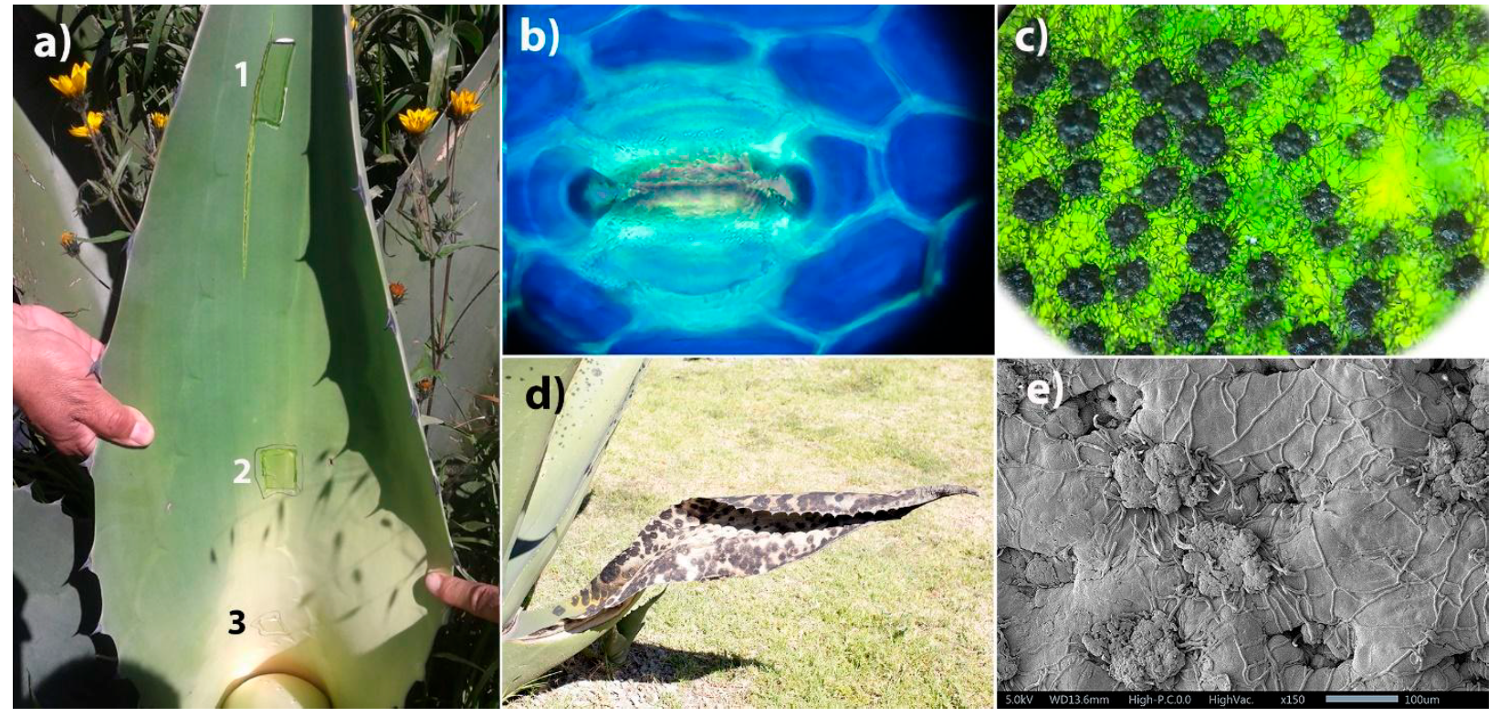
Figure 1 Cuticular layer of Agave salmiana Otto ex Salm-Dyck ssp. salmiana, (a) Extraction of the cuticular layer in three areas of the adaxial face: 1) distal area, 2) middle area and 3) proximal area; (b) Adverse of stomata, guard cells and epidermal cells; (c) appressoria observed in black spot (4X), (d) dehydrated penca due to invasion of the bold and (e) appressoria obstructing the stomata (SEM).
Stomatal index (SI) and stomatal density (SD) were determined with an optical microscope (Zeigen, ZB-7300). Cuticles were stained with methylene blue for better definition. The SI was calculated according to what was recommended by Sosa et al. (2014) from the following formula:
Where:
NE = Number of stomata per observation field
CE = Number of epidermic cells in the same observation field
SD was determined by calculating the number of stomata found in 19.635 mm-2, equivalent to the field observed (40X) in a 2.5 mm radius. The SI and SD results were analyzed with ANOVA and the significant differences (P < 0.05) between the groups with the Bonferroni test (GraphPad Prism Software version 5.00 for Windows, GraphPad Software Inc.).
For the analysis of the microstructure, the cuticular layers were dehydrated in a vacuum oven (Shel-Lab, Mod. 1407) at 40 °C and 45 kPa. Subsequently, they were fixed with carbon tape to an aluminum slide, covered with gold-palladium and observed with a scanning electron microscope (SEM; Model JEOL JMS-5600LV).
The distribution of the stomata in the cuticular layer of the abaxial face showed homogeneity in the analyzed areas. In the micrographs of the cuticular layer of A. salmiana ssp. salmiana with a 121 ± 2.8 mmascomata thickness, the presence of waxes, epidermal cells and stomata was recognized. The average thickness recorded for the cuticle of A. tequilana F. A. C. Weber is 8.67 μm for the abaxial face (Hernández-Valencia et al., 2003).
SD showed homogeneity in the analyzed areas. The tetracytic-type stomata, the size of the ostioles of 57.9 mm ± 5 long and 23.75 mm ± 1.25 wide, and the isodiametric polygonal epidermal cells (Figure 1b) are similar to those reported by Vargas-Rodríguez et al. (2017) and by Chávez-Güitrón et al. (2019). The difference in the size of the ostioles is attributed to variations in the thickness and distribution of the cuticular wax, to the species and to the environmental conditions in which the plants grow (García, 2007; Bernardino-Nicanor et al., 2012).
Macroscopically, bold disease is seen as gray spots that turn black over time (Figure 1d). Inside the stain, the existence of ascomata with hyphae specialized in the invasion of plant tissue is identified (Figure 1c) that accumulate in the Agave leaf and obstruct the stomata; in addition, other hyphae adhered to the cuticular layer are observed (Figure 1e).
SD (22.67 to 27.67 stomata mm-2) did not show significant differences between the areas of the adaxial face of the analyzed leaves or between municipalities (Figure 2a); the values (mm-2) were similar to those recognized for other Agave species, such as: A. mapisaga Trel in L. H. Bailey (20-50 stomata; Nobel, 1994), A. deserti Engelm. (34 stomata; Gentry and Sauck, 1978) and A. atrovirens Karw. Ex Salm-Dyck (29-30 stomata; Bernardino-Nicanor et al., 2012); but they differ from A. promontorii Trel. (18 stomata) and A. sisalana Perrine (11 stomata; Neto and Martins, 2012).

Densidad estomática = Stomatal density; Índice estomático = Stomatal index; Apical = Apical; Media = Middle; Basal =Base.
Figure 2 Index and stomatal density in the cuticular layer of Agave salmiana Otto ex Salm-Dyck ssp. salmiana in different municipalities.
SI for A. salmiana ssp. salmiana is 10.61-14.15 (Figure 2b) while for A. tequilana it is between 5.72 and 6.68 stomata/epidermal cells (Hernández-Valencia et al., 2003). Both parameters (IE and SD) are influenced by some environmental and nutritional conditions (Wilkinson, 1979; Roth et al., 1986).
In the paradermal and transverse sections, it was noted that the cuticular layer acts as a mechanical barrier and prevents the passage of the reproductive structures of the pathogenic fungus into the interior of the Agave leave. With the SEM micrographs, it was detected that the hyphae and appressoria of the phytopathogen only appear on the obverse side of the cuticular layer since they could not be identified on the reverse side. In addition, it was observed that the appressoria are located mainly in the stomata, which suggests that the mechanism of invasion of this fungus consists of drying the leaves through the modulation of the stomata, by preventing them from completing the cycle (day-night) in a normal way for affected plants.
SEM micrographs show the importance of the cuticular layer as a mechanical barrier by preventing hyphae from entering the innermost layers of the maguey leaf. Therefore, the infection is restricted to the obverse side of the cuticular layer.
On the other hand, SD coincides with the records for other species of the Agave genus. However, this is the first report on the importance of the cuticular layer of Agave salmiana ssp. salmiana during its interaction with the phytopathogenic fungus responsible for the bold disease. Complementary studies are necessary to propose alternatives to prevent or control fungal invasion.
Referencias
Bernardino-Ninacor, A., R. Mora-Escobedo, J. L. Montañez-Soto, S. Filardo-Kerstupp and L. González-Cruz. 2012. Microstructural differences in Agave atrovirens Karw leaves and pine by age effect. African Journal of Agricultural Research 7(24):3550-3559. Doi:10.5897/ AJAR11.1185. [ Links ]
Biswapriya, B. M., R. A. Biswa, D. Granot, S. M. Assmann and S. Chen. 2015. The guard cell metabolome: functions in stomatal movement and global food security. Frontiers in Plant Science 6:1-13. Doi:10.3389/fpls.2015.00334. [ Links ]
Bury, M., A. Andolfi, B. Rogister, A. Cimmino, V. Mégalizzi, V. Mathieu, O. Feron, A. Evidente and R. Kiss. 2013. Fusicoccin A, a phytotoxic carbotricyclic diterpene glucoside of fungal origin, reduces proliferation and invasion of glioblastoma cells by targeting multiple tyrosine kinases. Translational Oncology 6(2):112-23. Doi: 10.1593/tlo.12409. [ Links ]
Comité Estatal de Sanidad Vegetal de Guanajuato A.C. (CESAVEG). 2008. Manual de plagas y enfermedades del Agave. Casa editorial. Irapuato, Gto., México. 28p. [ Links ]
Chávez-Güitrón, L. E., F. C. Salinas-Pérez, E. A. Pérez-Salinas, J. Caballero, A. Vallejo-Zamora y E. Sandoval-Zapotitla. 2019. Variación de caracteres epidérmico-foliares de Agave salmiana subsp.salmiana (Asparagaceae) en el centro de México. Botanical Sciences 97 (4): 711-724. Doi: 10.17129/botsci.2159. [ Links ]
García M., A. 2007. Los agaves de México. Ciencias 87:14-23. https://www.redalyc.org/pdf/644/64408704.pdf (20 de enero de 2021). [ Links ]
Gentry, H. S. and J. R. Sauck. 1978. The stomatal complex in Agave: groups Deserticolae, Campaniflorae, Umbelliflorae. Proceedings of the California Academy of Sciences series 41:371-387. https://ia800206.us.archive.org/0/items/biostor-78394/biostor-78394.pdf (25 de octubre de 2020). [ Links ]
Gudesblat, E. G., S. P. Torres and A. A. Vojnov. 2009. Stomata and pathogens: warfare at the gates. Plant Signaling & Behavior 4(12):1114-1116. Doi: 10.4161/psb.4.12.10062. [ Links ]
Hernández-Valencia, R. E. M., R. López-Franco and A. Benavides-Mendoza. 2003. Micromorfología de la epidermis foliar de Agave tequilana Weber. Agrofaz 3(2):387-396. https://www.researchgate.net/publication/283995003 (15 de octubre de 2020). [ Links ]
Lee S., H. Choi, S. Suh, I.S. Doo, K. Y. Oh, E. J. Choi, A. T. S. Taylor, P. S. Low and Y. Lee. 1999. Oligogalacturonic acid and chitosan reduce stomatal aperture by inducing the evolution of reactive oxygen species from guard cells of tomato and Commelina communis. Plant Physiology 121:147-52. Doi:10.1104/pp.121.1.147. [ Links ]
Lee, J. S. 2010. Stomatal opening mechanism of CAM plants. Journal of Plant Biology 53:19-23. Doi:10.1007/s12374-010-9097-8. [ Links ]
Mendgen, K., M. Hahn and H. Deising. 1996. Morphogenesis and mechanisms of penetration by plant pathogenic fungi. Annual Review of Phytopathology 34:367-86. Doi: 10.1146/annurev.phyto.34.1.367. [ Links ]
Neto, I. L. C. and F. M. Martins. 2012. Anatomia dos órgãos vegetativos de Agave sisalana Perrine ex Engelm (Agavaceae). Revista Caatinga 25(2):72-78. https://www.redalyc.org/pdf/2371/237123825011.pdf (12 de febrero de 2021). [ Links ]
Nobel, P.S. 1994. Remarkable Agaves and Cacti. Cambridge University Press. New York, NY, USA. 166 p. [ Links ]
Pérez-España, V. H., J. A. Cuervo-Parra, C. Paz-Camacho, M. A. Morales-Ovando, C. A. Gómez-Aldapa, G. C. Rodríguez-Jimenes, V. J. Robles-Olvera, P. A. López-Pérez and T. Romero-Cortés. 2019. General characterization of cuticular membrane isolated from Agave salmiana. International Journal of Bio-resource and Stress Management 10(1):046-052. Doi:10.23910/IJBSM/2019.10.1.1950. [ Links ]
Reina-Pinto, J. J. and A. Yephremov. 2009. Surface lipids and plant defenses. Plant Physiology and Biochemistry 47(6):540-549. Doi:10.1016/j.plaphy.2009.01.004. [ Links ]
Roth I., T. Merida y H. Lindorf. 1986. Morfología y anatomía foliar de plantas de la Selva Nublada de Rancho Grande. Parque Nacional “Henry Pittier”. El ambiente físico, ecología general y anatomía vegetal. Fondo Editorial Acta Científica Venezolana. Caracas, Venezuela. pp. 205-241. [ Links ]
Sosa, C. M., G. S. Alemán, H. Y. Pérez, C. E. Abreu, C. D. Sosa and O. G. González. 2014. Caracterización de la lámina foliar de plantas de Agave fourcroydes Lem. obtenidas por propagación asexual. Biotecnología Vegetal 14(1):37-44. https://revista.ibp.co.cu/index.php/BV/article/view/40/433 (12 de febrero de 2021). [ Links ]
Staiger, S., P. Seufert, K. Arand, M. Burghardt, C. Popp and M. Riederer. 2019. The permeation barrier of plant cuticles: uptake of active ingredients is limited by very long-chain aliphatic rather than cyclic wax compounds. Pest Management Science 75(12):3405-3412. Doi: 10.1002/ps.5589. [ Links ]
Staples, R. C. and V. Macko. 1980. Formation of infection structures as a recognition response in fungi. Experimental Mycology 4(1):1-15. Doi:10.1016/0147-5975(80)90045-6. [ Links ]
Tafolla-Arellano, J. C., A. González-León, M. E. Tiznado-Hernández, L. Zacarías G. and R. Báez-Sañudo. 2013. Composición, fisiología y biosíntesis de la cutícula en plantas. Revista Fitotecnia Mexicana 36(1):3-12. http://www.scielo.org.mx/scielo.php?script=sci_arttext&pid=S0187-73802013000100001&lng=es&tlng=es (20 de noviembre de 2020). [ Links ]
Vargas-Rodríguez, L., M. I. García-Vieyra, B. I. León-Bata and P. Lozano-Sotomayor. 2017. Physical properties and microscopic structure of the Agave salmiana cuticle (mixiote). Revista Chapingo Serie Zonas Áridas 17(2):1-9. Doi: 10.5154/r.rchsza.2017.12.017. [ Links ]
Wilkinson, H. 1979. The plant surface (mainly leaf). In: C. R. Metcalfe and L. Chalk (eds.). Anatomy of Dicotiledons. Oxford Claredous Press. London, UK. pp. 97-16. [ Links ]
Zeng, W. and S. Y. He. 2010. A prominent role of the flagellin receptor FLAGELLIN-SENSING2 in mediating stomatal response to Pseudomonas syringae pv tomato DC3000 in Arabidopsis. Plant Physiology 153:1188-1198. Doi: 10.1104/pp.110.157016. [ Links ]
Received: August 17, 2021; Accepted: February 22, 2022
 Este es un artículo publicado en acceso abierto bajo una licencia Creative Commons
