


Services on Demand
Journal
Article
 text in
  English (pdf)
 Article in xml format
 Article references
 Send this article by e-mail
Indicators
-
 Cited by SciELO
 -
 Access statistics
Related links
-
 Similars in
SciELO
Similars in
SciELO
Share

 Permalink
Revista mexicana de ciencias agrícolas
Print version ISSN 2007-0934
Rev. Mex. Cienc. Agríc vol.10 n.7 Texcoco Sep./Nov. 2019 Epub Dec 04, 2020
https://doi.org/10.29312/remexca.v10i7.1827
Essays
Main diseases in citrus
1División de Estudios de Postgrado e Investigación-Facultad de Ingeniería y Ciencias-Universidad Autónoma de Tamaulipas. Centro Universitario Adolfo López Mateos, Cd. Victoria, Tamaulipas, México. Tel. 834 3181721, ext. 2111. (alhe-saenz@outlook.com; benestrada@docentes.uat.edu.mx; wpoot@docentes.uat.edu.mx; rdelgado@docentes.uat.edu.mx).
2Departamento de Investigación en Alimentos-Facultad de Ciencias Químicas-Universidad Autónoma de Coahuila. Boulevard Venustiano Carranza esquina con Ing. José Cárdenas Valdés, Col. República, Saltillo, Coahuila, México. CP. 25280. Tel. 844 4161238. (raul.rodriguez@uadec.edu.mx).
Citrus fruits are one of the most important crops worldwide, as it is produced in 140 countries. One of the limitations for its production are diseases, they represent considerable damages in the orchards and due to this they diminish the production and cause millionaire losses in the citrus sector. The main diseases are: Phytophthora spp., Mycosphaerella citri, Lasiodiplodia theobromae, Candidatus Liberibacter spp. and citrus sadness virus (VTC). Pathogens can occur at any phenological stage of the crop and throughout the plant, whether in leaves, stems, roots or fruits. These microorganisms cause symptoms of chlorosis, mottling, less development and growth, deficiencies in the absorption of water and nutrients, rot, necrosis and in more extreme cases the death of the plant. For the management of phytopathogens there are different alternatives, the application of agrochemicals based on sulfur and copper (oxychloride and sulfate) are efficient in 70%, in addition, the use of resistant varieties (C. volkameriana, C. aurantium, among others), cultural measures such as thermotherapy, weed control and good drainage in the garden. On the other hand, to a lesser extent biological control is implemented, the application of beneficial microorganisms (Trichoderma, Bacillus subtilis) have demonstrated effectiveness up to 80%. As well as the release of parasitoids and predators such as Tamarixia radiata, Cycloneda sanguinea and Harmonia axyridis, which represent a good alternative for vector control. Therefore, the objective of this review is to describe the impact and status of the main citrus diseases present in Mexico.
Keywords: control; epidemiology; pathogen; symptomatology
Los cítricos son uno de los cultivos de mayor importancia a nivel mundial, ya que se produce en 140 países. Una de las limitantes para su producción son las enfermedades, representan daños considerables en las huertas y debido a esto disminuyen la producción y provocan pérdidas millonarias en el sector citrícola. Las principales enfermedades son: Phytophthora spp., Mycosphaerella citri, Lasiodiplodia theobromae, Candidatus Liberibacter spp. y virus de la tristeza de los cítricos (VTC). Los patógenos pueden presentarse en cualquier etapa fenológica del cultivo y en toda la planta, ya sea en hojas, tallos, raíces o frutos. Dichos microorganismos causan síntomas de clorosis, moteado, menor desarrollo y crecimiento, deficiencias en la absorción de agua y nutrientes, pudrición, necrosis y en casos más extremos la muerte de la planta. Para el manejo de los fitopatógenos existen diferentes alternativas, la aplicación de agroquímicos a base de azufre y cobre (oxicloruro y sulfato) son eficientes en 70%, además, el uso de variedades resistentes (C. volkameriana, C. aurantium, entre otros), medidas culturales como termoterapia, control de malezas y buen drenaje en el huerto. Por otra parte, en menor medida se implementa el control biológico, la aplicación de microorganismos benéficos (Trichoderma, Bacillus subtilis) han demostrado efectividad hasta 80%. Así como la liberación de parasitoides y depredadores como Tamarixia radiata, Cycloneda sanguínea y Harmonia axyridis, los cuales representan una buena alternativa para el control de vectores. Por lo anterior, el objetivo de esta revisión es describir el impacto y estatus de las principales enfermedades de cítricos presentes en México.
Palabras clave: control; epidemiología; patógeno; sintomatología
The Citrus genus is one of the most important crops worldwide, it is distributed in tropical and subtropical regions of more than 140 countries (Vu et al., 2018). This crop has a world production of more than 124 million tons, the main producing countries are China, Brazil, India, United States of America, Spain and Mexico (FAO, 2017). In Mexico, currently the cultivated area of citrus is 590 thousand h and a production volume of 8 million tons (SIAP, 2019). However, citrus production has been affected by the damage of pests and diseases (Zhang et al., 2012), which translates into large economic losses.
To counteract this problem, it is necessary to monitor the culture and detect the pathogens in the first phase of infection to reduce their incidence (Martinelli et al., 2015). Citrus diseases are mainly caused by fungi, viruses and bacteria. Concerning fungi, these microorganisms make up the majority of the phytopathogenic agents in the Citrus genus (Baraona and Sancho, 2000) and their damage can manifest in roots, trunks, branches, leaves and fruits (Zhao et al., 2015; Showler, 2017).
Among the most important cryptogamic diseases in citrus fruits, there are: Phytophthora spp., Mycosphaerella citri and Lasiodiploida theobromae (Zhao et al., 2015; García-Martin et al., 2018). In general, these pathogens cause lesions and stem rot (Yan et al., 2017), necrotic spots on leaves (Silva et al., 2015), defoliation of the tree (Picos-Muñoz et al., 2015), rot of fruits and roots, decrease the vigor and production of the tree (Showler, 2017) and finally death (Graham et al., 2013).
On the other hand, viruses depend completely on a host to survive, as are some species of plants, they spread through insects, nematodes, mites and propagation of plant material, the main viral diseases are: exocortis, excretion of the citrus woody veins and gills, citrus sadness virus and leprosis (Agusti, 2010). Bacterial diseases decrease production and in more severe cases the death of trees (Hernández et al., 2013).
As with the bacterium Candidatus Liberibacter spp., which causes huanglongbing or citrus greening, which is currently considered the most destructive citrus disease, since its appearance in China at the end of the 20th century until 2017 has caused the death of more than 60 million trees (Zhang et al., 2013; Wang et al., 2017). Based on the above, the objective of this review is to summarize and discuss the impact and status of the main citrus diseases present in Mexico.
Gummosis (Phytophthora spp.)
Economic importance: the pathogen is present in most citrus orchards in Brazil, California and Florida, in the last two locations the pathogen reduces production 46% and 8%-20% of the total cultivated area respectively, this is It translates into losses ranging from 30 to 60 million dollars (Graham and Feichtenberger, 2015). Similarly, in Tabasco, Mexico presents an incidence of the disease of 10%, which represents losses of 730 thousand tons.
Etiology and epidemiology: the mycelium morphology are cenocitic with white cottony colonies (Figure 1a) (Alvarez-Rodríguez et al., 2016). The ontogeny of the sporangiophore is simple and the sporangia are papillated, deciduous, fusiformly with long and off-center pedicel insertion (Figure 1b (Hanumanthappa et al., 2018). Various factors influence the occurrence of the disease, rain splashes, runoff, sprinkler irrigation systems, water stagnation, susceptible varieties and grafts close to the ground (Baraona and Sancho, 2000).

Figure 1 a) growth of Phytophthora spp.; b) mycelium and sporangium of Phytophthora.; c) rubber on the trunk; and d) tree with symptoms of gummosis.
Similarly, for the multiplication and spread of spores of the pathogen, a relative humidity greater than 80% and temperatures of 28 °C to 32 °C are required (Vicent, 2011). The infection begins at ground level by zoospores that are transported through water and extends through the trunk and down to the roots (Srinivasulu et al., 2018).
Symptomatology: stem gummosis manifests near the ground, at the junction of the rootstock and the variety (Figure 1c), in the same way, the pathogen rings the stem, affects the root cortex until the fibrous roots break down (Yan et al., 2017). Therefore, the absorption of water and nutrients such as nitrogen, phosphorus, potassium, calcium, iron and magnesium decreases. Consequently, the tree shows chlorosis, defoliation, lower vegetative growth and fruit production (Figure 1d) (Tanoi and Kobayashi, 2015; Srivastava and Shirgure, 2018).
Control methods: due to its high toxicity against the pathogen, low cost and prolonged residual effects (Lamichhane et al., 2018), the most common control method is the use of chemical fungicides such as phosphites and copper sulfate (Graham and Feichtenberger, 2015). For example, quarterly applications during a year via trunk injection of fosetil aluminum in conjunction with propamocarb (2.8 L ha-1), show twice the efficacy in the attack of the disease (Pabon-Villalobos and Cataño-Zapata, 2015).
In the same way, the biological control shows an efficiency of 45% with Trichoderma applications to the tree stem at intervals of 21 days during a year (Adedeji et al., 2010). Finally, the use of resistant patterns such as Citrange Troyer, Citrange Carrizo, Swingle Citrumelo CPB 4475, Poncirus trifoliata and Citrus aurantium represent a good option for pathogen control (Lucas and Beltrán, 2004).
Greasy spot (Mycosphaerella citri)
Economic importance: he greasy spot of citrus is caused by the fungus Mycosphaerella citri, it is considered the most important foliar fungal disease in Florida, Texas, eastern Mexico, Central America and the Caribbean basin (Baraona and Sancho, 2000; Showler, 2017). While Ghana, Africa, the greasy spot is the most important fruit disease, and can cause a 22% loss in citrus production (Brentu et al., 2012). In Mexico it was first detected in 1980 in the states of Chiapas and Tabasco, later in Veracruz. The combat of this fungus represents 35 to 45% of the total cost of production (Orozco-Santos et al., 2012).
Etiology and epidemiology: the fungus Mycosphaerella citri is characterized by its greenish hyphae, which make up the conidiophores with integrated conidiogens that expand near the apex, the scars can be generally pigmented and dark. On the other hand, conidia are formed individually or in short chains. The shape of the ascospores varies from cylindrical to fusiform, warts, obovate to obconical, subhyaline to pigmented, 0-pluri-septate, with conspicuous refractory yarn, slightly pigmented, thickened (Crous et al., 2009).
Fallen leaves are the main source of inoculum for this disease, during the winter it is present as pseudotecios and once the ascospores are mature they spread more easily by air currents (Showler, 2017), the relative humidity close to 100% and high temperatures (35 °C) for prolonged periods favor the manifestation of the disease (Silva et al., 2015).
Symptomatology: the disease manifests itself mainly in leaves and to a lesser extent in fruits, the symptoms in leaves are expressed in the underside of mature leaflets and slightly elevated lesions of different shades appear (Figure 2a and 2b). In the early stages of infection, the color begins yellow, then brown in an intermediate advance and ends with black “necrotic” or chlorotic spots with oily margins (Silva et al., 2015).
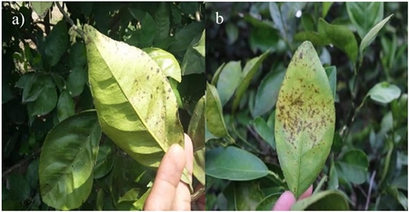
Figure 2 a) and b) The symptoms of Mycosphaerella citri manifest with necrotic lesions on the underside of the leaf.
After the necrosis, the plant presents early defoliation in winter, so that the vigor and the yield of the fruits decrease between 20% and 45%. In the fruits, tiny necrotic spots appear in the shell and thereby detract from the commercial value of the product (Silva et al., 2015; Showler, 2017).
Control methods: the use of cupric fungicides and oils represent a control solution against the fungus; however, its effectiveness is not reliable (Abbas and Fares, 2009). Since, the excess of applications in the same cycle causes resistance of the fungus, consequently, there is a high presence of inoculum and decreases the effectiveness by 45% (Rodríguez et al., 2011).
On the other hand, they concluded that the application of fungicides Frutriafol and Trifloxistrobim significantly reduce the incidence and severity of the pathogen by up to 70%. However, the efficacy in the control of the pathogen is greater with the joint application of Difeconazole and Azoxidifem (87.5%) and copper oxychloride (75%). Similarly, Showler (2017) mentioned that the use of vegetable oils or fish oil in combination with organic mixtures such as molasses, humic acid and cornmeal reduces foliage damage by up to 25%.
Neck rot (Lasiodiplodia theobromae)
Economic importance: the fungus Lasiodiplodia theobromae is of great economic importance (Sathya et al., 2017). In Mexico, this pathogen is reported in cocoa, avocado and papaya crops; however, in the available literature there is no study that quantifies the attack and monetary losses of this pathogen to citrus cultivation (Picos et al., 2015).
Etiology and epidemiology: the causative agent, Lasiodiplodia theobromae, shows pycnidia (fruiting bodies) in the form of a dark flask, in advanced stages of maturation they have a hollow, long and neck-shaped structure, by this same, through a pore the conidia of globose appearance and light brown coloration are released, as they mature, a longitudinal tabulation and striation appears, therefore, they usually have a size of 31.3 - 42.9 × 15.6 - 19.5 μm (Netto et al., 2014).
Additionally, the description of this pathogen is based on the sequencing of the intergenic spacer regions of the rDNA (ITS) and the elongation factor 1 alpha (EF-1) (Picos et al., 2015). For the growth of this pleomorphic and ubiquitous fungus, temperature of 28 °C with 75% relative humidity are optimal. Infection of the fungus begins with the production and development of pycnidia in dead or senescent leaves.
Subsequently, the conidia disperse through water or wind and enter the plant through wounds caused by insects, pruning or natural causes. Consequently, the fungus colonizes endophitically the tissue of the branches prior to inflorescence, days after flowering the pathogen reaches the pedicel of the fruit (Noriega-Cantu, 2017). Finally, the pathogen enters through the spongy central axis of the fruit and infects the calyx and the floral disk, therefore, this latent pathogen is considered, since it causes an endophytic infection (Sathya et al., 2017).
Symptomatology: the symptoms are expressed in stem rot, the cortex softens at the periphery of the button and acquires a brown coloration on the infected surfaces (Figure 3a). Therefore, the pathogen is more easily dispersed to other plant organs (Zhao et al., 2016). Under the cuticle the tissue is necrotic and mummifies the fruit; however, in the leaves there are spots in more advanced stages causing senescence and with it the descending death (Figure 3b) (Picos-Muñoz et al., 2015).

Figure 3 Damage from Lasiodiplodia theobromae. a) stem rot; and b) descending death, the tree presents total defoliation.
Control methods: the common control method is using chemicals, derivatives of copper and sulfur. For example, applications of benomyl and copper oxychloride in different phenological phases of the crop are effective in combating this fungus. As a biological control of the pathogen, Segura-Contreras et al. (2017), mention the extract of Schinus molle, at a concentration of 30% of it can inhibit 90% the presence of the fungus.
Huanglongbing (Candidatus Liberibacter spp.)
Economic importance: huanglongbing (HLB) or yellow dragon, is considered one of the most devastating diseases of citrus worldwide because of its rapid spread and devastation, since it reduces crop yields and causes the death of the tree (Wang et al., 2017). Its distribution covers countries of the Asian, African and American continent (Gottwald, 2010). In Mexico, the decrease in production per year is around 25% (1.84 million t) due to this disease.
Also, Salcedo et al. (2010) stated that HLB damage can reach up to 41% of production (three million tons) under a high intensity epidemic scheme. On the other hand, the states most affected by the pathogen in the Mexican Republic are Colima and Yucatan, with a harvest decrease of 17.3% and 62%, respectively (Mora-Aguilera et al., 2016). On the contrary, the citrus region of the state of Sonora is free of the disease.
Etiology and epidemiology: HLB are associated with the bacterium Candidatus Liberibacter, present in infected plant material and insects such as Trioza erytreae and Diaphorina citri (Zhang et al., 2014), the latter being considered the main vector of the causative agent of the disease (Hall, 2018). Based on phylogenetic studies it has been detected in the 16S rRNA region that Ca. Liberibacter belongs to the α-2 proteobacteria and is a large negative bacterium. In addition, the bacterium has a diameter of 0.2 to 0.3 μm and a membrane that contains a layer of peptidoglycan (Camacho-Tapia et al., 2016).
The species of Ca. Liberibacter that have been identified by their pathogenicity exclusively in citrus fruits are: Candidatus Liberibacter asiaticus (CaLas), Candidadus Liberibacter africanus (CaLaf), and Candidatus Liberibacter americanus (CaLam) (Camacho-Tapia et al., 2016). The bacterium is located in the hemolymph and salivary glands of insect vectors, enters the plant through the stylus of the psyllid and travels; through the phloem sieve tubes until reaching the root system, it is here that the bacteria replicates and distributes irregularly to the rest of the plant (Johnson et al., 2013).
Symptomatology: the infection in trees presents with a bulge in the middle lamella of the leaves, in more advanced cycles collapses of the phloem cells occur, consequently, the vascular tissue becomes necrotic and blocks the flow and translocation of nutrients; this leads to anatomical changes in the leaves such as: yellowing of the ribs and the appearance of irregular spots ranging from yellow to dark green tones (Figure 4a and 4b) (Folimonova and Achor, 2010), this condition can be confused with zinc deficiency (McCollum et al., 2016). Some leaves have the symptom of ‘rabbit ears’, which consists of the vertical growth of new shoots with compressed internodes (Tolba and Soliman, 2015).

Figure 4 Symptomatology of HLB. a and b) irregular spotted on the leaves; c) color inversion; and d) deformed fruit with thickening of the pericarp.
In fruits, it is manifested in asymmetry and smaller size (Figure 4d), thickening of the pericarp, seed abortion, yellow staining of the vascular region, color inversion (Figure 4c) and reduction of soluble solids and Brix grades, this last characteristic demerits organoleptic quality, therefore, is not possible for use in industry (Bove, 2006). It should be noted that the disease in more advanced stages causes severe defoliation in the tree, abortion of fruits and finally death (Wang et al., 2017).
Control methods: there is currently no effective cure or treatment for HLB, however, preventive disease management is carried out through combat and eradication of the psyllid by chemical and biological control (Hernández et al., 2013). In Mexico, insecticide applications are made such as: argenomine, azadarictine, imidacloprid, zetacipermethrin, in addition, the use of antibiotics such as oxytetracilin, ampicillin, streptomycin, tetracycline and penicillin (Zhang et al., 2014).
Another option to combat the bacteria is the nutritional programs based on zinc, copper and manganese (Gottwald et al., 2012). Similarly, thermotherapy with a range of 40 °C- 42 °C has 90% effectiveness (Fan et al., 2016). Biological control is carried out by the release of Tamarixia radiata which reduces the populations of the Diaphorina citri psyllid. Finally, the use of certified plant material tolerant to HLB is essential (Mora-Aguilera et al., 2016). Currently, ‘Lise’ a variety of Mexican lemon developed at the National Institute of Forestry, Agricultural and Livestock Research (INIFAP) of Tecoman, Colima offers greater resistance to the HLB attack. Also, it can be successfully grown in the states of Michoacán, Oaxaca and Guerrero with an average yield of 40 t per hectare per year (SADER, 2018).
Citrus sadness virus (VTC)
Economic importance: it is one of the viral diseases in citrus of major economic importance in the world. In the decade of the 30’s, the VTC caused the death of more than 50 million trees in an epidemic that spread in Brazil and Argentina. Similarly, California, Florida, Spain, South Africa, among other countries, present the death of millions of orange and tangerine trees grafted on sour orange, as well as Mexican lemon (Agusti, 2010).
In Mexico, the presence of the VTC pathogen and the vector (Toxoptera citricide) were identified in 20 and 10 citrus producing states, respectively (Villegas and Mora, 2011). In Tamaulipas, 150 trees were registered that tested positive for the virus; likewise, 80% of citrus plantations in the country have as their rootstock the sour orange (Citrus aurantium), a pattern highly susceptible to VTC, therefore, the risks of loss are very high (Ruiz-García et al., 2009).
Etiology and epidemiology: the virion is filamentous and flexible, has a size of 20 kilobases of ssRNA genome (ingle-stranded RNA), presents a helical shape with a primary helix passage in a range of 3.4-3.8 nm, each of they have 10 protein subunits that show a central hole 3-4 nm, the diameter of this phytopathogen comprises 12 nm and its length range extends from 650 nm to more than 2 000 nm.
On the other hand, their covers are mostly formed by protein components. Also, the main layer protein (CP, encodes the p25 gene) and represents 95% of the virus and the minor coat protein (CPm, encodes the p27 gene) contributes to the rest of the virus. The virus is dispersed by plant material and aphids, such as the brown aphid (Toxoptera citricida) that develops at temperatures close to 25 °C (Lomeli-Flores et al., 2013). Once the pathogen is present, the growth of the plant decreases and consequently the yields decrease.
Symptomatology: the disease manifests with chlorosis in most of the leaves and a slight stem bite, then 50% of the tree has defoliation and the stem bite is more noticeable and covers a larger area, the leaves show yellow halos for both sides, the fruits are smaller and of a pale green color, therefore, the production of citrus is very little or zero and the tree dies.
Control methods: the chemical control consists of the application of malathion (1 000 g L-1), naled (900 g L-1), diazinon (400 g kg-1) and dimethoate (400 g L-1) against the vectors Toxoptera citricida, Aphis Gossypii, Aphis citrícola and Toxoptera aurantii (Abubaker et al., 2017). Due to the danger that these vectors represent, in Mexico a monitoring and detection campaign has been implemented since 2014 through the norm NOM-031-FITO2000 (SENASICA, 2014). In addition, biological control; through the release in the field of species such as Cycloneda sanguinea (L.) and Harmonia axyridis, they represent a viable alternative to combat VTC vectors (Reyes-Rosas et al., 2013).
Moreover, applications of 20% Tephrosia vogelii ethanolic extract have shown effectiveness in combating the population of Toxoptera citricide (Rahman et al., 2016). While the use of VTC-resistant patterns, for example: FA5, F418, Citrus volkameriana, Citrumelo CPB 4475, sweet orange, common tangerine and Cleopatra, plants and graft buds certified free of the virus are efficient alternatives (Agusti, 2010).
Conclusions
Mexico is one of the main citrus producers worldwide, however, the presence of diseases has a strong agronomic and economic impact, with losses of 10% to 80%. The main pathogens in the country are: Phytophthora spp., Mycosphaerella citri, Lasiodiplodia theobromae, Candidatus Liberibacter spp. and Citrus sadness Virus.
However, the different control methods are insufficient in citrus fruits, if there is no integrated pest management. Therefore, diseases such as the HLB and the VTC represent a serious problem in Mexico in the future, since, in other countries, the serious damages that they cause, both economically and in cultivation, have been seen. In other words, more than 80% of the surface of the national territory destined for citrus fruits runs the risk of reducing their production and reducing the number of healthy plants.
It is necessary to have an integrated pest management that allows the producer to make the correct decisions to monitor, prevent, control or eradicate the different phytopathogens and their vectors without these representing considerable economic damages and losses.
Acknowledgments
To the staff of CESAVETAM, for its disposition and advice, to the National Council of Science and Technology (CONACYT), for the scholarship for the first author.
REFERENCES
Abbas, F. and Fares, A. 2009. Best management practices in citrus Production. Tree Fores. Sci. Biotechnol. 3(1):1-11. doi: 10.1007/978-94-007-4171-3-26. [ Links ]
Abubaker, M. Y. A.; Elhassan, S. M. and Irabi, A. I. A. 2017. First report of Citrus tristeza virus (CTV) disease in commercial citrus Orchards in Sudan. Asian Res. J. Agric. 3(1):1-11. doi: 10.9734/ARJA/2017/31045. [ Links ]
Adedeji, A. R.; Odebode, A. C.; Sanusi, R. A. and Olaiya, A. O. 2010. Comparative efficacy and economic viability of Trichoderma strains as bio-control agents for the control of Phytophthora pod rot of cocoa in Nigeria. African Research Review. 4(3):349-366. doi: 10.4314/afrrev.v4i3.60199. [ Links ]
Agustí, M. 2010. Citricultura 2a (Ed.). Mundi Prensa. Madrid, España. 507 p. [ Links ]
Álvarez-Rodríguez, B.; Carillo-Fasio, J. A.; García-Estrada, R. S. y Allende-Moral, R. 2016. Caracterización de Phytophthora nicotianae causante de tizón de vinca en áreas urbanas y viveros de ornamentales en Culiacán, México. Rev. Mex. Fitopatol. 34(3):35-50. [ Links ]
Baraona, M. y Sancho, E. 2000. Cítricos fruticultura. Especial 1 2a Ed. San José, Costa Rica. Universidad Estatal a Distancia. 96 p. [ Links ]
Bové, J. 2006. Huanglongbing: a destructive, newly-emerging, century-old disease of citrus. J. Plant Pathol. 88(1):7-37. doi: 10.4454/jpp.v88i1.828. [ Links ]
Brentu, F. C.; Oduro, K. A.; Offei, S. K.; Odamtten, G. T.; Vicent, A.; Peres, N. A. and Timmer, L. W. 2012. Crop loss, aetiology, and epidemiology of citrus black spot in Ghana. Eur. J. Plant Pathol. 133(3):657-670. [ Links ]
Camacho-Tapia, M.; Rojas-Martínez, R. I.; Rebollar-Alviter, A.; Aranda-Ocampo, S. and Suárez-Espinosa, J. 2016. Biological, ecological, epidemiological and management aspects of Candidatus Liberibacter. Rev. Chapingo Ser. Hortic. 22(1):5-16. . doi: 10.5154/r.rchsh.2015.09.021 [ Links ]
Crous, P. W.; Summerell, B. A.; Carnegie, A. J.; Wingfield, M. J.; Hunter, J. C.; Burgess, T. I.; Andjic. V.; Barber, P. A. and Groenewald, J. Z. 2009. Unravelling Mycosphaerella: do you believe in genera? Persoonia. 23(1):99-118. doi: 10.3767/003158509X479487. [ Links ]
Fan, G. Ch.; Xia, Y. L.; Lin, X. J.; Hu, H. Q.; Wang, X. Da.; Ruan, Ch. Q.; Lu, L. M. and Liu, B. 2016. Evaluation of thermotherapy against huanglongbing (citrus greening) in the greenhouse. J. Integ. Agric. 15(1):11-119. doi: /10.1016/S2095-3119(15)61085-1. [ Links ]
FAO. 2017. Citrus fruit - fresh and processed statistical bulletin 2016. http://www.fao.org/3/a-i8092e.pdf. [ Links ]
Folimonova, S. Y. and Achor, D. S. 2010. Early events of citrus greening (huanglongbing) disease development at the ultrastructural level. Phytopathology. 100(9):949-958. doi: 10.1094/PHYTO-100-9-0949. [ Links ]
Garcia-Martin, J. F.; Olmo, M. and García, J. M. 2018. Effect of ozone treatment on postharvest disease and quality of different citrus varieties at laboratory and at industrial facility. Postharvest Biol. Technol. 137(3):77-85. doi: 10.1016/j.postharvbio.2017.11.015. [ Links ]
Gottwald, T. R. 2010. Current epidemiological understanding of citrus huanglongbing. Phytopathol. 48:119-39. doi: 10.1146/annurev-phyto-073009-114418. [ Links ]
Gottwald, T. R.; Graham, J. H.; Irey, M. S.; McCollum, T. G. and Wood, B. W. 2012. Inconsequential effect of nutritional treatments on huanglongbing control, fruit quality, bacterial titer and disease progress. Crop Protection. 36:73-82. doi: 10.1146/annurev-phyto-073009-114418. [ Links ]
Graham, J. H. and Feichtenberger, E. 2015. Citrus phytophthora diseases: management challenges and successes. J. Cutaneous Pathol. 2(1):1-11. [ Links ]
Graham, J. H.; Johnson, E. G.; Gottwald, T. R. and Irey, M. S. 2013. Presymptomatic fibrous root decline in citrus trees caused by huanglongbing and potential interaction with Phytophthora spp. Plant Dis. 97(9):1195-1199. doi:10.1094/pdis-01-13-0024-re. [ Links ]
Hall, D. G. 2018. Incidence of ʻCandidatus Liberibacter asiaticusʼ in a Florida population of Asian citrus psyllid. J. Appl. Entomol. 142(1):97-103. doi: 10.1111/jen.12466. [ Links ]
Hanumanthappa, P. V.; Hegde, V.; Mahalingeshwara, S. K.; Krishna, R. M.; Edathil, R. K. and Pallem, C. H. 2018. Differentiation of Phytophthora species associated with plantation crops using PCR and high-resolution melting curve analysis. J. Plant Pathol. 100(2):233-240. doi:10.1007/s42161-018-0065-3. [ Links ]
Hernández, M. B. 2013. Enfermedades bacterianas asociadas a cítricos. Rev. Mex. Fitopatol. 31(1):61-62. doi: 10.29312/remexca. v4i5.1168. [ Links ]
Johnson, E. G.; Wu, J.; Bright, D. B. and Graham, J. H. 2013. Root loss on presymptomatic huanglongbing affected trees is preceded by Candidatus Liberibacter asiaticus root infection but not phloem plugging. Plant Pathol. 63(2):290-298. doi:10.1111/ppa.12109 10.1111/ppa.12109. [ Links ]
Lamichhane, J. R.; Osdaghi, E.; Behlau, F.; Köhl, J.; Jones, J. B. and Aubertot, J. N. 2018. Thirteen decades of antimicrobial copper compounds applied in agriculture. A review. Agronomy for Sustainable Development. 38(28):1-18. doi: 10.1007/s13593-018-0503-9. [ Links ]
Lomelí-Flores, J. R.; Peña-Martínez, R.; Trejo-Loyo, A. G. y Villegas-Jiménez, N. Enemigos naturales del pulgón verde de los cítricos Aphis spiraecola patch (Hemiptera: Aphididae). Vedalia. 14 (1):43-53. [ Links ]
Lucas, E. A. y Beltrán, P. C. 2004. Phytophthora en cítricos, un problema de difícil solución. Vida Rural. 56-62 pp. [ Links ]
Martinelli, F.; Scalenghe, R.; Davino, S.; Panno, S.; Scuderi, G.; Ruisi, P. and Dandekar, A. M. 2015. Advanced methods of plant disease detection. A review. Agron. Sustainable Development. 35(1):1-25. doi:10.1007/s13593-014-0246-1. [ Links ]
McCollum, G.; Hilf, M.; Irey, M.; Luo, W. Q. and Gottwald, T. 2016. Susceptibility of sixteen citrus genotypes to ʻCandidatus Liberibacter asiaticusʼ. Plant Dis. 100(6):1080-1086. doi:10.1094/pdis-08-15-0940-re. [ Links ]
Mora-Aguilera, G.; Robles-García, P.; López-Arroyo, J. I.; Flores-Sánchez, J.; Acevedo-Sánchez, G.; Domínguez-Monge, S. and González-Gómez, R. 2016. Situación actual y perspectivas del manejo del hlb de los cítricos. Rev. Mex. Fitopatol. 32(2):108-119. [ Links ]
Netto, M. S. B.; Assuncao, I. P.; Lima, G. S. A.; Marques, M. W.; Lima, W. G.; Monteiro, J. H. A. and Camara, M. P. S. 2014. Species of Lasiodiplodia associated with papaya stem-end rot in Brazil. Fungal Diversity. 67(1):127-141. doi: 10.1007/s13225-014-0279-4. [ Links ]
Noriega-Cantú, D. H. 2017. Efecto de factores climatológicos sobre la fluctuación de esporas en árboles de mango cv. Ataulfo en Guerrero, México. Rev. Mex. Fitopatol. 35(2):227-241. doi: 10.18781/R.MEX.FIT.1610-1. [ Links ]
Orozco-Santos, M.; Farías-Larios, J.; Manzo-Sánchez, J. and Guzmán-González, J. 2012. Black Sigatoka disease (Mycosphaerella fijiensis Morelet) in Mexico. Infomusa. 10(1):33-37. [ Links ]
Pabón-Villalobos, J. and Castaño-Zapata, J. 2015. Evaluación de productos químicos y uno biológico para el manejo de Phytophthora spp. en naranjo ‘Salustiana’ injertado en portainjerto Sunki. Revista UDCA Actualidad y Divulgación Científica. 18(2):339-349. [ Links ]
Picos-Muñoz, P. A.; García-Estrada, R. S.; León-Felix, J.; Sañudo-Barajas, A. and Allende-Molar, R. 2015. Lasiodiplodia theobromae en cultivos agrícolas de México: Taxonomía, hospedantes, diversidad y control. Rev. Mex. Fitopatol. 33(1):54-74. doi: 10.1007/s13225-014-0279-4. [ Links ]
Rahman, S.; Biswas, S. K.; Barman, N. C. and Ferdous, T. 2016. Plant extract as selective pesticide for integrated pest management. Biotec. Res. J. 2(1):6-10. [ Links ]
Reyes-Rosas, M. A.; Loera-Gallardo, J. y López-Arroyo, J. I. Comparación de control natural y químico del psílido asiático de los cítricos Diaphorina citri. Rev. Mex. Cienc. Agríc. 4(4):495-501. [ Links ]
Rodríguez, da S. V. A.; Mazza, J. S. M.; Avanza, A. M. M. y Bertucci, B. S. M. 2011. Evaluación de fungicidas y momentos de aplicación para el control de sarna en mandarinos Clemenules y Otsikus. Fitosanidad. 15(4):223-230. [ Links ]
Ruíz-García, N.; Mora-Aguilera, G.; Rivas-Valencia, P.; Góngora-Canul, C.; Loeza-Kuk, E.; Ochoa-Martínez, D.; Ramírez-Valverde, G.; Gutiérrez-Espinoza, M. A. y Álvarez-Ramos, R. 2009. Sensibilidad de inmunoimpresión-elisa y das-elisa en el diagnóstico y muestreo del virus de la tristeza de los cítricos en huertos comerciales de Tamaulipas, México. Rev. Chapingo Ser. Hortic. 15(1): 41-47. [ Links ]
SADER. 2018. Secretaría de Agricultura y Desarrollo Rural. http://www.sicde.gob.mx/portal/bin/nota.php?from=30&accion=buscar&subrutina=pagina-1&column=2&busqueda=&orderBy=Notas.FechaNota&order=ASC&fecha=¬aId= 15151141575b9bd275832cc. [ Links ]
Salcedo, D.; Hinojosa, R.; Mora, G.; Covarrubias, I.; DePaolis, F.; Cíntora, C. y Mora, S. 2010. Evaluación del impacto economico de la enfermedad de los citricos huanglongbing (HLB) en la cadena citricola mexicana. IICA, México. 120 p. [ Links ]
Sathya, K.; Parthasarathy, S.; Thiribhuvanamala, G. and Prabakar, K. 2017. Morphological and molecular variability of Lasiodiplodia theobromae causing stem end rot of mango in Tamil Nadu, India. Inter. J. Pure Appl. Bio. 5(6):1024-1031. doi: 10.18782/2320-7051.5892. [ Links ]
Segura-Contreras, S.; Rodríguez-Espejo, M. and Chico-Ruiz, J. 2017. Actividad antifúngica del extracto etanólico de las hojas de Schinus molle sobre el crecimiento de Lasiodiplodia theobromae en condiciones de laboratorio. Revista Científica de la Facultad de Ciencias Biológicas. Universidad, Nacional de Trujillo. 35(2):47-52. [ Links ]
SENASICA. 2014. Servicio Nacional de Sanidad, Inocuidad y Calidad Agroalimentaria. Pulgón café de los cítricos Toxoptera citricida (Kirkaldy). Ficha técnica núm. 37. 1-28 pp. [ Links ]
Showler, A. T. 2017. Suppression of greasy spot disease caused by Mycosphaerella citri Whiteside on grapefruit trees in an organic orchard using an aqueous organic mixture of composted cornmeal, humic acid, molasses, and fish oil versus vegetable oil. Crop Protection. 99(9):137-143. doi:10.1016/j.cropro.2017.05.016. [ Links ]
SIAP. 2019. Servicio de Información Agroalimentaria y Pesquera. https://nube.siap.gob.mx/cierreagricola/. [ Links ]
Silva, S. X. B.; Soares, A. C. F.; Almeida, D. O.; Santos, H. P. and Laranjeira, F. F. 2015. Temporal patterns of citrus greasy spot-induced defoliation of sweet orange cultivars in Brazil. Annals Appl. Biol. 167(1):55-62. doi: 10.1111/aab.12208. [ Links ]
Srinivasulu, A.; Pavan K. S.; Raja B. K. and Kumar K. A. 2018. Integrated nutrient & disease management in citrus. International Journal of Chemical Studies. 6(6):2315-2321. [ Links ]
Srivastava, A. K. y Shirgure, P. S. 2018. Nutrient diagnostics and fertilizer prescription in Citrus: a perspective analysis. J. Eco-friendly Agric. 13(2):1-17. [ Links ]
Tanoi, K and Kobayashi, N. I. 2015. Leaf senescence by magnesium deficiency. Plants. 4(4):756-772. doi: 10.3390/plants4040756. [ Links ]
Tolba, I. H and Soliman, M. 2015. Citrus huanglongbing (greening disease) in Egypt: symptoms documentation and pathogen detection. American-Eurasian J. Agric. Environ. Sci. 15(10):2045-2058. doi: 10.5829/idosi.aejaes.2015.15.10.12794. [ Links ]
Vicent, A.; Bassimba, D. D. M. and Intrigliolo, D. S. 2011. Effects of temperature, water regime and irrigation system on the release of ascospores of Mycosphaerella nawae, causal agent of circular leaf spot of persimmon. Plant Pathol. 60(5):890-898. [ Links ]
Villegas, M. A. y Mora, A. A. 2011. Avances de la fruticultura en México. Rev. Bras. Frutic. 179-186 pp. [ Links ]
Vu, T. X.; Ngo, T. T.; Mai, L. T. D.; Bui, T. T.; Le, D. H.; Bui, H. T. V. and Tran, V. T. 2018. A highly efficient Agrobacterium tumefaciens-mediated transformation system for the postharvest pathogen Penicillium digitatum using DsRed and GFP to visualize citrus host colonization. J Microbiol. Methods. 144(1):134-144. doi: 10.1016/j.mimet.2017.11.019. [ Links ]
Wang, N.; Pierson, E. A.; Setubal, J. C.; Xu, J.; Levy, J. G.; Zhang, Y. Z. and Martins, J. 2017. The Candidatus Liberibacter-host interface: insights into pathogenesis mechanisms and disease control. Ann. Rev. Phytopathol. 107(4):451-482. doi: 10.1146/annurev-phyto-080516-035513. [ Links ]
Yan, H. X.; Zhong, Y.; Jiang, B.; Zhou, B. R.; Wu, B. and Zhong, G. G. 2017. Guanggan (Citrus reticulata) shows strong resistance to Phytophthora nicotianae. Scientia Horticulturae. 225(12):141-149. doi: 10.1016/j.scienta.2017.06.068. [ Links ]
Zhang, M. Q.; Powell, C. A.; Guo, Y.; Doud, M. S. and Duan, Y. P. 2012. A Graft-based chemotherapy method for screening effective molecules and rescuing huanglongbing-affected citrus plants. Phytopathology. 102(6):567-574. doi: 10.1094/phyto-09-11-0265. [ Links ]
Zhang, M.; Guo, Y.; Powell, C. H. A.; Doud, M. S.; Yang, C. H. and Duan, Y. 2014. Effective antibiotics against ‘Candidatus Liberibacter asiaticus’ in hlb-affected citrus plants identified via the graft-based evaluation. PLoS ONE. 9(11):1-11. doi: 10.1371/journal.pone.0111032. [ Links ]
Zhao, W.; Bai, J.; McCollum, G. and Baldwin, E. 2015. High incidence of preharvest colonization of huanglongbing-symptomatic citrus sinensis fruit by Lasiodiplodia theobromae (Diplodia natalensis) and exacerbation of postharvest fruit decay by that fungus. Appl. Environ. Microbiol. 81(1):364-372. doi: 10.1128/aem.02972-14. [ Links ]
Zhao, W.; Gottwald, T.; Bai, J. H.; McCollum, G.; Irey, M., Plotto, A. and Baldwin, E. 2016. Correlation of Diplodia (Lasiodiplodia theobromae) infection, huanglongbing, ethylene production, fruit removal force and pre-harvest fruit drop. Sci. Hortic. 212(15):162-170. doi:10.1016/j.scienta.2016.09.032. [ Links ]
Received: August 01, 2019; Accepted: September 01, 2019
 Este es un artículo publicado en acceso abierto bajo una licencia
Creative Commons
Este es un artículo publicado en acceso abierto bajo una licencia
Creative Commons
