


Services on Demand
Journal
Article
 text in
  English (pdf)
 Article in xml format
 Article references
 Send this article by e-mail
Indicators
-
 Cited by SciELO
 -
 Access statistics
Related links
-
 Similars in
SciELO
Similars in
SciELO
Share

 Permalink
Revista odontológica mexicana
Print version ISSN 1870-199X
Rev. Odont. Mex vol.20 n.1 Ciudad de México Jan./Mar. 2016
https://doi.org/10.1016/j.rodmex.2016.02.005
Case reports
Implant treatment for a patient with aggressive periodontitis associated to diabetes mellitus. Clinical case report. Surgical phase
a Graduate. Prosthetic and Surgical Oral Implantology Specialty, National University of Mexico (UNAM).
b Professor Prosthetic and Surgical Oral Implantology Specialty, National University of Mexico (UNAM).
c Coordinator. Prosthetic and Surgical Oral Implantology Specialty, National University of Mexico (UNAM).
Diabetes mellitus is a chronic degenerative disease characterized by a set of metabolic disorders and presence of hyperglycemia. When analyzing the effect of diabetes on implants, bone remodeling process alteration and deficient mineralization have been observed: these factors result in poorer bone integration. The aim of the present study was to describe surgical and implant treatment conducted in order to re-establish function and esthetics of a patient's periodontal and general circumstances. Resulting ridge (flange) preservation as well as elevation of the maxillary sinus achieved a suitable flange to place implants, by means of a tomographic surgical guide in preparation for further rehabilitation of implant-supported prostheses. As a conclusion we might propose that late diagnosis of aggressive periodontitis can lead to edentulism in young patients, which could be solved with endo-osseous implants.
Keywords Diabetes mellitus; aggressive periodontitis; bone integration; tomographic guide
La diabetes mellitus es una enfermedad crónica degenerativa que se caracteriza por un conjunto de trastornos metabólicos y la presencia de hiperglicemia. Al analizar el efecto de la diabetes sobre los implantes se ha demostrado una alteración en los procesos de remodelación ósea y una deficiente mineralización, que se traduce en una menor oseointegración. El objetivo de este estudio es describir el manejo quirúrgico e implantológico que se llevó a cabo para restablecer la función y estética de una paciente con estas condiciones sistémicas y periodontales. Los resultados de las preservaciones de reborde y elevaciones de piso de seno maxilar lograron un reborde adecuado para la colocación de los implantes por medio de una guía quirúrgica tomográfica para su posterior rehabilitación con prótesis fijas implantoretenidas. Como conclusión tenemos que el diagnóstico tardío de la periodontitis agresiva puede llevar al edentulismo en pacientes jóvenes lo que puede ser solucionado con los implantes endoóseos.
Palabras clave Diabetes mellitus; periodontitis agresiva; oseointegración; planeación tomográfica
INTRODUCTION
Diabetes mellitus (DM) is a systemic disease with many complications which affects the integrity of the human body along its life span. Periodontal disease is one of these complications.1 Generalized aggressive periodontitis is characterized by affecting subjects under 30 years of age, and causing pronounced natural episodes of alveolar bone destruction. It exhibits generalized proximal insertion loss, affecting at least three teeth other than the third molars and incisors, and it presents a poor serum response of antibodies to infectious agents.2 A direct relationship has been established between metabolic disorder and periodontitis incidence and severity.3 The aforementioned conditions have been considered a relative contraindication for placement of tooth implants. In many research projects it has been mentioned that DM patients exhibit greater tendency to infection and delayed healing. This has been associated to poor glycemic control and hyperglycemia, which exert negative effect on bone formation, causing increased resorption, affecting thus bone integration processes.4,5
CLINICAL CASE
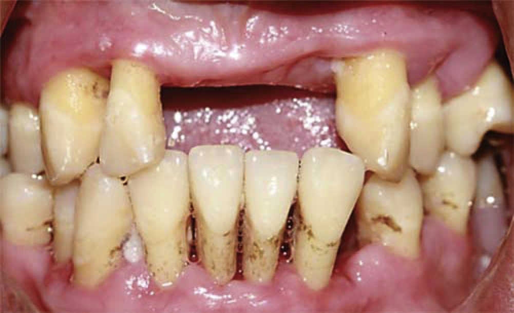
A 25 year old female attended the implantology service. As personal history the patient reported suffering type 2 Diabetes Mellitus for three years, controlled with metformin. Oral examination revealed a partially edentulous upper jaw as well as generalized pathological migration, with calculi deposits, purulent exudate, evident gingival inflammation as well as grade Ii and III mobility. Periodontal probing revealed insertion loss greater than 5 mm (Figure 1A). Radiographic studies showed severe bone resorption with multiple bone defects (Figure 1 B). Based on these facts, the patient received a diagnosis of severe generalized aggressive periodontitis associated to diabetes mellitus, with unfavorable prognosis for all remaining teeth.

After analyzing several treatment alternatives, two surgical phases and one prosthetic phase were devised. In the first phase it was decided to conduct multiple extractions with flange preservation and elevation of bilateral maxillary sinus floor. The second phase was planned once bone graft integration times were fulfilled. This second phase consisted on placement of 12 endo-osseous implants: 6 in the upper jaw, and 6 in the lower jaw. Finally, in the third rehabilitation phase, two fixed implant-supported prostheses were placed.
PREOPERATIVE MEASURES
Before initiating surgical procedure the patient's glycemic level was ascertained with the help of glycated hemoglobin (HbA1c lesser than 7%). Amoxicillin was chosen as antibiotic (12 g per mouth 1 hour before surgery and 500 mg every eight hours for eight days) as well as mouthwash with 0.12% chlorhexidine 3 times a day for 15 days. Amoxicillin was selected as antibiotic since it is a wide-spectrum drug and acts against pathogen agents (streptococci, Gram-positive and Gram-negative anaerobes) which most frequently cause post-operative complications after implant placement.6 Antibiotic prophylaxis and use of chlorhexidine 0.12% mouthwash proved to be clearly beneficial to reduce failure rates from 13.5 to 4.4% in type 2 diabetic patients, in a 36 month follow-up period.7
SURGICAL TREATMENT
The patient was anesthetized with mepivacaine with 2% epinephrine. Multiple extractions with osteoplasty were conducted, after this procedure, de-mineralized cortical bone allograft and collagen membrane were placed in both arches; the membrane was stabilized with tacks (studs). Healing by first intention was observed in the primary closure of surgical wounds (Figure 2A-C).

Immediate full prostheses were manufactured to be then placed in a passive and transitory manner. After this, six months were allowed to elapse in order to achieve healing of soft and hard tissues.
In a second surgical stage, a bilateral maxillary sinus floor elevation was executed with the lateral window technique first described by Taum8 and modified by Boyne and James.9
Maxillary sinuses were approached by means of an oval osteotomy measuring approximately 15 × 10 mm; the cut bone fragment was removed. During approach to the right side a small perforation was produced (Figure 3A-B), therefore, a collagen barrier was placed in order to repair the perforation and protect access to the sinus.10,11 It was decided to place bovine bone graft since it has been shown that this type of bone, due to its slow resorption, favors implant integration (Figure 3C-D).12,13

TOMOGRAPHIC AND SURGICAL PLANNING
After the six month healing period, a diagnostic waxup was performed, since guided bone regeneration and elevation of maxillary sinus floor presented favorable changes for implant planning and placement (Figure 4 A-C).

Figure 4 A-C Evolution observed six month after surgery of the flange preservation and maxillary sinus elevation in order to place endoosseous implants.
Prosthetic phase was initiated with a diagnostic wax-up (DW) which was replicated in clear acrylic so as to conduct an esthetic test and decide on rehabilitation type, since the inter-phase between flange and diagnostic wax-up is the most important aspect in problem identification and expectations for the final result of the prosthesis (Figure 5).

With the esthetic test it could be determined that rehabilitation should be by means of two implantsupported, hybrid, fixed dentures. Once patient and specialists were satisfied with the projected result, a contrast medium was placed into the DW so as to assess with computerized axial tomography (CAT) the areas of implant placement.
Tomographic analysis showed a suitable flange for implant placement. The silhouette of the teeth of the diagnostic guide was observed (Figure 6). The function of this guide was to establish bony tissue characteristics and its direct relation with the type of planned restoration, establishing thus the angle present between residual crest with respect to the final restoration's proposed axial profile. This is a parameter that can be determined by this kind of device, and thus achieve a tomographic guide. Once the study was completed, data were digitally uploaded in order to be processed with a third dimension program (Figure 7).


IMPLANT PLACEMENT
The suitable glycemic control exhibited by the patient allowed us to conduct implant placement surgery with the aforementioned pre-operative care. This is to say, a full-thickness flap was executed in the lower jaw. Settling of tomographic surgical guide was assessed, said guide was stabilized with fixation screws. Burr-use protocol was initiated in order to achieve surgical beds. Six external connection implants were placed, implants were of regular diameter and were strategically distributed in the lateral, first premolar and first molar zones they were placed with a torque greater than 35N. Flaps were approximated with 4-0 Vycril suture (Figure 8A-B). One month later, the same procedure was executed in the upper arch.

A control panoramic X-ray of the patient was taken in order to assess that implant position corresponded to tomographic surgical planning (Figure 9).

DISCUSSION
After review of articles published in the last ten years, it can be reported that survival rate of dental implants in diabetic patients ranged from 88.8 to 97.3% one year after implant insertion. Ranges of 85.6 to 97.6% were found one year after functional load with prosthesis.7,14
Fiorellini, published a retrospective review of 215 dental implants in 40 diabetic patients: 31 implants failed, out of which 24 (11.2%) failed in the first year of functional load. This analysis shows a survival rate of 85.6% after 6.5 years.
Olson, JW et al also conducted a prospective study with 89 type 2 diabetic patients; they studied 178 implants in the lower jaw. In this study they reported an early failure rate of 2.2% (4 implants) and a later failure rate of 7.3% (one year after prosthesis placement).15
The fact that most failures occur after the second surgical phase and during the first year of functional load could indicate that micro-vascular affectation is one of the factors playing a role in the failure of diabetic patients implants.16,17
Morris HF and Farzard P concluded that, although there is greater failure risk in a diabetic patient, preservation of suitable levels of glucose in the blood along with other measures, improve implant survival percentages in these patients.7,17
CONCLUSIONS
Late diagnosis of aggressive periodontitis can lead to edentulism in young patients and thus decrease their quality of life.
Suitable hygiene, rigorous glycemic control as well as maintenance program will help decrease the risk of suffering peri-implantitis, which would lead to implant loss.
REFERENCES
1. A. Mellado-Valero, J.C. Ferrer-García, A. Herrera-Ballester, C. La-baig-Rueda. Effects of diabetes on the osseointegration o dental implants Med Oral Patol Oral Cir Bucal. 2007, 12: E38-E43 [ Links ]
2. G.C. Armitage. Development of a classification system for periodontal disease and conditions. Ann Periodontol. 1999; 4:1p [ Links ]
3. A.M. Iacopino. Periodontitis and diabetes interrelationships: role inflammation. Ann Periodontol. 2001; 6:125p [ Links ]
4. M. McCracken, J.E. Lemons, F. Rahemtulla, C.W. Prince, D. Feldman. Bone response to titanium alloy implants placed in diabetic rats. Int J Oral Maxillofac Implants. 2000, 15: 345-354 [ Links ]
5. M.L. Nevins, N.Y. Karimbux, H.P. Weber, W.V. Giannobile, J.P. Fiorellini. Wound healing around endosseous implants in experimental diabetes. Int J Oral Maxillofac Implants. 1998; 13:620p [ Links ]
6. T. Beikler, T.F. Flemming. Antimicrobials in implant dentistry. Antibiotic and antimicrobial use in dental practice. Chicago: Quintessence. 2001. 195p [ Links ]
7. H.F. Morris, S. Ochi, S. Winkler. Implant survival in patients with type 2 diabetes: placement to 36 months. Ann Periodontol. 2000, 5: 157-165 [ Links ]
8. D. Emmerich, W. Att, C. Stappert. Sinus floor elevation using osteotomes: a systematic review and meta-analysis. J Periodontol. 2005; 76:1237p [ Links ]
9. P.J. Boyne, R.A. James. Grafting of the maxillary sinus floor with autogenous marrow and bone. J Oral Surg. 1980, 38: 613-616 [ Links ]
10. A. Sculean, D. Nikoliadis, F. Schwarz. Regeneration of periodontal tissues: combinations of barrier membranes and grafting materials-biological foundations and preclinical eviden a systematic review. J Clin Periodontal. 2008, 35: 106-116 [ Links ]
11. H. Agis, M. Magdalenko, K. Stögerer, G. Watzek, R. Gruber. Collagen barrier membranes decrease osteoclastogenesis in murine bone marrow cultures. Clin Oral Implants Res. 2010; 21:656p [ Links ]
12. P. Valentini, D.J. Abensur. Maxillary sinus grafting with anorganic bovine bone:a clinical report of longterm results. Int J Oral Maxillofac Implants. 2003; 18:556p [ Links ]
13. H. Browaeys, P. Brouvry, H. De Bruyn. A literature review on biomaterials in sinus augmentation procedures. Clin Impl Dent Relat Res. 2007; 9:166p [ Links ]
14. J.P. Fiorellini, P.K. Chen, M. Nevins, M.L. Nevins. A retrospective study of dental implants in diabetic patients. Int J Periodontics Restorative Dent. 2000; 20:366p [ Links ]
15. J.W. Olson, A.F. Shernoff, J.L. Tarlow, J.A. Colwell, J.P. Scheetz, S.F. Bingham. Dental endosseous implant assessments in a type 2 diabetic population: a prospective study. Int J Oral Maxillofac Implants. 2000, 15:811-818 [ Links ]
16. P. Farzad, L. Andersson, J. Nyberg. Dental implant treatment in diabetic patients. Implant Dent. 2002; 11:262p [ Links ]
17. M. Peled, L. Ardekian, N. Tagger-Green, Z. Gutmacher, E.F. Matchei. Dental implants in patients with type 2 diabetes mellitus: a clinical study. Implant Dent. 2003; 12:116p [ Links ]
*This article can be read in its full version in the following page: http://www.medigraphic.com/facultadodontologiaunam
Received: January 2015; Accepted: May 2015
 Este es un artículo publicado en acceso abierto bajo una licencia Creative Commons
