


Services on Demand
Journal
Article
 text in
  English (pdf)
 Article in xml format
 Article references
 Send this article by e-mail
Indicators
-
 Cited by SciELO
 -
 Access statistics
Related links
-
 Similars in
SciELO
Similars in
SciELO
Share

 Permalink
Revista odontológica mexicana
Print version ISSN 1870-199X
Rev. Odont. Mex vol.15 n.3 Ciudad de México Jul./Sep. 2011
Original research
Presence of bacterial enzymatic activity in patients with periodontal pockets in the city of Corrientes (Argentina)
Miguel Acuña,* Javier Monzon,§ Ernesto Canga,II Ricardo Diez,¶ Elías Azzi**
* Assistant Instructor Periodontics and Physicis-Biological Chemistry.
§ Assistant Professor-Periodontics.
II Professor and Head of Periodontics Chair.
¶ Professor and Head of Physics and Biological Chemistry Chair.
** Intern-Initiation in Research.
FOUNNE.
Received: 17 September 2009.
Accepted: 5 March 2010.
ABSTRACT
This research has the purpose of determining enzymatic activity found in periodontal pockets in one sector of the population of the City of Corrientes. This task will be undertaken applying enzymatic biochemical diagnostic test or reaction of N&-benzoyl-DL-arginine-2-naphthyl-amide (BANA) as an auxiliary to conventional clinical diagnosis. We worked with 62 patients whose pocket depth was greater than 3 mm. A control group was established with ten patients whose pocket depth had a maximum of 2 mm 72 patients were appraised and a total of 124 sites were examined. On these sites enzymatic activity was manifested in the following manner: 6 (six) sites tested positive for the BANA test (more than 500,000 anaerobic CFU at the sample site). 80 periodontal pocket samples yielded in the BANA test a result of slightly positive. This indicates periodontal bacteria in a range of 100,000 to 500,000 anaerobic CF U in all sampled sites. 18 sampled sites tested negative. This indicates that bacterial presence at the sampled site did not reach 100,000 CFU. When 20 sites of the 10 patients in the control group were examined, no enzymatic activity was observed. Through results gathered from this research, we can emphasize the benefit of including Non Conventional Diagnostic Techniques such as the BANA hydrolysis test (N-&-benzyl-DL-arginine-2-Naphthylamide) to periodontal clinical examinations. This represents an adjunct for dentists in need to emit an early diagnosis of this disease.
Key words: Periodontal pocket, BANA test, periodontal disease diagnosis.
INTRODUCTION
In general, the term periodontal disease is used to describe inflammatory and infectious lesions that originate and may persist in the periodontium. The clinical classification of these processes is based on the topographic area of the inflammatory response. When located in the gums, the lesion is called gingival disease. When the damaging process affects the support system (cement, periodontal ligament and alveolar bone) the condition is called periodontal disease. To determine periodontal disease the most important anatomical and clinical feature is the presence of a periodontal pocket which is defined as a pathological deepening of the gingival sulcus.
Clinical diagnosis of periodontal disease is achieved by measuring the connective tissue loss of insertion to the tooth surface (loss of clinical attachment) and loss of alveolar bone (alveolar bone loss).
At this point in time, dentists count with a variety of methods to diagnose this disease. To begin with, they can take a medical and dental history, as well as careful exploration of the patient. This includes taking a medical and dental history, a clinical examination and an updated radiographic analysis. This diagnostic procedure is not always as accurate a method as a diagnostic test within an actual clinical situation. Tests provide a correct answer when they are positive for the presence of disease, and negative for the absence of it. However, this diagnostic tool can be misleading when it yields the result of positive in absence of disease (false positive) or negative in the presence of the same (false negative).1 Conventional diagnostic procedures, such as probing tests, Loe/Silness) gingival index, O'Leary's Index and radiographic studies (Long Cone technique) measure only the resultant periodontal destruction, and do not indicate the cause of the disease, the patient's sensitivity to the disease and whether the condition is in progress or remission. New procedures have been developed to improve the diagnosis. These new procedures include subtraction radiography, nuclear medicine techniques computarized tomography DNA probings, enzymatic methods for bacterial identification (BANA hydrolisis test) higher values of serum antibodies against periodontal bacterial and crevicular fluid sampling. All these procedures are focused to determine sensitivity of the host and periodontal disease activity.2 In view of the difficulty the professional encounters to promptly determine activity or inactivity in periodontal pockets, new diagnostic procedures are emerging as a viable solution to this reality.
BANA hydrolysis test is designed to tackle the synthesis of a tripsinoid enzyme produced by three possible pathogens: Polyphyromonas gingivalis, tanerella forsythensis and treponema denticola this enzyme not only degrades the extracellular matrix proteins of the host, it is also able of hydrolyzing the synthetic peptide N-&-benzyl-DL-arginine-2-naphtylamide, or BANA. This peptide is colorless, but its hydrolysis releases the chromophore beta-naphthylamide, which, together with another substance, aniline black evans (fast black) elicits a shift towards bluish hue. Both the substrate BANA and the fast black enhance the reactive Ora Tec Card, which is the specific equipment required to perform this diagnostic procedure.3-9
The purpose of this research is to determine enzymatic activity of periodontal pockets in a sector of the population of the city of Corrientes, through application of the Enzymatic biochemical diagnostic test or through reaction of the N-&-benzoyl–DL-arginine-2-naphthyl-amide (BANA) as an adjuvant to conventional clinical diagnosis.
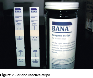
MATERIALS AND METHODS (Figures 1-10 (2, 3, 4, 5, 6, 7, 8, 9)


Sample characteristics
The sample comprised 72 patients who presented periodontal pocket depths of 3 mm patients were of both sexes, with ages ranging from 20 (twenty) to 50 (fifty) years. Sampling was carried out in the Clinical Sector, in Emergency Services, and patients referred from the School of Dentistry linked to the UNNE.
For the control group, 10 patients with 2 mm pocket depth were selected.
Criteria for inclusion of patients in this research project was as follows: not having been subject to previous periodontal treatment, not having ingested antibiotics or any other medication six months prior to the appointment, and not presenting a compromised systemic involvement or having undergone orthodontic treatment consisting of fixed appliances.
Selected patients had to agree to an inform consent form to validate their participation in the project.
Patient management
a) Medical and dental history
A clinical medical history was taken to provide all medical and systemic information of the patient. A simple questionnaire was elaborated, to confirm or rule out situations which might present medical risks (heart diseases, allergies, transplants, etc).
b) Periodontal examination
The attached gingival tissue in both jaws was examined and palpated; color, shape and consistency were described. The mucogingival line was located and its location was plotted into the periodontogram. The amount of attached gingival was assessed and note was taken of teeth presenting a small amounts of gingival tissue. A periodontal probe placed at the cement and enamel line measured the degree of gingival recession. The teeth positions were established, indicating any tilting towards lingual or vestibular direction. Teeth relationships with the interproximal papillae were also established in cases of crowding, diastema, or tooth rotation.
c) Application of BANA test
• Sampling
A BANA Test strip was taken marking date and tooth where the bacterial plaque sample was to be collected. When present, supragingival plaque was removed, prior to taking the final sample. Using Gracey 5/6 curettes subgingival plaque samples were obtained from sites of deeper periodontal pockets.
The extracted subgingival plaque sample was placed on the lower section of the BANA Test strip.
• Incubation of samples
The upper section of the Test strip was moistened with a sterile cotton swab impregnated with distilled water. Both ends of the strip were joined to attain contact and placed in the BANA incubator. Plaque samples were incubated at 55 degrees for 15 minutes. This corresponds to level number 3 of the incubator. The incubator starts working when the light comes on and it stops when the light goes off. This determines when the strip 's incubation begins and ends. Upon completion of the process, the strip extremities were separated and its readings were marked.
Results were read using the reactive matrix according to W Loesche (Loesche et al, 1990)9 which are as follows: Positive: change of color to bright blue, Slight positive: change of color to light blue, Negative: no color change.
When the sample is positive, the more intense is the color, greater is the number of bacterial colonies present in the sampled plaque.
RESULTS
In the 72 patients, a total of 124 sites were examined. The enzymatic activity of these sites was classified in the following fashion:
In the 62 patient group with periodontal pocket depth of 3 mm:
• 6 (six) sites resulted positive in the BANA test (more than 500,000 anaerobic CFU at the site of the sample).
• 80 periodontal pocket samples resulted slight positive in the BANA test. This indicates presence of periodontal pathogenic bacteria, ranging from 100,000 to 500,000 anaerobic CFU in each sampled site.
• 18 samples resulted negative. This indicates that bacterial presence in the sample site does not reach 100,000 CFU.
In the 10 patient group taken as control with pocket depth of 2 mm, 20 sites proved negative in the BANA Test (Figure 11).

DISCUSSION
This report studies the presence of bacterial activity found in 3 mm periodontal pockets. When applying the BANA enzymatic test to a total of 124 samples, 80 sites showed weak positive reaction and six sites positive reaction. This shows a moderate risk of periodontal disease in the absence of clinical features. This concurs with work carried out by Loesche et al10 where the weak positive reaction of the enzymatic BANA test manifests itself as a pale blue color over the area in contact with plaque, which can be very small, or of the total area contact. The meaning of this color intensity in a patient free of clinical symptoms of periodontal disease, indicates the presence of pathogenic bacteria in a range of 100,000 to 500,000 anaerobic CFU at the site where the sample was taken, this determines that the patient presents medium risk of periodontal disease.
Studies conducted by Grizzi et al11 report different results. They found a high frequency of BANA test positive results (66.1%) in sites with depths ranging from 2 to 3 mm. Nevertheless, in the present study, the results of the BANA test for weak positive enzyme presence show they appear most often in 3 mm periodontal pockets, thus demonstrating possible activity of subgingival plaque bacteria.
Garcia Echevarria et al2 argue that conventional periodontal diagnostic procedures, measure only the results of periodontal destruction, and do not indicate activity or remission of the same. We fully agree with this concept, since in our study 86 of a total 124 sites tested presented active periodontal disease in absence of clinical symptoms.
Negative results observed in this study are probably below the detection limit of the BANA test, as proposed in the study by Loesche et al in 1989.12,13
CONCLUSIONS
Through results obtained from the present study, we can emphasize the value of including non conventional diagnosis techniques such as the hydrolysis BANA test (N-&-benzyl-DL-arginine-2-naphthylamide) in periodontal clinical examinations, This arises due to the need dentists face to carry out an early diagnosis of this disease. Periodontal disease is the main cause of tooth loss, this is probably due to the fact that traditional or conventional diagnosis techniques such as the conventional periodontal probe do not provide effective information. These traditional techniques yield a rather objective information which brings as a consequence difficulties to clinically diagnose bacterial activity, and thus achieve dispense proper treatment at the right moment.
REFERENCES
 Mailing address:
Miguel Acuña
Faculty of Dentistry–UNNE–
Avenida Libertad 5450,
Corrientes 3400
E-mail: odontoacuna@gmail.com
Note
This article can be read in its full version in the following page: http://www.medigraphic.com/facultadodontologiaunam
1. Carranza, Newman. Periodontología Clínica. Octava edición. McGraw-Hill Interamericana Editores, S.A. de C.V. 1998: 836. [ Links ]
2. Grisi MFM, Correa FTA, Fanganiello CLS, Martins Jr W, Silva-Neto CR, Salvador SL. Relación entre la presencia o ausencia de sangrado gingival y el test enzimático de BANA. Braz Dent J 2001; 12 (1): 23-26. [ Links ]
3. Ledesma MC, Miñarro RJ, Gacés OM, Martínez AG, Rodríguez MA. La reacción BANA como método de diagnóstico de periodontitis activa. Rev ADM 1995; 52 (5): 251-254. [ Links ]
4. Grisi MFM, Ito IY, Novaes AB. BANA hydrolysis and probing depth to monitor periodontal treatment. J Dent Res 1991 70 (special issue): 320 (abstract). [ Links ]
5. Bretz WA, Lopatin D, Hujoel P, Taylor C, Löesche WJ. BANA hydrolysis and T. denticola and/or B. gingivalis in periodontal plaques. In: Annual Session of IADR 18. San Francisco, 1989. Apud J Dent Res 1989 68(sp. issue): 241 (abstract 481). [ Links ]
6. Schmidt EF, Bretz WA, Hutchinson RA, Löesche WJ. Correlation of the hydrolysis of Benzoyl-Arginine-Naphthylamide (BANA) by plaque with clinical parameters and subgingival levels of spirochetes in periodontal patients. J Dent Res 1988; 67: 1505-1509. [ Links ]
7. Bretz WA, Löesche WJ. Characteristics of trypsin-like activity in subgingival plaque samples. J Dent Res 1987; 66: 1669-1672. [ Links ]
8. Löesche WJ, Syed SA, Stoll J. Trypsin-like activity in subgingival plaque. A diagnostic marker for spirochetes and periodontal disease? J Periodontol 1987; 58: 266-273. [ Links ]
9. Mühlemann HR, Son S. Gingival sulcus bleeding a leading symplom in initial gingivitis. Helvetica Odontológica Acta 1971; (15): 107-113. [ Links ]
10. Löesche WJ, Bretz WA, Kerschensteiner D, Stoll J, Socransky SS, Hujoel PP, Lopatin DE. Development of hydrolysis of Benzoyl-DL-Arginine-Naphthylamide. J Clin Microbiol 1990; 28: 1551-1559. [ Links ]
11. Grisi MFM, Novaes AB, Ito IY, Salvador SL. Relationship between clinical probing depth and reactivity to the BANA test of samples of subgingival microbiota from periodontally involved patients. Braz Dent J 1998; 9: 77-84. [ Links ]
12. Acuña M, Monzón J, Caramello CR, Diez R. Estudio comparativo entre la prueba enzimática BANA y el índice gingival de Löe en pacientes de la Provincia de Corrientes, Argentina. R Cúspide 2006; 13: 22-25. [ Links ]
13. Löesche WJ, Bretz W, Killoy W, Rau C, Weber HP, Lopatin D. Detection of T. denticola and B. gingivalis in plaque with perioscan. In: Annual Session of IADR, 18. San Francisco. Apud J Dent Res 1989; 68 (special issue): 241 (abstract 482). [ Links ]

