SciELO - Scientific Electronic Library Online

 
vol.25 issue1Stroke-induced epilepsia partialis continuaParkinson's disease-associated pain in a Mexican Institute author indexsubject indexsearch form
Home Pagealphabetic serial listing  

Services on Demand

Journal

Article

Indicators

Related links

  • Have no similar articlesSimilars in SciELO

Share


Revista mexicana de neurociencia

On-line version ISSN 2604-6180Print version ISSN 1665-5044

Rev. mex. neurocienc. vol.25 n.1 Ciudad de México Jan./Feb. 2024  Epub Apr 16, 2024

https://doi.org/10.24875/rmn.23000062 

ORIGINAL ARTICLES

Location and morphology of cortical lesions in multiple sclerosis

Localización y morfología de las lesiones corticales en la esclerosis multiple

Nancy R. Bertado-Ramírez1 

José A. García-Santiago1 

José P. Calderón-Cánovas2 

Yasmín Juárez-Mora2 

Arturo García-Galicia3  * 

Álvaro J. Montiel-Jarquín4 

Jorge Loría-Castellanos5 

1Jefatura de División de Educación en Salud, Unidad Médica de Alta Especialidad Hospital de Especialidades de Puebla, Centro Médico Nacional "Gral. de Div. Manuel Ávila Camacho", Instituto Mexicano del Seguro Social (IMSS), Puebla

2Servicio de Imagenología y Radiodiagnóstico, Unidad Médica de Alta Especialidad Hospital de Especialidades de Puebla, Centro Médico Nacional "Gral. de Div. Manuel Ávila Camacho", IMSS, Puebla

3Jefatura de División de Investigación en Salud, Unidad Médica de Alta Especialidad Hospital de Especialidades de Puebla, Centro Médico Nacional "Gral. de Div. Manuel Ávila Camacho", IMSS, Puebla

4Dirección de Educación e Investigación en Salud, Unidad Médica de Alta Especialidad Hospital de Especialidades de Puebla, Centro Médico Nacional "Gral. de Div. Manuel Ávila Camacho", IMSS, Puebla

5Coordinación de Proyectos Especiales en Salud, IMSS, Ciudad de México. Mexico


Abstract

Objective:

Multiple sclerosis (MS) is a chronic inflammatory demyelinating disease of the central nervous system. Diagnosis is based on the Mc-Donalds criteria, magnetic resonance imaging (MRI) studies, and the expanded disability status scale (EDSS) which assesses disease progression. These criteria do not include the recently described cortical lesions. The aim of the study was to describe the most frequent location and morphology of cortical lesions in patients with MS in Puebla, Mexico.

Methods:

A descriptive, retrospective, cross-sectional, and analytical study was conducted on patients with MS at a tertiary care hospital. Patients diagnosed with relapsing-remitting, secondary-progressive, and progressive-relapsing MS variants with cranial magnetic resonance imaging were included in the study. Age, sex, MS variant, EDSS score, cognitive impairment, annual relapse rate, morphology, location, and number of cortical lesions were evaluated. Descriptive statistics were used. To compare features between groups, the χ2 test was used, and for correlations, the Spearman's Correlation Coefficient was used. A p ≤ 0.05 was considered significant.

Results:

Twenty-five patients met the selection criteria. The most frequent location of cortical lesions was the parietal region 84%, and the second was the temporal region 16%. The most common morphology was juxtacortical at 64% and mixed at 36%. The most frequent variant of MS was relapsing-remitting, present in 92%, and 8% had the secondary progressive variant. In the EDSS scale, the scores most frequently observed were 0.0 and 3.5.

Conclusions:

The most frequent location of cortical lesions was in the parietal region, and the most common morphology was juxtacortical.

Keywords Multiple sclerosis; Cortical lesions; Neurology; Magnetic resonance imaging

Resumen

Objetivo:

La esclerosis múltiple es una enfermedad desmielinizante inflamatoria crónica del sistema nervioso central. El diagnóstico se basa en los criterios de Mc-Donalds, los estudios de Resonancia Magnética y la Escala Expandida del Estado de Discapacidad (EDSS) que evalúa la progresión de la enfermedad. Estos criterios no incluyen las lesiones corticales descritas recientemente. Describir la localización y morfología más frecuente de lesiones corticales en pacientes con esclerosis múltiple en Puebla, México.

Métodos:

Se realizó un estudio descriptivo, retrospectivo, transversal y analítico en pacientes con esclerosis múltiple en un hospital de tercer nivel de atención. Se incluyeron pacientes con diagnóstico de Esclerosis Múltiple con variantes Remitente-Recurrente, Secundaria-Progresiva y Progresiva-Recurrente con resonancia magnética de cráneo. Se evaluó edad, sexo, variante de esclerosis múltiple, puntaje de EDSS, deterioro cognitivo, tasa de recaída anual, morfología, ubicación y numero de lesiones corticales. Se utilizó estadística descriptiva. Para comparar características entre grupos se utilizó χ2, para correlaciones se utilizó Coeficiente de Correlación de Spearman. Una p ≤ 0.05 se consideró significativa.

Resultados:

25 pacientes cumplieron con criterios de selección. La localización más frecuente de lesiones corticales fue la región parietal 84% y la segunda temporal 16%. La morfología más frecuente fue la yuxtacortical en un 64%, y la mixta en 36%. La variante más frecuente de esclerosis múltiple fue Remitente-Recurrente presente en 92%, y 8% la variante Secundaria Progresiva. En la escala de EDDS la puntuación con mayor frecuencia fue 0.0 y 3.5.

Conclusiones:

La localización más frecuente de las lesiones corticales fue en región parietal y la morfología más frecuente la yuxtacortical.

Palabras clave Esclerosis múltiple; Lesiones corticales; Neurología; Resonancia magnética

Introduction

Multiple sclerosis (MS) is a chronic inflammatory demyelinating disease of the central nervous system. It is most common in adults and affects more than two million people worldwide. It follows a variable progression, leading to disability and high costs for healthcare systems1,2. Diagnosis is based on the Mc-Donalds criteria, the expanded disability status scale (EDSS) which assesses disease progression and magnetic resonance imaging (MRI). MRI plays an important role in clinical practice by enabling accurate diagnosis. It helps to understand the evolution of the disease and to evaluate treatments by monitoring response to treatment3-5. All of this is based on the McDonald criteria, but until now, these criteria have not included the cortical lesions that have been recently described6-9.

Cortical lesions are more frequently observed in primary and secondary progressive forms, affecting the cerebral and cerebellar cortex, particularly in the hippocampus and cerebellum10,11. They are less common in acute and remitting forms but tend to be larger in size. Considered an additional pathological substrate, they contribute to cognitive decline in these patients. Cortical lesions are significant in the neurodegenerative phase of recurring variants2,12,13. Magnetic resonance imaging assessment of cortical lesions in MS patients improves disease monitoring and diagnosis and enables precise treatment targeting by providing a detailed understanding of lesion location and morphology7,12,14-16.

The purpose of this study was to provide a description of the most common location and morphology of cortical lesions among MS patients receiving treatment at a tertiary care hospital at Puebla, Mexico.

Materials and methods

A retrospective, descriptive study was conducted on patients with MS who received treatment at a tertiary care hospital of the Mexican Social Security Institute (IMSS). Patients diagnosed with relapsing-remitting, secondary-progressive, and progressive-relapsing MS by cranial magnetic resonance imaging were included in the study.

The McDonald criteria are used by neurologists to make a diagnosis of MS9,13. Based on the current classification of progressive, non-progressive, activity-including, and activity-excluding variants, patients meeting the operational definitions outlined below were included in the study13,17.

The MS subtypes were defined as:

  • – Relapsing-remitting: Clearly defined episodes of new or increasing neurological symptoms, such as vision problems, vertigo, generalized weakness, ataxia, and loss of bladder control, are followed by periods of recovery or remission13,17.

  • – Secondary-progressive: characterized by evidence of disability accumulation over time, with or without relapses or new activity observed in MRI images13,17.

  • – Progressive-relapsing: Deterioration in neurological function or accumulation of disability from the onset of symptoms, with initial periods of relapse and/or remission13,17.

Procedure

The diagnosis of the subtype of MS was provided by the neurology service, in addition, age, sex, EDSS score, and annual relapse rate were evaluated, which were data collected during outpatient visits; the Montreal Cognitive Assessment (MOCA) was used to evaluate cognitive impairment, taking < 26 points as cognitive impairment, according to the recommendations of the MACFIMS consensus18.

The morphology, location, and number of cortical lesions were taken from the final report of the radiodiagnostic and imaging service.

Statistical analysis

Data analysis was conducted using descriptive statistics. The χ2 test was used to compare features between groups of cortical lesions and morphology. Spearman's test was employed for correlations. A p-value of ≤ 0.05 was deemed significant.

Ethical aspects

The Local Health Research Committee No. 2101 of the IMSS has approved this study. All participants signed an informed consent chart, and their anonymity was preserved throughout the study. The data were utilized solely for scientific purposes and for this study.

Results

A total of 25 patients were included, 10 (40%) were men and 15 (60%) were women.

The most common MS subtype was relapsing-remitting, found in 23 (92%) patients, while secondary-progressive was less common, found in 2 (8%) patients. Cognitive impairment was observed in only 5 (20%) patients.

The most common location of cortical lesions was the parietal region (Fig. 1), found in 21 (84%) cases, followed by the temporal location, found in 4 (16%) cases.

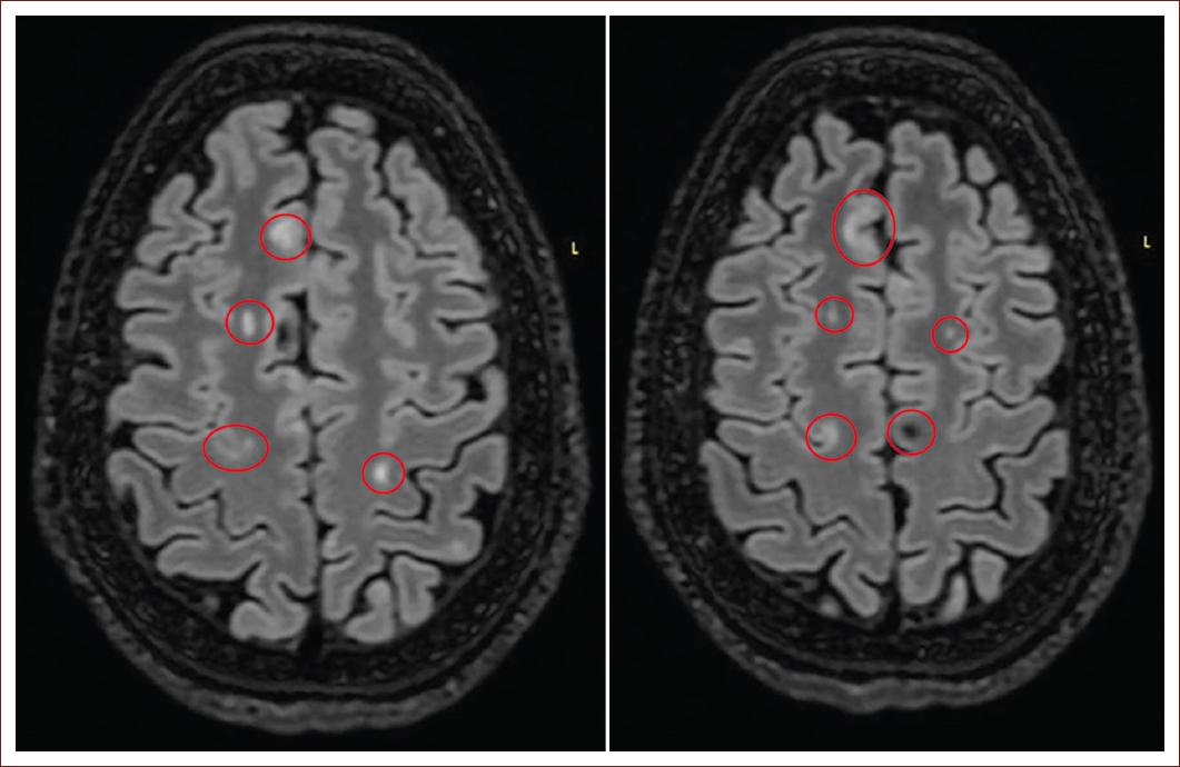
Figure 1 Fluid attenuated inversion recovery enhanced sequence, axial plane, subcortical, and juxtacortical frontoparietal demyelinating plaques. 

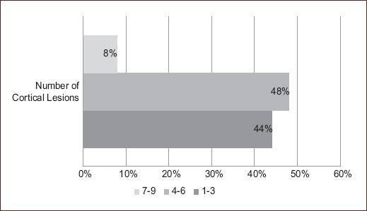
The most common morphology was juxtacortical (Fig. 1), found in 64%, and mixed, found in 36%. On the EDSS scale, the most frequently observed scores were 0.0 and 3.5 (Fig. 2). According to the number of cortical lesions, 4-6 (48%) were the most common (Fig. 3).

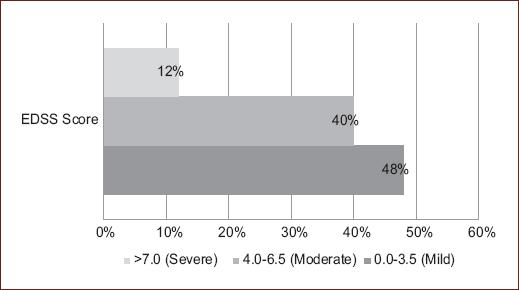
Figure 2 Disability in patients with multiple sclerosis (expanded disability status scale scale score) (n = 25). 

Figure 3 Number of cortical lesions in multiple sclerosis (n = 25). 

The predominant number of relapses was 1 (52%), followed by 0 relapses 40%, the correlation of the number of lesions with relapses and with EDSS was 0.58 (p = 0.02) and 0.678 (p < 0.0001), respectively. The association between lesion count and EDSS score showed statistically significant differences. (Fig. 4)

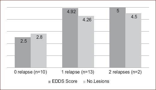
Figure 4 Distribution of number of lesions and disability (expanded disability status scale score) by number of relapses in patients with multiple sclerosis (p < 0.0001). 

Cognitive impairment was more prevalent in women with 80% (n = 4) compared to men with 20% (n = 1) (p = 0.6).

The number of lesions and cognitive impairment did not showed a significative correlation, with an asymptotic Pearson Chi-squared value of 0.464 and a likelihood ratio of 0.391.

The association of lesion morphology with the EDSS scale did not show a correlation, with an asymptotic likelihood ratio of 0.112 and a Chi-squared of 0.311.

Discussion

The predominance of the variable sex, age, and type of MS is consistent with previous reports in the Mexican population15.

It is noteworthy that the percentage of cognitive impairment does not agree with previously reported studies, in our study, it was only present in 20%, while in others, it is variable (40-70% and 42-52.5%)19,20.

Cognitive impairment does not correlate with physical disability and can be present in early stages of the disease. The most frequently affected areas are information processing speed, working memory, visual and verbal memory, verbal fluency, learning, word retrieval, and executive functions18,20. Because a neuropsychological evaluation was not conducted to assess a baseline state of cognitive impairment or any previously established alterations in mental functions, these aspects remain unclarified19,21.

The predominance in our study of the MS subtype was relapsing-remitting, having concordance with other previous imaging and histopathology studies22,23.

The average number of lesions is relatively higher in our study (4.01) compared to the population of Harrison et al. (3.38)24. However, both coincide closely in the intrinsic gradual relationship with the presence of high EDSS scores. The greater the number of lesions, the higher the EDSS score.

Pareto et al. describe only the relationship of the presence of juxtacortical lesions in patients with relapsing remitting MS describing that there is cortical thinning and loss of subcortical gray matter25.

This study complements the previous line of research by also describing the location and making correlations with other variables, finding that the most common morphology was juxtacortical lesions and the most common location was the parietal region. These findings cannot be compared because there is no existing literature that reports the morphology and location of lesions by brain lobe, making this the first studies of its kind in Mexico26.

It was observed that the number of lesions was associated with the number of relapses, and in turn, if the patient presented one or more relapses, was associated with intermediate EDSS scores (both p < 0.001). This finding may be due to the small sample size. It is already known from previous literature that these variables are not always directly proportional21,26,27.

Aldrete et al suggest that the number of lesions is associated with greater cognitive impairment; their study was also in Mexican population. In this study, no association was found between the number of lesions and cognitive impairment or between lesion morphology and EDSS scale. Several factors, including biological, medical, and psychosocial factors, may contribute to cognitive impairment in MS. These findings need to be investigated in future studies with larger sample sizes21.

The use of high detail resonators is recommended for the identification of more cortical lesions, for this study, 3-T MRI was used28.

Perhaps multicenter studies are needed to increase the sample size. This would allow us to draw conclusions. There is also a need for further monitoring of this research topic to identify areas for better utilization in both clinical and diagnostic settings.

Conclusions

The most common location of cortical lesions in MS was in the parietal region, and the most common morphology is juxtacortical. Studies with larger sample sizes are needed to improve the diagnosis, treatment, and prognosis of patients with MS.

References

1. Reich DS, Lucchinetti CF, Calabresi PA. Multiple sclerosis. New Engl J Med. 2018;378:169-80. [ Links ]

2. Dimitriou NG, Meuth SG, Martinez-Lapiscina EH, Albrecht P, Menge T. Treatment of patients with multiple sclerosis transitioning between relapsing and progressive disease. CNS Drugs. 2023;37:69-92. [ Links ]

3. Filippi M, Preziosa P, Arnold DL, Barkhof F, Harrison DM, Maggi P, et al. Present and future of the diagnostic work-up of multiple sclerosis:the imaging perspective. J Neurol. 2023;270:1286-99. [ Links ]

4. Tartaglino LM, Friedman DP, Flanders AE, Lublin FD, Knobler RL, Liem M. Multiple sclerosis in the spinal cord:MR appearance and correlation with clinical parameters. Radiology. 1995;195:725-32. [ Links ]

5. Lövblad KO, Anzalone N, Dörfler A, Essig M, Hurwitz B, Kappos L, et al. MR imaging in multiple sclerosis:review and recommendations for current practice. AJNR Am J Neuroradiol. 2010;31:983-9. [ Links ]

6. Konen FF, Schwenkenbecher P, Wattjes MP, Skripuletz T. Performance of the revised 2017 Mcdonald criteria. Nervenarzt. 2023;94:538-45. [ Links ]

7. Mainero C, Treaba CA, Barbuti E. Imaging cortical lesions in multiple sclerosis. Curr Opin Neurol. 2023;36:222-8. [ Links ]

8. Peterson JW, BöL, Mörk S, Chang A, Trapp BD. Transected neurites, apoptotic neurons, and reduced inflammation in cortical multiple sclerosis lesions. Ann Neurol. 2001;50:389-400. [ Links ]

9. Filippi M, Rocca MA, Ciccarelli O, De Stefano N, Evangelou N, Kappos L, et al. MRI criteria for the diagnosis of multiple sclerosis:MAGNIMS consensus guidelines. Lancet Neurol. 2016;15:292-303. [ Links ]

10. Kutzelnigg A, Faber-Rod JC, Bauer J, Lucchinetti CF, Sorensen PS, Laursen H, et al. Widespread demyelination in the cerebellar cortex in multiple sclerosis. Brain Pathol. 2007;17:38-44. [ Links ]

11. Preziosa P, Pagani E, Bonacchi R, Cacciaguerra L, Falini A, Rocca MA, et al. In vivo detection of damage in multiple sclerosis cortex and cortical lesions using NODDI. J Neurol Neurosurg Psychiatry. 2022;93:628-36. [ Links ]

12. Ziemssen T, Bhan V, Chataway J, Chitnis T, Cree BA, Havrdova EK, et al. Secondary progressive multiple sclerosis:a review of clinical characteristics, definition, prognostic tools, and disease-modifying therapies. Neurol Neuroimmunol Neuroinflamm. 2023;10:e200064. [ Links ]

13. Eshaghi A, Young AL, Wijeratne PA, Prados F, Arnold DL, Narayanan S, et al. Identifying multiple sclerosis subtypes using unsupervised machine learning and MRI data. Nat Commun. 2021;12:2078. [ Links ]

14. Landete L, Casanova B. Cognitive impairment, clinic forms and progression in multiple sclerosis. Rev Neurol. 2001;32:884-7. [ Links ]

15. Bertado-Cortés B, Villamil-Osorio L, Carrera-Pineda R, Martínez-Cortés C, Guerrero-Cantera J. Aportaciones originales características clínicas y demográficas de los pacientes con esclerosis multiple. [Clinical and demographic characteristics of patients with multiple sclerosis]. Rev Med Inst Mex Seguro Soc. 2016;54:S186-90. [ Links ]

16. Tuñón T, Ayuso T. Aspectos patofisiológicos de la esclerosis múltiple. Rev Esp Escler Mult. 2015;36:5-12. [ Links ]

17. Lin X, Zhang X, Liu Q, Zhao P, Zhong J, Pan P, et al. Social cognition in multiple sclerosis and its subtypes:a meta-analysis. Mult Scler Relat Disord. 2021;52:102973. [ Links ]

18. Benedict RH, Fischer JS, Archibald CJ, Arnett PA, Beatty WW, Bobholz J, et al. Minimal neuropsychological assessment of MS patients:a consensus approach. Clin. 2002;16:381-97. [ Links ]

19. Custodio N, Montesinos R, López-Góngora M. Deterioro cognitivo en pacientes con esclerosis múltiple. An Fac Med. 2018;79:338-45. [ Links ]

20. Katsari M, Kasselimis DS, Giogkaraki E, Breza M, Evangelopoulos ME, Anagnostouli M, et al. A longitudinal study of cognitive function in multiple sclerosis:is decline inevitable?J Neurol. 2020;267:1464-75. [ Links ]

21. Aldrete Cortez VR, Duriez-Sotelo E, Carrillo-Mora P, Pérez-Zuno JA. Correlación entre las lesiones desmielinizantes y el deterioro de las funciones ejecutivas en una muestra de pacientes mexicanos con esclerosis múltiple. Neurología. 2013;28:394-9. [ Links ]

22. Calabrese M, Agosta F, Rinaldi F, Mattisi I, Grossi P, Favaretto A, et al. Cortical lesions and atrophy associated with cognitive impairment in relapsing-remitting multiple sclerosis. Arch Neurol. 2009;66:1144-50. [ Links ]

23. Kutzelnigg A, Lucchinetti CF, Stadelmann C, Brück W, Rauschka H, Bergmann M, et al. Cortical demyelination and diffuse white matter injury in multiple sclerosis. Brain. 2005;128:2705-12. [ Links ]

24. Harrison DM, Roy S, Oh J, Izbudak I, Pham D, Courtney S, et al. Association of cortical lesion burden on 7-T magnetic resonance imaging with cognition and disability in multiple sclerosis. JAMA Neurol. 2015;72:1004-12. [ Links ]

25. Pareto D, Sastre-Garriga J, Auger C, Vives-Gilabert Y, Delgado J, TintoréM, et al. Juxtacortical lesions and cortical thinning in multiple sclerosis. AJNR Am J Neuroradiol. 2015;36:2270-6. [ Links ]

26. Toro J, Cárdenas S, Fernando Martínez C, Urrutia J, Díaz C. Multiple sclerosis in Colombia and other Latin American countries. Mult Scler Relat Disord. 2013;2:80-9. [ Links ]

27. Martínez HR, Arredondo-Estrada JH, Rangel-Guerra R, Onofre-Castillo J. Esclerosis múltiple con lesiones en sustancia gris. Evaluación por resonancia magnética. Rev Mex Neurociencia. 2001;2:177-9. [ Links ]

28. Quattrocchi CC, Cherubini A, Luccichenti G, Grasso MG, Nocentini U, Beomonte Zobel B, et al. Infratentorial lesion volume correlates with sensory functional system in multiple sclerosis patients:a 3.0-Tesla MRI study. Radiol Med. 2010;115:115-24. [ Links ]

FundingThe authors declare that this work was carried out with the authors' own resources.

Ethical disclosures

Protection of human and animal subjects. The authors declare that no experiments were performed on humans or animals for this study.

Confidentiality of data. The authors declare that they have followed the protocols of their work center on the publication of patient data.

Right to privacy and informed consent. The authors have obtained the written informed consent of the patients or subjects mentioned in the article. The corresponding author is in possession of this document.

Use of artificial intelligence for generating text. The authors declare that they have not used any type of generative artificial intelligence for the writing of this manuscript nor for the creation of images, graphics, tables, or their corresponding captions.

Received: September 22, 2023; Accepted: December 16, 2023

* Correspondence: Arturo García-Galicia E-mail: neurogarciagalicia@yahoo.com.mx

Conflicts of interest

The authors declare that they have no conflicts of interest.

Creative Commons License Instituto Nacional de Cardiología Ignacio Chávez. Published by Permanyer. This is an open ccess article under the CC BY-NC-ND license