


 text new page (beta)
 English (pdf)
 Article in xml format
 Article references
 Send this article by e-mail
 Cited by SciELO
  Similars in
SciELO
Similars in
SciELO 
 Permalink
Pulmonary hypertension (PH) is a syndrome with multiple etiologies. The WHO classifies it into five different groups based on its pathophysiological mechanisms, clinical presentation, hemodynamic characteristics, and therapeutic management.1
The last Pulmonary Hypertension Symposium, held in Nice, established an elevation of mPAP > 20 mmHg as a new diagnostic value for HP. Furthermore, the isolated precapillary pulmonary hypertension phenotype requires a pulmonary capillary pressure (PCP) ≤15 mmHg and a pulmonary vascular resistance (PVR) ≥3 wood units (WU).1,2
We present the case of a 35-year-old patient with neurological maturation retardation and a history of bicuspid aortic valve and subaortic membrane, who had started 15 days earlier with progressive dyspnea of functional class II to III-IV, orthopnea and angina in the last 24 hours. She had edema in the lower limbs, crackles in the lung bases, first and second tones present, and a tricuspid systolic murmur of 3/6 intensity with Rivero Carvallo's sign. The chest X-ray revealed cardiomegaly and dilatation of the pulmonary artery (Figure 1). Transthoracic echocardiography showed a preserved ejection fraction, systolic flattening of the interventricular septum, slightly dilated right ventricle with a 19 mm TAPSE, bicuspid aortic valve with preserved opening, with a small subaortic membrane, pulmonary, tricuspid, mitral and mild aortic aortic insufficiencies, PSAP 67mmHg, without intracardiac shunts. Acute pulmonary embolism was excluded by pulmonary angiography. The tomography showed a dilation of all the branches of the pulmonary artery (Figures 2 and 3). The right heart catheterization showed a mean pulmonary pressure (PAPm) of 52 mmHg, pulmonary capillary pressure (PCP) 13mmHg, pulmonary vascular resistance (PVR) 6 wood units, and cardiac index of 3.53 liters/minute/m2. Diuretic treatment with furosemide generated clinical improvement. Sildenafil and bosentan were subsequently started.
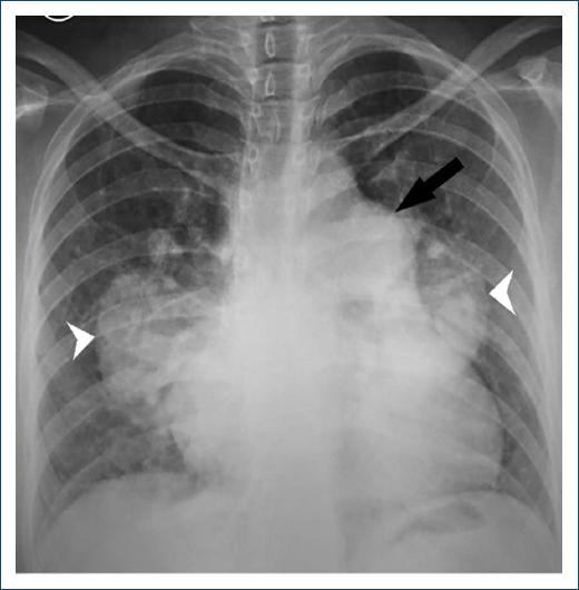
Figure 1 X-ray of the chest. Cardiothoracic index> 0.5, prominent main pulmonary artery (arrow) and dilated right and left pulmonary arteries (arrowhead).

Figure 2 Three axial slices at three different levels of a pulmonary angiography. 48mm pulmonary artery trunk, AP/Ao ratio: 1.7, dilation of the interlobar pulmonary arteries.
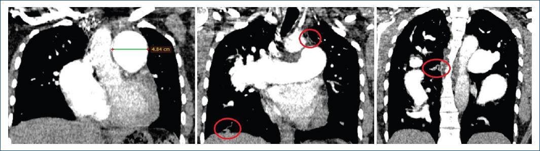
Figure 3 Three coronal sections of the pulmonary angiography. 48mm pulmonary trunk; red circle shows neovascularization.
This case is classified as a severe pulmonary hypertension precapillary phenotype of group 1.4.4 associated with congenital heart disease. The great dilation of the pulmonary artery and its branches led us to present chest radiography and pulmonary angiography images.

