


 text new page (beta)
 English (pdf)
 Article in xml format
 Article references
 Send this article by e-mail
 Cited by SciELO
  Similars in
SciELO
Similars in
SciELO 
 Permalink
We present the case of a term newborn, with no significant perinatal history, who was taken to the emergency room at 18 days old for intermittent episodes of cyanosis, with no signs of respiratory distress, oxygen saturation of 85%, arterial gases with moderate hypoxemia, and normal chest X-ray. The transthoracic echocardiogram (TTE) revealed a large undulating membrane that divided the right atrium into two chambers and partially prolapsing through the tricuspid valve (Fig. 1A-D and Video 1). The caval veins drain into the posteromedial chamber, with part of its flow redirected by the membrane to the left atrium through the foramen ovale (Fig. 2A-D and Video 2). Contrast with an agitated saline solution was performed, observing initial accumulation of microbubbles in the posteromedial chamber (Fig. 3A-B).
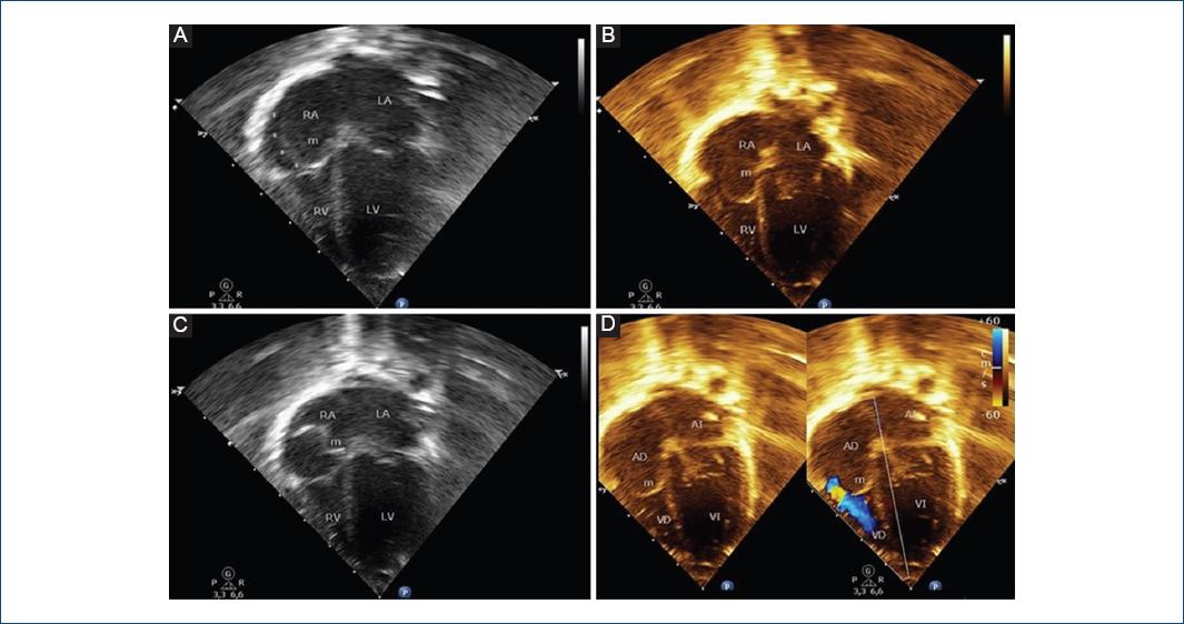
Figure 1 Apical four-chamber transthoracic echocardiographic images. A-C: Illustrating a membrane (m) that divides the right atrium into two chambers and partially prolapsing through the tricuspid valve. D: Echocardiographic image with color Doppler shows eccentric flow through the tricuspid valve from the outer part of the right atrium to right ventricle (with no significant gradient). RA: right atrium; LA: left atrium; RV: right ventricle; LV: left ventricle; M: membrane; FO: foramen ovale; SVC: Superior vena cava.
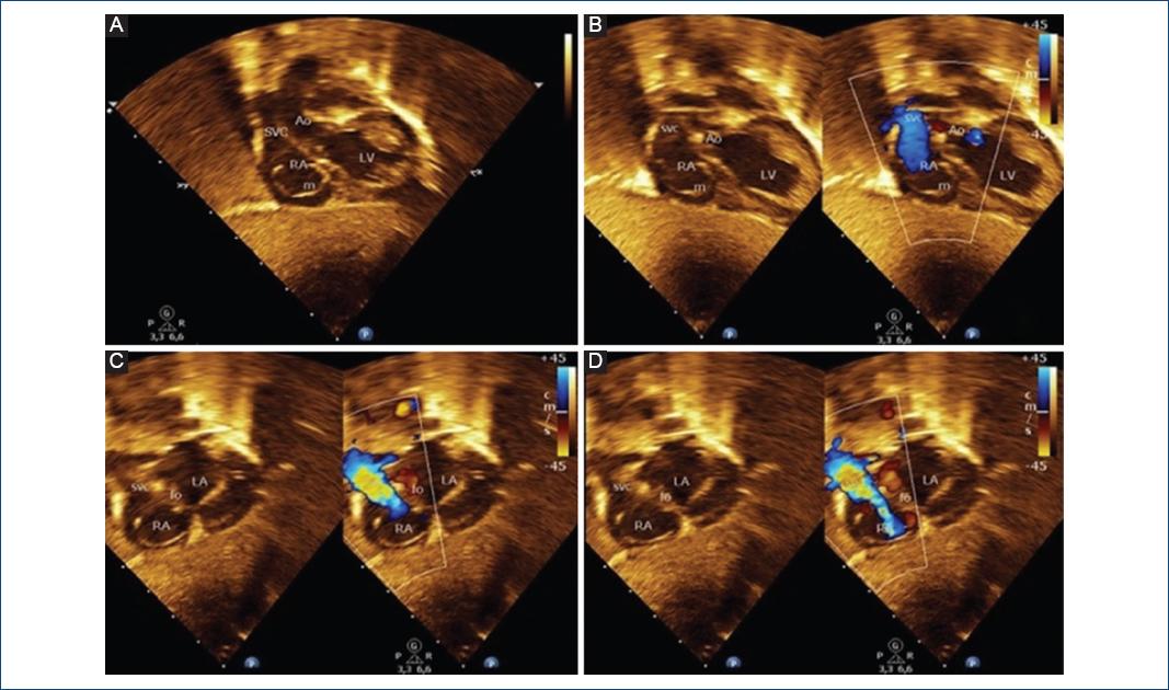
Figure 2 A: Two-dimensional and B-D: Two-dimensional color Flow Doppler echocardiography images from the subcostal window. A: The Cor-Triatriatum Dexter membrane (m) that separates the right atrium into two chambers. B-D: The inner part of right atrium receives the systemic venous blood from superior vena cava which is directed by the Cor-triatriatum membrane to the left atrium through the foramen ovale.
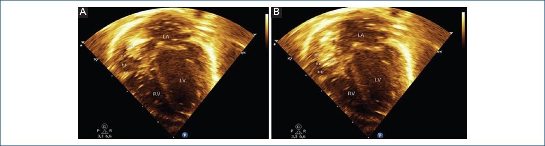
Figure 3 A-B: Contrast echocardiography showing the initial accumulation of microbubbles in the posteromedial chamber.
Cor triatriatum dexter is a rare congenital heart disease (0.025% of all congenital heart defects) characterized by the presence of a large membrane that divides the right atrium into two chambers: an posteromedial chamber that receives the flow of the caval veins, and an anterolateral chamber related to the tricuspid valve and right atrial appendage. This membrane represents the persistence of the right valve of sinus venosus, an early embryonic structure that involutes between the 9th and 15th weeks of gestation. The diagnosis can be properly established with a detailed TTE, observing a large linear echogenic structure that divides the right atrium into two chambers, with insertion to the roof of the atrium (crista terminalis) and systemic venous flow partial obstruction. The differential diagnosis must be established with the Chiari network, which has the echocardiographic appearance of a floating network of fibers, with multiple insertions in the upper part of the right atrium, and with the eustachian valve located at the orifice of the inferior vena cava, with little mobility1-3.

