


Servicios Personalizados
Revista
Articulo
 Inglés (pdf)
 Artículo en XML
 Referencias del artículo
 Enviar artículo por email
Indicadores
-
 Citado por SciELO
 -
 Accesos
Links relacionados
-
 Similares en
SciELO
Similares en
SciELO
Compartir

 Permalink
Revista latinoamericana de química
versión impresa ISSN 0370-5943
Rev. latinoam. quím vol.39 no.1-2 Naucalpan de Juárez 2011
Bioactive constituents and biochemical composition of the egyptian brown alga Sargassum Subrepandum (Forsk)
Ghada S. E. Abou-El-Wafaa, Khaled A. Shaabanb, Mohamed E. E. El-Naggarb, Mohamed Shaabanb, c, *
a Department of Botany, Faculty of Science, Mansoura University, Algomhuria st.60, El-Mansoura 35516, Egypt.
b Institute of Organic and Biomolecular Chemistry, University of Göttingen, Tammannstrasse 2, D-37077 Göttingen, Germany.
c Chemistry of Natural Compounds Department, Division of Pharmaceutical Industries, National Research Centre, El-Behoos st. 33, Dokki-Cairo 12622, Egypt. *Corresponding author: M. Shaaban, Chemistry of Natural Compounds Department, Division of Pharmaceutical Industries, National Research Centre, El-Behoos st. 33, Dokki-Cairo 12622, Egypt. Tel: +202-33371010/int-2609, Fax: +202-33370931. E-mail: mshaaba_99@yahoo.com.
Received September 2011.
Accepted November 2011.
ABSTRACT
During our search program for bioactive compounds from Egyptian marine sources, we isolated a spatane diterpene tetraol (1), fucosterol (2) and linoleic acid (3) from the brown alga Sargassum subrepandum (Forsk) C.Ag. In addition to those compounds, the four hydrocarbons were detected by using GC-MS named heptadecane, 2,6,10,14-tetramethyl-hexadecane, nonadecane and heneicosane. The chemical structure of 1 is assigned here definitely to first time by spectroscopic analyses including mass spectrometry (EI-MS, HR/EIMS), 1D and 2D NMR experiments. Phytochemical study of the unsaponified fraction of the algal extract and analysis by GC-MS confirmed the existence of nine compounds, among them 4,4,7a-trimethyl-5,6,7,7a-tetrahydrobenzofuran-2[4H]-one (6). In this article, we report as well the biochemical composition of Sargassum subrepandum (including ash, fibre, lipid, carbohydrate, total protein, and amino acids compositions) together with biological activities of its extract and the isolated compounds using diverse assays.
Keywords: Brown alga; Sargassum subrepandum (Forsk); Spatane diterpene; GC-MS; Biochemical composition; Biological activity.
RESUMEN
Dentro de nuestro programa de compuestos bioactivos de fuentes marinas Egipcias, en este trabajo aislamos tetraol (1 ), fucosterol (2) y ácido linoleico (3) del alga café Sargassum subrepandum (Forsk) C.Ag. También detectamos por CG-EM cuatro hidrocarburos llamados heptadecano, 2,6,10,14-tetrametil-hexadecano, nonadecano y heniocosano. La estructura química definitiva de 1 se asignó aquí por primera vez por el análisis espectrométrico (EM-IE, AD/EMIE) y espectroscópico de RMN con experimentos de1D y2D. El estudio fitoquímico de la fracción no saponificable del extracto de la alga y el análisis por CG-EMconfirmó la existencia de nueve compuestos, entre ellos4,4,7a-trimethyl-5,6,7,7a-tetrahydrobenzofuran-2(4H)-one (6). En este artículo también reportamos la composición bioquímica de Sargassum subrepandum (que incluye cenizas, fibra, lípidos, carbohidratos proteínas totales y composición de aminoácidos) y la actividad biológica de sus extractos y compuestos aislados utilizando diferentes ensayos.
Palabras clave: Brown alga; Sargassum subrepandum (Forsk); CG-EM; composición bioquímica; actividad biologica.
INTRODUCTION
In the 1940s, natural product research interest was directed towards marine flora as potential sources in the production of metabolic constituents possessing antimicrobial, antiviral and antifungal properties (Baslow, 1969; Perez et al., 1990), besides their effective pharmaceutical and economic properties (Ali & Pervez, 2003). Marine brown algae are prolific producers of interesting secondary metabolites, consisting of C11-acetate derived compounds (Moore, 1978), sesquiterpenoids, diterpentoids (Gerwick et al., 1981) and compounds of mixed biosynthesis origin (Fenical et al., 1973). The diterpenoids from these algae are particularly unique, as the novel ring systems produced represent unconventional diterpenoid cyclizations, which have not yet observed from terrestrial sources (Gerwick et al., 1980; Gerwick & Fenical, 1983).
Brown algae are also frequently encountered as the major vegetation in shallow-water tropical and subtropical habitats, even though herbivorous predators are plentiful. Hence, the correlation between secondary metabolite synthesis within this family and predator avoidance seems to be pronounced (Gerwick et al., 1981). In the littoral zone of the Egyptian coast, brown algae are currently the most dominant group. Members of Sargassum genus represent valuable sources of a wide spectrum of complex lipids, essential fatty acids and amino acids (Hossain et al., 2003). Sargassum subrepandum (Forsk) C.Ag. is quite common in the Egyptian Red Sea coast (El-Naggar et al., 1995), however, the bioactive constituents and biochemical composition of this alga have never been reported.
The marine brown alga Sargassum subrepandum (Forsk) was collected from Ras Gharib in the Egyptian Red Sea coast. The genus of Sargassum can be distinguished from its leaves and stem parts. Examination of the organic extract of the two parts of Sargassum subrepandum (Forsk) using TLC was visualized by spraying reagents, revealed the alga to contain a unique complex mixture of several components, including fatty acids, steroids and diterpenoids. Therefore, the whole alga was extracted with dichloromethane using a soxhlet. The afforded extract was then subjected for isolation using a series of chromatographic techniques. As a result, three major components were isolated named tetraol (1), fucosterol (2) (Popov et al., 1985; Frost & Ward, 1968; Falsone et al., 1994; Zhang et al., 2007; Tang et al., 2002; Sheu et al., 1999) and linoleic acid (3) (Shaaban, 2004). On other hand, the less polar fractions were subjected to GC-MS, establishing the existence of heptadecane, 2,6,10,14-tetramethyl-hexadecane, no-nadecane and heneicosane. Moreover, the biochemical composition of the alga was studied including the nutritional properties and the amino acids compositions of the alga.
The biological activity of the algal extract and its isolated compounds were as well evaluated against pathogenic microorganisms, and brine shrimp and tumor cell line MCF7 for cytotoxicity.
RESULTS AND DISCUSSION
ISOLATION AND STRUCTURE ELUCIDATION
Tetraol (1) was reported previously, however, with no proton-carbon assignments (Gerwick et al., 1981). Therefore, we discuss the structure of 1 here in details, including a full assignment for its structure. Compound 1 was isolated as colourless oil and on TLC shown a weak UV absorption. The spot turned dark violet when treated with anisaldehyde/sulphuric acid spraying reagent. The molecular weight of 1 was deduced as 336 amu according to DCIMS, and the corresponding molecular formula, C20H32O4, was proved by HRESIMS, possessing five degrees of unsaturation.
The 1H NMR/HMQC spectra of 1 showed two signals for doublet 1H at δ 5.79 (δC 137.7) of a Trans-olefinic proton (J~15.6, 3.9 Hz), and its vicinal 1H as multiplet (δ 5.60, δC 131.9). Two 1H signals for an exo methylene (δ5.30, 4.91, δC 108.2) and two oxy-methines were visible at δ 4.38 (δC 75.1) and δ 3.63 (δC 80.7). Signals for an oxy-methylene (δH 3.39, δC 70.6), five multiplet methines (δH 2.92~1.80; δC 44.7~37.8) and three methylenes (δH 2.28~1.70; δC 37.8~28.8) were observed. Three methyl signals were further visible, one of them was doublet at δ 0.93 (δC 14.9), while the remaining two methyls were singlets at δ 1.23 (δC 24.5) and 0.95 (δC 13.8).
The 13C NMR/HMQC data of 1 displayed 20 carbon signals located in the sp2 and sp3 regions, including three Cq, among them one sp2 (δ 151.1), one oxygenated sp3 (δ 73.7), while the third was at δ48.2. Base on the HMQC spectra, the respite 17 carbons were belonging to 9 CH, five methylenes and three methyl signals.

COSY analysis revealed the connectivity of H-17 (δ5.79)/H-16 (δ5.60), H-16 (δ 5.60)/H-15(δ4.38) as shown in Figure 2A. Another COSY analysis showed the connectivity of H-5 (δ3.63)/H-6a or b (δ 2.28 or 1.66), H-6a or b (δ 2.28 or 1.66)/H-7 (δ2.92), H-7 (δ2.92)/H-8 (δ 1.96), H-8 (δ 1.96)/H-9 (δ 2.04), H-9 (δ 2.04)/H-10 (δ 1.96), H-10 (δ 1.96)/H-3a or b (δ 1.70 or 1.4), H-3 a or b (δ1.70 or 1.4)/H-2 (δ1.76), H-2 (δ1.76)/H-1 (δ1.80), H-1 (δ1.80)/H-11 (δ0.93), H-1(δ1.80)/H-9 (δ1.80) as shown in figure 2. (δC 137.7) of a Trans-olefinic proton (J~15.6, 3.9 Hz), and its adjacent partner at δ 5.60 (δC 131.9) was fixed according to COSY experiment (Figure 2). Three additional signals were located at δ 5.30, 4.91 and 4.38, the first two of them were attributed to an exo methylene (δC 108.2), and the third was of an oxygenated methine (δC 75.1). The latter (δ4.38) displayed a 3J COSY correlation with the olefinic methine at δ5.60, recognizing their neighbourhood.
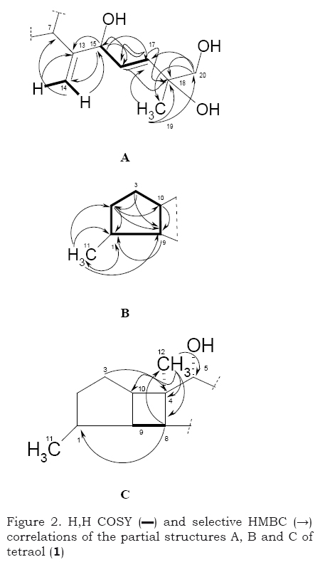
To assign the final structure of 1, the HMBC experiment was used. In accordance, the exo-olefinic methylene protons H-14a and/or H-14b (δ5.30 or 4.91) displayed three relevant correlations towards quaternary carbon C-13 (151.1), hydroxy-methine C-15 (75.1) and CH-7 (43.8) constructing an isobut-2-ene-1-ol moiety. The last fragment was in turn combined with the trans olefinic double bond via a mentioned COSY correlation between H-15 and H-16 beside to the clear HMBC correlation from H-17 (δH 5.79, δC 137.7) to C-15, and vice versa. Alternatively, the singlet methyl H3-19 (δ 1.23) displayed three critical correlations; two among them were towards C-17 and the oxy-methylene C-20 (δ 70.6), beside to its directly attached C-18 (δ73.7). This assignment was further recognized as the H2-20 showed two correlations at C-17 and C-18, respectively, confirming the partial structure A.
In addition to exhibited COSY coupling between the doublet methyl H3-11 (δ 0.93) and H-1 (δ1.80), this methyl showed rather three HMBC correlations at CH-1 (δ37.8), CH2-2 (δ36.3) and CH-9 (δ38.7).
The methylene CH2-2 (δ1.76) displayed two HMBC correlations to CH-10 (δ44.1) and CH-9 (δ38.7), beside to a visible COSY at H-3a (δ1.70 and H-3b (δ1.40), recognizing a 1-methyl-2,3-disubstituted cyclopentane B. Proton signal (δ0.95) of the third methyl CH3-12 exhibited four further correlations at C-4 (δ 48.2), CH-10 (δ44.1), CH-8 (δ 44.7) and the oxy methine C-5 (δ80.7). The last methine CH-8 (δ 1.96) was confirmed to be directly connect with CH-9 (δ 2.04) according to a shown COSY correlation. In the HMBC spectra, the methylene protons H-3a,b ((δ1.70, 1.40) displayed correlation to C-4 (δ48.2) confirming a fusion between cyclobutane and ring B via C-4 and CH-8, affording fragment C. According to COSY, H-5/H2-6/H-7, were confirmed to be directly attached, constructing a spatane moiety as shown in partial structure A (Figure 2).
In accordance, the final structure of 1 was recognized as (E)-6-(3-Hydroxy-3a,6-dimethyl-decahydro-cyclobutadicyclopenten-1-yl)-2-methyl-hepta-3,6-diene-1,2,5-triol; tetraol; a spatane diterpene (Figure 3). As the insufficiency of the compound's (1) amount, it was not able to fix its absolute configuration. Spatanes are 5-4-5 membered tricyclic diterpenes, which have not been reported from terrestrial sources. Spatol (4) the first member of this class of diterpenes, was isolated from the brown alga Spatoglossum schmittii (Gerwick et al., 1980; Venkateswarlu & Biabani, 1995) and subsequently from Stoechospermum maroinatum (Gerwick et al., 1981; Rao et al., 1987). Some of this class of diterpenes showed cytotoxic (Gerwick et al., 1980) and antibacterial (De Silva et al., 1982) activities.

The unpolar fractions of the algal extract were deduced to contain several bands of extremely confused components; however, it was not able to purify them by the usual techniques. So, they were being applied to detection on the bases of GC-MS. In accordance, four hydrocarbons were fixed; heptadecane (Rt = 18.30 min) (Acevedo et al., 2010), 2,6,10,14-tetramethyl-hexadecane (Rt = 18.37 min) (Watanabe et al., 2008), nonadecane (Rt = 20.33 min) (Meng et al., 2008) and heneicosane (Rt = 22.19 min) (Jerkovicet & Marijanovic, 2009 ).
Lipids form large group of natural compounds, which are water insoluble, but very soluble in hydrocarbons and ether. Lipids are mostly glyceride esters of long chain carboxylic acid, fatty acids, which are usually unbranched, oils and fats. The most important members of this class are widely distributed throughout animal and vegetable kingdom. An application of the ethereal extract of the desired marine alga to saponification and working up, the obtained unsaponified fraction from Surgassum subrepandum (0.05 g, 0.5 %) was estimated on the bases of GC-MS analyses. In accordance, nine hydrocarbons were detected, 2-ethyl-1-hexanol (Rt = 8.13), 4,4,7a-trimethyl-5,6,7,7a-tetrahydrobenzofuran-2(4H)-one (6) (Rt = 15.53), heptadecane (Rt = 17.20), nonadecane (Rt = 18.28), 2,6,10,14-tetramethyl-hexadecane (Rt = 18.35), 4,6,10-trimethyl-2-pentadecanone (Rt = 18.76), 6,10,14-trimethylpentadecan-2-ol (Rt = 18.83), heneincosane (Rt = 20.32) and heptacosane (Rt = 21.27).
CHEMICAL COMPOSITION OF SARGASSUM SUBREPANDUM
Nutritional properties of Sargassum subrepandum
Generally, the nutritional properties of seaweeds are usually determined from their biochemical composition such as protein, carbohydrates, crude fibre, lipids (fats) and ash content (McDermid & Stuercke, 2003). Biochemical composition of Sargassum subrepandum is shown in Table 1. Ash content was the most abundant component of dried material in Sargassum subrepandum (29.49 %) as similar to those reported (Matanjun et al., 2009).

From Table 1, it is apparent that crude fibre composition in Sargassum subrepandum had markedly high content (7%) (Rupèrez & Saura-Calixto, 2001). The fibre content of seaweed varieties is higher than those found in most fruits and vegetables. The human consumption of algal fibre has been proven to be health-promoting and its benefits (Dawczynski et al., 2007). Dietary fibre consumption promotes the growth and protection of the beneficial intestinal flora. Moreover, its consumption, in combination with high glycemic load foods reduces the overall glycemic response, greatly increases stool volume and reduces the risk of colon cancer (Goni et al., 2000; Guidel-Urbano & Goni, 2002; Jimènez-Escrig & Sànchez-Muniz, 2000).
Lipid content of marine macroalgae accounts for 1-6 g/100g d.w. (Fleurence et al., 1994; Herbreteau et al., 1997). The lipid content of Sargassum subrepandum was presently of 3.61 %. Carbohydrate contents in S. subrepandum represented a ratio of 10.21 %. Burtin (2003) stated that most of those seaweeds contain large amounts of polysaccharides, which are mostly concentrated in algal cell walls, e.g. alginates predominate in brown seaweeds. Burtin (2003) revealed that the protein content of brown seaweeds is generally small (average: 5-15 % of the dry weight). In Sargassum subrepandum, the examined total protein content her was found to be 3.2 % (Table 1). This low ratio of protein content is mostly seasonal affected (Galland-Irmouli, 1999).
Amino acid composition of Sargassum subrepandum
Concentrations of protein amino acids in Sargassum subrepandum are listed in Table 2. In accordance, glutamic acid (8.86 mg/g), valine (7.89 mg/g) and aspartic acid (7.44 mg/g) represent the most predominant amino acids in this alga. Furthermore, the results indicated that methionine was present in low concentration (1.79 mg/g). Moreover, a total amount of the protein amino acids represent a large amount in Sargassum subrepandum (78.99 mg/g), indicating the high tendency of these taxa (species) to accumulate the amino acids in their tissues.
BIOLOGICAL ACTIVITIES
Patterns of activity against set of microorganisms namely; Bacillus subtilis, Staphylococcus aureus, Streptomyces viridochromogenes (Tü 57), Escherichia coli, Candida albicans, Mucor miehi, Chlorella vulgaris, Chlorella sorokiniana, Scenedesmus subspicatus, Rhizoctonia solani and Pythium ultimum have been carried out for crude extract of S. subrepandum (Forsk.) C.Ag. (40 microg/disc, ∅ 9 mm) indicating its moderate activity against the Gram positive Staphylococcus aureus (14 mm). Moreover, the algal extract was found to reveal a potent antitumor activity at IC50 of 0.94 microg/mL against the breast carcinoma tumor cell line MCF7 in comparison with doxorubicin (IC50 0.7 microg/mL). In contrast, the extract exhibited a very weak cytotoxic activity against the brine shrimp (1.7 %o, 100 microg/mL). Inversely, compounds 1 -3 showed no cytotoxic or antimicrobial activity against any of the mentioned test organisms, indicating that the activity of the crude extract was due to some minor components which was not be able to isolate in sufficient amounts for analysis.
EXPERIMENTAL
GENERAL EXPERIMENTAL PROCEDURES
The NMR spectra were measured on a Bruker AMX 300 (300.135 MHz), a Varian Unity 300 (300.145 MHz) and a Varian Inova 600 (150.820 MHz) spectrometer. ESI MS was recorded on a Finnigan LCQ with quaternary pump Rheos 4000 (Flux Instrument). EI mass spectra were recorded on a Finnigan MAT 95 spectrometer (70 eV). DCI mass spectra were recorded on Finnigan MAT 95 A (200 eV) using reactant gas NH3. GC-MS was used a Trace GC-MS
Thermo Finnigan, ionization mode EI eV 70, instrument equipped with a capillary column CP-Sil 8 CB for amines (length: 30 m; inside diameter: 0.25 mm; outside diameter: 0.35 mm; film thickness: 0.25 μm). The analysis was carried out at a programmed temperature: initial temperature 40°C (Kept for 1 min), then increasing at a rate of 10°C/min and final temp 280 °C (kept for 10 min), Injector temp was 250 °C and detector (mode of ionization: EI) temp at 250 °C, He as a carrier gas at flow rate 1 mL/min, total run time 27 min and Injection volume 0.2 μL. Flash chromatography was carried out on silica gel (230-400 mesh). Rf-values were measured on Polygram SIL G/UV254 (Macherey-Nagel & Co.). Size exclusion chromatography was done on Sephadex LH-20 (Pharmacia).
COLLECTION AND TAXONOMY OF THE MARINE ALGA
The alga S. subrepandum (Forsk.) C.Ag. was collected from Ras Gharib on Suez-Gulf, Red Sea, Egypt. The identification was carried out according to method reported previously (Nasr, 1940; Abou-ElWafa, 2005). A voucher reference specimen (no. 4200) of the alga is deposited at the Department of Botany, Faculty of Science, Mansoura University, Egypt.
PREPARATION OF SAMPLES
The collected samples were cleaned up from epiphytes and non-living matrix in running water, and rinsed many times in distilled water. The samples were then spread on string nets and allowed to dry in air. The air-dried samples were ground and stored in suitable closed bottles at room temperature.
EXTRACTION AND ISOLATION OF THE ACTIVE CONSTITUENTS
The air dried leaves (c.a.100 g) and stems (c.a. 30 g) of S. subrepandum were individually applied to extraction by soxhlet using CH2Cl2 for ~ 12 hrs. The afforded extracts were filtered off and concentrated in vacuo at 40°C to yield greenish brown crude extracts (2.24 g and 0.37 g) from leaves and stems, respectively. As the identical TLC for both extracts, they were combined (2.61 g) and applied to Sephadex LH-20 (MeOH). TLC monitoring, visualized under UV light and spraying with anisaldehyde/ sulphuric acid furnished three fractions. Fraction I (1.5 g) was mostly constructed from undesired pigments, including chlorophyll and fatty acids. The fraction was applied to silica gel column eluted with C6H12-CH2Cl2 gradient, and the delivered unpolar components were combined and submitted to GC-MS analysis. Fraction II (0.71 g) was applied to Sephadex LH-20 column (CH2Cl2/40% MeOH) lead to three sub-fractions, FIIa (0.29 g), FIIb (0.18 g), and FIIc (0.28 g). Sub-fraction FIIa was applied to silica gel column (C6H12-CH2Cl2) giving unpolar mixture of oil, KSSR5 (120 mg), which was detected by GC-MS analysis. The two sub-fractions FIIa and FIIb were combined (0.46 g) and applied to silica gel column (CH2Cl2-MeOH) affording two components, fucosterol (2, 27 mg) and linoloeic acid (3, 18 mg). FIII (0.39 g) was purified on silica gel column (CH2Cl2-CH3OH) to obtain a colourless oil of 1 (3 mg).
Tetraol; 5(R),15,18(R),19-Tetrahydroxy-penta-13,16(E)-diene (1): C20H32O4 (336); an UV faint absorbing colourless oil, turned dark violet with anisaldehyde/sulphuric acid and heating. - DCI MS (NH4): m/z = 354 ([M+NH4]+). - HRESI MS: 359.21921 (calc. 359.21927 for C20H32O4Na).- 1H NMR (CDCl3, 300 MHz), δ (ppm): 1.80 (m, 1H, H-1), 1.76 (m, 1H, H2-2), 1.70 (m, 1H, H-3a), 1.40 (m, 1H, H-3b), 3.63 (d, J = 4.1 Hz, 1H, H-5), 2.28 (ddd, J = 13.0, 13.0, 4.3 Hz, 1H, Ha-6), 1.66 (m, 1H, Hb-6), 2.92 (m, 1H, H-7), 1.96 (m, 1H, H-8), 2.04 (m, 1H, H-9), 1.96 (m, 1H, H-10), 0.93 (d, J = 6.0 Hz, 3H, H3-11), 0.95 (s, 3H, H3-12), 5.30 (d, J = 1.3 Hz, 1H, Ha-14), 4.91 (s, 1H, Hb-14), 4.38 (d, J = 8.6 Hz, 1H, H-15), 5.60 (m, 1H, H-16), 5.79 (dd, J = 15.6, 3.9 Hz, 1H, H-17), 1.23 (s, 3H, H3-19), 3.39 (s, 2H, H2-20). -13C NMR (CDCl3, 150 MHz), δ(ppm): 37.8 (CH-1), 36.3 (CH2-2), 28.8 (CH2-3), 48.2 (Cq-4), 80.7 (CH-5), 37.8 (CH2-6), 43.8 (CH-7), 44.7 (CH-8), 38.7 (CH-9), 44.1 (CH-10), 14.9 (CH3-11), 13.8 (CH3-12), 151.1 (Cq-13), 108.2 (CH2-14), 75.1 (CH-15), 131.9 (CH-16), 137.7 (CH-17), 73.7 (Cq-18), 24.5 (CH3-19), 70.6 (CH2-20).
Fucosterol (2): C29H48O1 (412); Colourless solid, turned blue on spraying with anisaldehyde/sulphuric acid and heating. - EI MS: m/z (%) = 413 ([M]+, 28), 399 ([M-CH2]+, 10), 314 (100), 299 (24), 271 (10), 255 (8), 229 (11), 211 (7), 145 (8), 119 (9), 81 (24), 69 (36), 55 (70), 44 (39), 41 (28). - 1H NMR (CDCl3, 300 MHz), δ(ppm): 5.37 (d, J = 4.7 Hz, 1H, H-6), 5.19 (q, 1H, J = 6.3 Hz, H-28), 3.51 (m, 1H, H-3), 2.27 (m, 1H, H-25), 2.22 (m, 1H, Ha-4), 2.10-1.83 (m, 4H, H2-7, H2-22), 1.58 (d, 3H, J = 6.1 Hz, CH3-29), 1.55-1.18 (m, 6H), 1.15-1.05 (m, 6H), 1.01 (s, 3H, CH3-19), 0.99 (6H, CH3-26, CH3-27), 0.94 (d, 3 H, J = 6.1 Hz, CH3-21), 0.93-0.80 (m, 8H), 0.68 (s, 3H, CH3-18). - 13C NMR (CDCl3, 125 MHz), δ (ppm): 36.5 (CH2-1), 31.7 (CH2-2), 71.8 (CH-3), 42.4 (CH2-4), 140.7 (Cq-5), 121.6 (CH-6), 31.9 (CH2-7), 31.9 (CH-8), 50.1 (H-9), 37.3 (Cq-10), 21.1 (CH2-11), 28.3 (CH2-12), 42.3 (Cq-13), 56.8 (CH-14), 24.4 (CH2-15), 39.8 (cH2-16), 55.8 (H-17), 13.2 (CH3-18), 19.5 (cH3-19), 36.4 (CH-20), 18.8 (CH3-21), 35.3 (cH2-22), 25.7 (CH2-23), 146.9 (Cq-24), 34.8 (CH-25), 22.2 (CH3-26), 22.3 (CH3-27), 115.5 (CH-28), 11.9 (CH3-29).
Linoleic acid; (9Z,12Z)-9, 12-octadecanoic acid (3): C18H32O2 (280.45); an UV absorbing colourless oil, turned to blue by anisaldehyde/sulphuric acid and heating.
- EI MS (70 eV): m/z (%) = 280 (80), 264 (28), 137 (10), 124 (15), 110 (28), 95 (60), 81 (84), 67 (100), 55 (92), 41 (92). - 1H NMR (CDCl3, 300 MHz), δ (ppm): 8.98 (s, br, 1 H, COOH), 5.43-5.28 (m, 4 H, 9,10,12,13-CH), 2.78 (t, 3J = 6.0 Hz, 2 H, 11-CH2), 2.38 (t, 3J = 7.2 Hz, 2 H, 2-CH2), 2.08 (m, 4 H, 8,14-CH2), 1.63 (m, 2 H, 3-CH2), 1.42-1.23 (m, 14 H, 4,5,6,7,16,17-CH2), 0.85 (m, 3 H, 18-CH3). - 13C/APT NMR (CDCl3, 50 MHz), δ (ppm): 180.1 (CO, Cq), 130.1 (CH-13), 129.9 (cH-9), 128.0 (CH-10), 127.8 (CH-12), 31.5 (cH2-2), 29.6 (CH2-16), 29.6 (CH2-11), 29.5 (cH2-14), 29.3 (CH2-8), 29.1 (CH2-7), 29.0 (CH2-6), 29.0 (CH2-5), 27.1 (CH2-4), 25.6 (cH2-3), 24.7 (CH2-15), 22.5 (CH2-17) 14.0 (CH2-18).
Phytosterols and hydrocarbons estimation
A powdered sample (10 g) of S. subrepandum was extracted with petroleum ether (60-80 °C) at room temperature. The afforded extract was concentrated in vacuo to give an oily residue, which was then treated with 50 mL of 10 % alcoholic KOH and refluxed in a water bath for 2 hrs. After cooling, 50 mL water was then added and the aqueous solution was extracted with chloroform. The organic layer was washed with water until it became alkali free, and dried over anhydrous Na2SO4. The solvent was distilled off to give the unsaponified fraction as an oily extract. Finally, the oily sample was applied to GC-MS analysis, and the retention time of their peaks was compared with reported data in literatures.
Chemical composition of S. Subrepandum
Ash content
The ash content of the alga was estimated according to Marsham, Scott & Tobin (2007). Four grams of dried algal material were added to a preweighed crucible. The algal contents were then inserted in a muffle furnace (Metrawatt GmbH, RO-8) at 550 °C for ~10 hrs. After cooling, the contents were kept in a desiccator (to avoid any atmospheric moisture) and reweighed.
Lipid content
Ten grams from the air dried alga were applied to extraction with petroleum ether 40-60 °C during Soxhlet extractor (Soxtec System HT6, Tecator, Hoganas, Sweden) for ~10 hrs. After concentration in vacuo, the obtained crude lipids were weighed gravimetrically according to Wong and Cheung (2000).
Protein content
0.5 Gram from the air dried alga was extracted with 10 mL phosphate buffered saline (pH = 7). After centrifugation, 0.3 mL of the supernatant was raised to 1 mL by addition of phosphate buffered saline and treated with 5 mL coomassie brilliant blue- G250. According to the Bradford method (1976), the afforded sample was then applied to colorimetric measurments at wavelength 595 nm in comparison with bovine serum albumin (BSA) as reference. This was carried out using ATI UNICAM UV/Vis Spectrometer UV.2 (England).
Carbohydrate content
Soluble carbohydrates were determined according to the colorimetric method of Yemm and Willis (1954). Known weight of dried algal sample (0.1 g) was hydrolyzed with 5 mL 2.5 N HCl for 3 hrs in a boiling water bath, cooling and neutralization with Na2CO3 till everffesence ceases. After neutralization; the volume was completed to 100 mL using distilled water and filtered. 0.5 mL of the supernatant was completed to 1 mL using distilled water and mixed with 4 mL of 0.1 % anthrone/sulphuric acid and heated for 10 min in a water bath. After cooling, the developed blue green colour was measured at 630 nm using a spectrophotometer ATI UNICAM UV/VIS Spectrometer UV.2 (England). Carbohydrate concentration was calculated in comparison with D-glucose as a reference sugar.
Crude fibre content
Crude fibre was estimated by acid and alkaline digestion. Two grams of defatted residue was boiled with 200 mL of 1.25% H2SO4 for 30 min, cooled, filtered and the residue was washed (3 x) with 50 mL of boiling water. The washed residue was digested by boiling in 200 mL of 1.25% NaOH for 30 min, cooled, filtered and washed (3 x) with 50 mL of boiling water and finally with 25 mL ethanol. The residue was dried in an oven at 110 °C to constant weight using a pre-weighed crucible, cooled in desiccator and weighed. The residue was ashed at 550 °C for 8 hrs, cooled in a desiccator and reweighed. Crude fibre content was expressed as percentage loss in weight on ignition (Nesamvuni, Steyn & Potgieter, 2001; AOAC, 2000).
Amino acid content
Three grams of dried seaweed sample were treated with 6N HCl containing one crystal of phenol (approx. 0.5 mg) in a pyrex tube (6 x 50 mm) and (100 μL) of 2-mercaptoethanol. After sealing the vial under vacuum, the reaction mixture was applied to hydrolysis at 110 °C for 24 hrs. The vial was then applied to evaporation in vacuo, and the residue was dissolved in methanol and applied for subsequent chromato-graphic separation. Amino acid mixture in the hydrolysates was determined by an amino acid analyzer LC 3000 eppendorf/ Biotronik using column type H 125 x 4 mm, pre-column type H 60 x 4 mm, eluents and reagent type H1 (4 buffer system) (Walker, 1996). Determination of tryptophan was carried out using method described by Miller (1967) after hydrolysis of samples with barium hydroxide.
BIOLOGICAL ACTIVITY
Antimicrobial activity
Antimicrobial assays were conducted utilizing the disc-agar method (Burkholder, Burkholder & Almodovar, 1960) against diverse sets of microorganisms. The Sargassum subrepandum extract was dissolved in CH2Cl2/10% MeOH at a concentration of 1 mg/mL. Aliquots of 40 μL were soaked on filter paper discs (9 mm , no. 2668, Schleicher & Schüll, Germany) and dried for 1 h at room temperature under sterilized conditions. The paper discs were placed on inoculated agar plats and incubated for 24 hrs at 38 °C for bacterial and 48 hrs (30 °C) for the fungal isolates, while the algal test strains were incubated at ~ 22 °C in day light for 8~10 days. The algal extract was examined against the following test microorganisms: Bacillus subtilis, Staphylococcus aureus, Streptomyces viridochromogenes (Tü 57), Escherichia coli, Candida albicans, Mucor miehi, Chlorella vulgaris, Chlorella sorokiniana, Scenedesmus subspicatus, Rhizoctonia solani and Pythium ultimum.
Brine shrimp microwell cytotoxic assay
The cytotoxic assay was performed according to Takahashi et al. method (Takahashi, Kurasawa, Ikeda, Okami & Takeuchi, 1989) and Sajid et al. screening (Sajid, Fondja Yao, Shaaban, Hasnain & Laatsch, 2009).
In vitro cytotoxic activity using SRB assay
In vitro cytotoxicity of the desired components were tested according to (Skehan, Storeng, Scudiero, Monks, McMahon, Vistica, Warren, Bokesch, Kenney & Boyd, 1990). Cells were plated in a 96-multiwell plate (104cells/well) for 24 hrs before treatment with the components to be tested to allow attachment of cells to the wall of the plate. Different concentrations of the components under test (0, 1, 2.5, 5, 10 μg/mL) were carried out and added to the cell monolayer. Triplicate wells were prepared for each individual dose. Monolayer cells were incubated with tested components for 48 hrs at 37 °C in an atmosphere with 5% CO2. After incubation, cells were fixed, washed and stained with Sulfo-Rhodamine-B pigment. Excess of the dye was washed off with acetic acid, while the adsorbed dye was dissolved with Tris-EDTA buffer. Colour intensity was measured in an ELISA reader. The relation between surviving fraction and drug conc. was plotted to get the survival curve of each tumor cell line after application of the specified compound.
ACKNOWLEDGEMENTS
The authors are deeply thankful to Prof. Dr. H. Laatsch for his kind support, laboratory facilities and spectroscopic measurements. The assistance of Miss F. Lissy and Mr. A. Kohl are also gratefully acknowledged. This work has been financed and assisted by the DAAD for fellowships.
REFERENCES
Abou-El Wafa, G. S. E. (2005) Studies on the Biological Activities of Some Species of Egyptian Marine Algae, M.Sc. Thesis Faculty of Science, Mansoura University, Egypt. [ Links ]
Acevedo, Cristian A., Sanchez, E. Y., Reyes, J. G., Young, M. E. (2010) Volatile profiles of human skin cell cultures in different degrees of senescence. Journal of Chromatography B 878: 449-455. [ Links ]
Ali, M. S., Pervez, M. K. (2003) Seco-Dolastanes from the Marine Brown Alga Dictyota dichotoma (Huds.) Lamour". Zeitschrif für Naturforschung 58b: 438-442. [ Links ]
AOAC (2000) Official methods of analysis, 15th edn. Association of Official Agricultural Chemists, Washington, DC. [ Links ]
Bradford, M. M. (1976) A Rapid and Sensitive Method for the Quantitation of Microgram Quantities of Protein Utilizing the Principle of Protein-Dye Binding. Analytical Biochemistry 72: 248-254. [ Links ]
Baslow, M. H. (1969) Marine Pharmacology. A Study of Toxins and Other Biologically Active Substances of Marine Origin. Williams and Willkins Co., Baltimore. [ Links ]
Burkholder, P. R., Burkholder, L. M., Almodovar, L. R. (1960) Antibiotic activity of some marine algae of Puerto Rico. Botanica Marina 2: 149-156. [ Links ]
Burtin, P. (2003) Nutritional value of seaweeds. Electronic Journal of Environmental, Agricultural and Food Chemistry 2: 498-503. [ Links ]
Dawczynski, C., Schubert, R., Jahreis, G. (2007) Amino acids, fatty acids, and dietary fibre in edible seaweed products. Food Chemistry 103: 891-899. [ Links ]
De Silva, S. S. M., Gamage, S. K. T., Kumar, N. S., Balasubramaniam, S. (1982) Antibacterial activity of extracts from the brown seaweed Stoechospermum marginatum. Phytochemistry 21: 944-945. [ Links ]
Dictionary of Natural Products on CD-ROM, Chapman & Hall Chemical Database, 2004. [ Links ]
El-Naggar, M. E. E., El-Shora, H. M., Shaaban-Dessouki, S. A., Zaid, A. M. (1995) Fatty acid composition of Sargassum denticulatum and S. latifolium as influenced by time of collection and plant organ. Qatar University Science Journal 15: 95-100. [ Links ]
Falsone, G., Cateni, F., Vrech, E. (1994) Triacylglycerols, fucosterol, diacylglyceroglycolipids and fucoxanthin from Fucus virsoides J. AG. Zeitschrift für Naturforschung 49b: 1297-1304. [ Links ]
Fenical, W., Sims, J. J., Squatrito, D., Wing, R. M., Radlick, P. J. (1973) Marine natural products. VII. Zonarol and isozonarol, fungitoxic hydroquinones from the brown seaweed Dictyopteris zonarioides. Journal of Organic Chemistry 38: 2383-2386. [ Links ]
Fleurence, J., Gutbier, G., Mabeau, S., Leray, C. (1994) Fatty acids from 11 marine macroalgae of the French Brittany coast. Journal of Applied Phycology 6: 527-532. [ Links ]
Frost, J., Ward, J. (1968) Stereochemistry of 7,24(28)-stigmastadien-3β-ol and the fucosterols, Tetrahedron Letters, 3779-3782. [ Links ]
Galland-Irmouli, A. V., Fleurence, J., Lamghari, R., Luçon, M., Rouxel, C., Barbaroux, O., Bronowicki, J. P, Vuillaume, C., Guèant, J. L. (1999) Nutritional value of proteins from edible seaweed Palmaria palmata (Dulse). The Journal of Nutritional Biochemistry 10: 353-359. [ Links ]
Gerwick, W. H., & Fenical, W. (1983) Spatane diterpenoids from the tropical marine algae Spatoglossum schmittii and Spatoglossum howleii (Dictyotaceae). Journal of Organic Chemistry 48: 3325-3329. [ Links ]
Gerwick, W. H., Fenical, W., Sultanbawa, M. U. S. (1981) Spatane diterpenoids from the tropical marine alga Stoechospermum marginatum (Dictyotaceae). Journal of Organic Chemistry 46: 2233-2241. [ Links ]
Gerwick, W. H., Fenical, W., Van Engen, D., Clardy, J. J. (1980) Isolation and structure of spatol, a potent inhibitor of cell replication from the brown seaweed Spatoglossum schmittii. Journal ofthe American Chemical Society 102: 7991-7993. [ Links ]
Goni, I., Valdivieso, L., Garcia-Alonso, A. (2000) Nori seaweed consumption modifies glycemic response in healthy volunteers. Nutrition Research 20: 1367-1375. [ Links ]
Guidel-Urbano, M., Goni, I. (2002) Effect of edible seaweeds (Undaria pinnitafida and Porphyra tenera) on the metabolic activities of intestinal microflora in rats. Nutrition Research 22: 323-331. [ Links ]
Herbreteau, F., Coiffard, L. J. M., Derrien, A., De Roeck-Holtzhauer, Y. (1997) The fatty acid composition of five species of macroalgae. Botanica Marina 40: 25-27. [ Links ]
Hossain, Z., Kurihara, H., Takahashi, K. (2003) Biochemical Composition and Lipid Compositional Properties of the Brown Alga Sargassum horneri. Pakistan Journal of Biological Sciences 6: 1497-1500. [ Links ]
Jimènez-Escrig, A., Sànchez-Muniz, F. J. (2000) Dietary fiber from edible Seaweeds: Chemical structure, physicochemical properties and effects on cholesterol metabolism. Nutrition Research 20: 585-598. [ Links ]
Jerkovic, I., Marijanovic, Z. (2009) Screening of volatile composition of Lavandula hybrida REVERCHON II honey using headspace solid-phase microextraction and ultrasonic solvent extraction. Chemistry & Biodiversity 6: 421-430. [ Links ]
Marsham, S., Scott, G.W., Tobin, M. L. (2007) Comparison of nutritive chemistry of a range of temperate seaweeds. Food Chemistry 100: 1331-1336. [ Links ]
Matanjun, P., Mohamed, S., Mustapha, N. M., Muhammad, K. (2009) Nutrient content of tropical edible seaweeds, Eucheuma cottonii, Caulerpa lentillifera and Sargassum polycystum. Journal of Applied Phycology 21: 75-80. [ Links ]
McDermid, K. J., Stuercke, B. (2003). Nutritional composition of edible Hawaiian seaweeds. Journal of Applied Phycology 15: 513-524. [ Links ]
Meng, S., Xiao, X., Wang, S., Luo, C., Liu, X. (2008) Liposoluble chemical constituents of Periplaneta americana by GC-MS. Zhongnan Yaoxue 6: 23-25. [ Links ]
Moore, R.E. (1978) Marine Natural Products. P. J. Scheuer, Ed.; Academic Press: New York, Vol. 1, Chapter 2. [ Links ]
Miller, E. L. (1967) Determination of tryptophan content in feeding stuffs with particular reference to cereals. Journal of the Science of Food and Agriculture 18: 381-386. [ Links ]
Nesamvuni, C., Steyn, N., Potgieter, M. (2001) Nutrients analysis of selected western African foods. South African Journal of Science 97: 51-54. [ Links ]
Nasr, A. H. (1940) The marine algae of Alexandria. 1- A report on some marine algae collected from the vicinity of Alexandria. Notes and memoirs No. 36, Government Press, Bulaq, Cairo, pp. 33 pl. VI. [ Links ]
Perez, R. M., Avila, J. G., Perez, S., Martinez, A., Martinez, G. (1990) Antimicrobial activity of some American algae. Journal of Ethnopharmacology 29: 111-116. [ Links ]
Popov, S., Marekov, N., Konaklieva, M., Panayotova, M., Dimitrova-Konaklieva, S. (1985) Sterols from some Black Sea Ulvaceae. Phytochemistry 24: 1987-1990. [ Links ]
Rao, C. B., Pullaiah, K. C., Surapaneni, R. K., Suryaprabha, R., Raju, V. S. N. (1987) A new spatane diterpenoid from Stoechospermum marginatum C. Agardh. Indian Journal of Chemistry 26: 79-80. [ Links ]
Rupèrez, P. Saura-Calixto, F. (2001) Dietary fibre and physicochemical properties of edible Spanish seaweeds. European Food Research and Technology 212: 349-354. [ Links ]
Sajid, I., Fondja Yao, C. B., Shaaban, K. A., Hasnain, S., Laatsch, H. (2009) Antifungal and antibacterial activities of indigenous Streptomyces isolates from saline farmlands: prescreening, ribotyping and metabolic diversity. World Journal of Microbiology and Biotechnology 25: 601-610. [ Links ]
Shaaban, M. (2004) Bioactive Secondary Metabolites from Marine and Terrestrial Bacteria: Isoquinolinequinones, Bacterial Compounds with a Novel Pharmacophor. PhD Thesis, University of Goettingen, Germany. [ Links ]
Sheu, J., Wang, G., Sung, P., Duh, C. (1999) New Cytotoxic Oxygenated Fucosterols from the Brown Alga Turbinaria conoides. Journal of Natural Products 62: 224-227. [ Links ]
Skehan, P., Storeng, R., Scudiero, D., Monks, A., McMahon, J., Vistica, D., Warren, J. T., Bokesch, H., Kenney, S., Boyd, M. R. (1990) New colorimetric cytotoxicity assay for anti-cancer-drug screening. Journal of the National Cancer Institute 82: 1107-1112. [ Links ]
Takahashi, A., Kurasawa, S., Ikeda, D., Okami, Y., Takeuchi, T. (1989) Altemicidin, a new acaricidal and antitumor substance. I. Taxonomy, fermentation, isolation and physico-chemical and biological properties. Journal of Antibiotics 32: 1556-1561. [ Links ]
Tang, H., Yi, Y., Yao, X., Xu, Q., Zhang, S., Lin, H. (2002) Bioactive steroids from the brown alga Sargassum carpophyllum. Journal of Asian Natural Products Research 4: 95-101. [ Links ]
Venkateswarlu, Y., Biabani, M. A. F. (1995) A Spatane Diterpene from the Brown Alga Stoechospermum Marginatum. Phytochemistry 40: 331-333. [ Links ]
Walker, J. M. (1996) The Protein Protocol Handbook. Tatowa, New Jersey, USA: Humana Press Inc, pp, (760): 45-47. [ Links ]
Watanabe, A., Ueda, Y., Higuchi, M., Shiba, N. (2008) Analysis of volatile compounds in beef fat by dynamic-headspace solid-phase microextraction combined with gas chromatography-mass spectrometry. Journal of Food Science 73: C420-C425. [ Links ]
Wong, K. H., Cheung, C. K. (2000) Nutritional evaluation of some subtropical red and green seaweeds Part I—proximate composition, amino acid profiles and some physico-chemical properties. Food Chemistry 71: 475-482. [ Links ]
Yemm, E.W., Willis, A. L. (1954) The estimation of carbohydrates in plant extracts by anthrone. Biochemical Journal 57: 508-514. [ Links ]
Zhang, X., Han, F., Gao, P., Yu, D., Liu, S. (2007) Bioassay-guided fractionation of antifertility components of castorbean (Ricinus communis L.) seed extracts. Natural Product Research 21: 982-989. [ Links ]

