


 text new page (beta)
 English (pdf)
 Article in xml format
 Article references
 Send this article by e-mail
 Cited by SciELO
  Similars in
SciELO
Similars in
SciELO 
 Permalink
INTRODUCTION
The foot is one of the most complex parts of the human body, its structure integrated by ligaments, tendons, muscles, and bones, allows it to meet the demands of support and locomotion of the human [1]. The vault of the feet plays an important role in gait biomechanics, it reduces the impact loads from the ground to the body and keeps the correct alinement of the foot. Therefore, the modification to this structure will modify these functions [2]. The collapse of this vault is known as flatfoot. All children are born with flat feet [3], the formation of the medial plantar arc occurs naturally and it is generally until 5 or 6 years of age when the medial arch is adequately defined [4]. However, in some cases, the flatfoot deformity can stay until the first decade of life or after [2] [5] [6]. The etiology or the specific causes that produce flatfoot are unknown, and it is still a topic of debate nowadays [2] [5] [6] [7]. It has not been found a specific age when the vault of the feet is completely formed. In addition, when the formation of the vault of the feet in children is delayed becomes a common concern of parents.
On the other hand, the cavus foot (hollow foot) is the enlargement of the vault of the feet, increasing the contact on the anterior and posterior regions of the foot. This deformity is not a common pathology in the population. However, it has been reported an increment in the prevalence of cavus foot in athletes, especially in women [8] [9] [10] [11].
Although there are different studies about the morphology of the soles of the feet [2] [12] [13] [14] [15] [16] [17], to the best of our knowledge there is no research about the type of feet in a Mexican population from an early stage to adulthood. The true prevalence of the morphology of the feet in a Mexican population of all ages is unknown. It is known that the modification of any part of the structure of the foot will modify its biomechanics and generate injuries in the same structure and other parts of the body [2] [18] [19]. Therefore, the objective of this study is to analyze the morphology of the soles of the feet and obtain the Chippaux-Smirak Index (CSI) in a Mexican population to identify the type of feet and the prevalence of musculoskeletal disorders of the foot.
Literature review of the type of foot in the Mexican population
In order to validate the lack of knowledge about the type of feet in the Mexican population from the early stage to adulthood, a search of information related to the type of feet in a Mexican population was conducted. Search engines such as, Google scholar (n= 157), Scopus (n= 0), ScienceDirect (n= 6), and Scielo (n= 0) were used with the following search equation in English and Spanish: "Type of foot" AND "Footprint" AND "Mexican". The literature review was performed up to the 11th of January 2023. The equation gave a total of 163 studies, but not all of them related to the analysis of the soles of the feet to detect the morphology of the foot in a Mexican population. After reviewing the articles, duplicated manuscripts, theses, books, studies from different countries, and conference proceedings were excluded from the analysis, having a final number of 9 articles, [2] [3] [8] [10] [14] [15] [20] [21] [22]. Most of the studies are performed in a limited period of time and most of them are performed in young population [2] [3] [10] [14] [15] [20].
It is known that the increase or reduction of the medial arch of the foot could affect the biomechanics of the foot. The maturation of the vault of the foot occurs naturally and most of the time this happens in the first decade of life. Moreover, it is a common concern of parents when they notice that the medial longitudinal arch of the foot remains collapsed after the first decade of life. In Mexico, different studies have been performed to identify the type of foot, but most of them were developed in a young population, 2-5 and 6-12 years [3], 6-14 years [14], 9-11 years [20], 6-15 years [15], 6-13 years [10], and 3-6 years [2]. Although there are different studies of the type of foot in the Mexican population, most of them are performed when the vault of the foot seems to be not completely matured. There were just two studies performed on athletes with a larger range of ages from 9 to 20 years old [8] [22]. In the study performed by Miguel et al., it was found a high prevalence of cavus foot in women [8].
MATERIALS AND METHODS
A database of images of the soles of both feet was analyzed. The database contained images of one thousand and fourteen persons between 2 and 73 years old, all of them from Guanajuato state, Mexico. The images were acquired between April 2017 and February 2022. The soles of the feet of the participants were digitized with a 2D foot scanner (Sensormedica, Guidonia Montecelio, Rome, Italy). There were 544 (53.6 %) females and 470 (46.4 %) males in the database.
To classify the type of foot, the CSI was obtained in both feet [23], as shown in Figure 1a. The digitized images of the soles of the feet were analyzed in MATLAB R2015a version 8.5.0.197613. Matlab specific tools (ginput, GUI, imread) were used to locate the coordinate points in the image plane [8] [24] [25]. This process was performed manually by the same author to secure repeatability.

Figure 1 Morphology of the soles of the feet: a) ChippauxSmirak Index and b) Distribution of participants by age.
Once the soles of the feet were measured (medial and forefoot region), they were classified based on the Chippaux-Smirak Index (CSI) into six different types of feet, cavus foot, extreme cavus, normal foot, flatfoot 1°, flatfoot 2° and flatfoot 3°. For the cavus foot, the CSI was 0 < CSI < 0.25, for the extreme cavus the CSI was 0, for the normal foot the CSI was 0.25 ≤ CSI < 0.45, for the flatfoot 1° the CSI was 0.45 ≤ CSI < 0.5, for the flatfoot 2° the CSI was 0.5 ≤ CSI ≤ 0.6 and for the flatfoot 3° the CSI was CSI > 0.6 [8] [13] [23] [26] [27].
The database images were classified into groups of three years starting from 2 years until 73 years, as shown in Figure 1b. The minimum number of participants per group was 22 and the maximum was 68.
RESULTS AND DISCUSSION
Footprint data analysis
The results show that the prevalence of flatfoot is higher during the first decade of life and decreases after that (see Figure 2). This information will be useful for orthopedic technicians and parents who are worried about the health of their children. Moreover, health professionals such as orthopedists could use the information to detect if the development of the foot structure is normal according to the age of the patient.
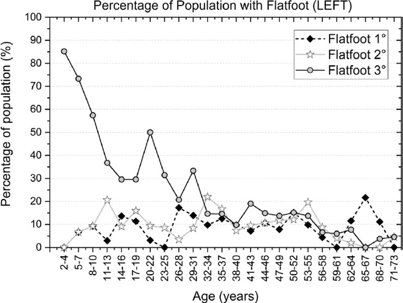
Figure 2 Prevalence of different types of flat feet in a Mexican population: Data from the left foot.
Figure 2 describes the different types of flatfoot (left foot), being the flatfoot 3° where the contact surface is the highest. The prevalence of the different types of flat feet from Figure 2 shows that most of the participants have flatfoot 3° until 10 years of age. From that age, the medial arc begins to elevate and the contact surface decreases. The flatfoot never completely disappears, there was a relatively small percentage of prevalence 10-20 % for the three types of flatfoot between the ages of 32 and 55 years. Between the ages of 65 and 67 years, there is a small increment of the prevalence of flatfoot 1°.
The CSI for the normal foot was considered between 0.25 and 0.45. A lower value of the CSI was considered as cavus foot, and when the contact surface of the medial region disappears the foot was categorized as extreme cavus (CSI = 0). Figure 3 presents the data distribution of the normal foot, cavus foot, and extreme cavus for all ages (left foot). From Figure 3, it can be seen that the normal foot begins with a low percentage of prevalence, and increases from 10 years of age. The cavus foot and extreme cavus stay most of the time below 10 % for all ages.
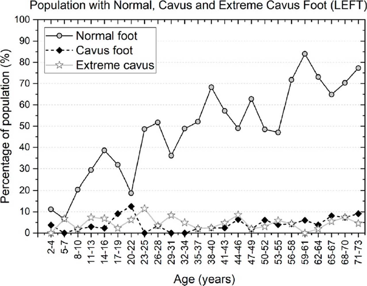
Figure 3 Prevalence of population with the normal foot, cavus foot, and extreme cavus: Data from the left foot.
Similarly, the analysis of the morphology of the soles of the feet in the right foot follows a comparable trend that the left foot. Figure 4 shows that the prevalence of flatfoot 3° stays high until 16 years, then it decreases until 19 years and stays close to 30 % until 40 years. This behavior is different from the left foot. Furthermore, flatfoot 1° and 2° stay most of the time below 20 % for all ages, as shown in Figure 4. The data from Figure 2 and 4 demonstrate that both feet (left and right feet) are not completely symmetrical.

Figure 4 Prevalence of different types of flat feet in a Mexican population: Data from the right foot.
Figure 5 shows the prevalence of normal foot, cavus foot, and extreme cavus of the right foot. The normal foot stays less than 10 % when the participants are less than 10 years old and increases beyond 20 % when the participants are older than 11 years old, as shown in Figure 5. The prevalence of cavus foot and extreme cavus is most of the time less than 10 % for all ages.

Figure 5 Prevalence of population with the normal foot, cavus foot, and extreme cavus: Data from the right foot
Finally, in order to validate about the prevalence of the type of foot for people over 17 and under 73 years, a statistical analysis using a z-test for proportions was performed for the morphologies of cavus and flat feet, in the left foot. It was considered a confidence interval of 95 % and a p-value of 0.05. For cavus foot, it was observed that less than 17 % of the population have this morphology (p = 0.018). For flatfoot 1 and flatfoot 2, the p-values were 0.0168 and 0.0065, respectively, proving that the proportion of the population with these two morphologies is less than 16 %. For flatfoot 3 the proportion was less than 25 %, obtaining a p-value of 0.0079. For the right foot, it was performed the same statistical analysis, obtaining p-values less than 0.05 for the same percentages in the cavus foot and the three types of flatfoot.
From the results and at least for the Mexican population, the idea that at 3-5 years [5], 5-6 years [4], 10 years [2] [6] or 12-13 years [28] the vault of the feet would already be formed seems to be inaccurate. From the results found in the analysis, the maturation of the vault of the feet in a Mexican population seems to be beyond the first decade of life. Moreover, there is a health problem related to the morphology of the feet in the adult Mexican population that needs to be carefully studied. Further research needs to be done to study the effect of these musculoskeletal disorders on the gait biomechanics and how the quality of life of the Mexican population is affected. It has been found that the morphology of the soles of the feet could have an effect on the plantar pressure distribution and this could be a risk factor for producing musculoskeletal disorders [22]. Furthermore, it has been reported that the deformities of flatfoot and cavus foot could negatively influence productivity in adult life [29].
This information will be useful to clarify that the medial arc of the foot could have a delay, and for the Mexican population, the formation of the vault takes more time than the already reported by other authors [2] [4] [5] [6]. Therefore, parents should not be worried if children present flatfoot pathology at an early stage unless the pathology produces pain or discomfort. Moreover, it has been reported that there is no treatment, orthopedic device, external force, or insoles that help to form the medial arc [6] [28] [30].
It has been found that flatfoot is related to overweight and obesity [13] [15] [20]. Mexico has serious problems with overweight and obesity. According to the National Survey of Health and Nutrition (ENSANUT Spanish abbreviation, Guanajuato 2006-2018), it was established that overweight and obesity in boys and girls between 5 and 11 years old have increased from 27.8 % in 2006 to 45.1 % in 2018 in the state of Guanajuato. In adolescents, between 12 and 19 years old, overweight and obesity increased from 34 % in 2006 to 38.1 % in 2018. Although some studies have found that the prevalence of flatfoot is related to overweight or obesity [15] [20], other studies have found that body weight does not affect plantar footprint alterations [10].
Although our results of the prevalence of cavus foot (less than 10 %) were not higher than the flatfoot in the young population, the values were similar to Aco Luna et al., [10]. However, our values of the cavus foot were lower than the values found by Gonzalez et al., [31] and Espinoza et al., [32]. Although it was found that cavus foot is a pathology with low prevalence, some studies have developed personalized insoles for the treatment of cavus foot and comorbidities [33]. Contrary to our results, other authors have found a high prevalence in cavus foot (hollow foot) in children between 3 to 13 years [34]. It is difficult to compare our results with other studies because they used a different methodology and/or the anthropometric features of the population could be different.
The results of flatfoot (10-20 %) in the adult population agree with the results found by other researchers [21] [35] and disagrees with others [36]. Moreover, it has been found that the prevalence of flatfoot decreases as the child grows [3] [13] [37]. It is important to mention that the classification of the foot types could be influenced by the method employed (CSI, Arch Index, Staheli Arc Index, so on), therefore the comparison among studies should be carefully performed [38]. Although there are other methods to evaluate the footprint, the SCI is one of the most used methods to describe the type of feet. Furthermore, although there is a good correlation among them, it has been reported that the other methods can produce different outcomes with slightly less accuracy in some cases [23] [26] [39].
Although there are several studies related to the foot type [2] [3] [10] [14] [15] [20], to the author’s knowledge, this is the first study developed in a Mexican population from 2 years to 73 years old. However, although this work is performed in a larger range of ages, the study is performed in a specific region of the country. Further studies should be performed in different regions of the country considering people of all ages.
One limitation of this study is that the analysis of the soles of the feet does not distinguish between flexible and rigid flatfoot. It is probable that the prevalence of flatfoot found in the first decade of life corresponds to flexible flatfoot as this type of foot disappears in adulthood. Moreover, most of the time the flexible flatfoot is asymptomatic and does not produce any pain. However, the prevalence of flatfoot that stays during adulthood could be the rigid type as this type of foot does not resolve with aging [4] [5] [12] [40] [41].
Another limitation of this study is that the results found cannot be generalized due to the sample population was from a limited region of the country. However, although there are some limitations of the study, the results clearly show the development and prevalence of the type of feet from the early stage to adulthood.
CONCLUSIONS
In conclusion, the analysis presented in this work shows that the formation of the medial arc could take more time than the reported by other authors. Moreover, it was found a relatively high prevalence of flatfoot in adult life should be studied. The cavus foot seems to be a pathology with low prevalence, lower than 10% for both feet, but it is something that should not be neglected.
Furthermore, as far as we know, this is the most completed study of the analysis of the morphology of the soles of the feet in a Mexican population from early stage to adulthood. This procedure to obtain the type of feet (flatfoot or cavus foot) is a secure method and does not represent a risk factor as they are not exposed to ionizing radiation, such as X-rays. The results found can be useful for orthopedists, physiotherapists, clinicians, and parents who are concerned about the foot health of their children.

