


 text new page (beta)
 English (pdf)
 Article in xml format
 Article references
 Send this article by e-mail
 Cited by SciELO
  Similars in
SciELO
Similars in
SciELO 
 Permalink
Introduction
In brown algae, fatty acids (FA) are known to have important properties. They constitute a source for pheromone biosynthesis and have antimicrobial (Rosell and Srivastava 1987), anti-inflammatory (Calder 2006, Kumari et al. 2010), and antioxidant (Kumar et al. 2011) properties. These metabolites are also known for their ability to prevent heart disease and inhibit tumor progression (Pereira et al. 2012). In situ, brown algae use FA-derived compounds as a chemical defense (Pohnert and Boland 2002, Barre et al. 2004), i.e., to deter herbivores (Schnitzler et al. 2001). Fatty acids (FAs) and lipid components have scarcely been studied for their chemotaxonomic implications in macroalgae (Khotimchenko et al. 2002, Galloway et al. 2012, Kumari et al. 2013).
Among brown algae, Cystoseira (Ochrophyta, Phaeophyceae) is one of the most widely distributed genera of the Fucales order in the Mediterranean (Amico 1995, Pellegrini et al. 1997). In the Mediterranean Sea, the Cystoseira are canopy-forming algae that dominate the rocky infralittoral zone. This genus comprises 42 species distributed mainly in the Mediterranean Sea and the adjacent Atlantic Ocean (Guiry and Guiry 2016).
A great diversity of Cystoseira is present along the Tunisian coast. According to Bouafif et al. (2014, 2016), 31 species are present on the Tunisian littoral, but little information about the chemical composition of the algae from this region has been published (Frikha et al. 2011). Moreover, it is hard to distinguish species and varieties of the genus Cystoseira due mainly to the high morphological plasticity of the taxa and to the active process of speciation of the genus (Ercegovič 1952, Amico 1995). Classification of this genus based on chemical compounds, such as sterols and terpenes, started in the 1990s (Piattelli 1990, Amico 1995). Metabolites isolated from the Cystoseira species have been used as chemotaxonomic markers (Amico 1995, Pellegrini et al. 1997). Total lipid and FA profiles vary significantly among Cystoseira species (Vizetto-Duarte et al. 2015) and might be useful for taxonomic purposes (Galloway et al. 2012). Moreover, the nutritional value of Cystoseira FAs has never been studied in Tunisia. However, some works have been done on the nutritional quality of FAs of some Cystoseira species in the Mediterranean (Ragonese et al. 2014), the Atlantic (Vizetto-Duarte et al. 2015), and the Indian Ocean (Kumari et al. 2010, Kumari et al. 2013).
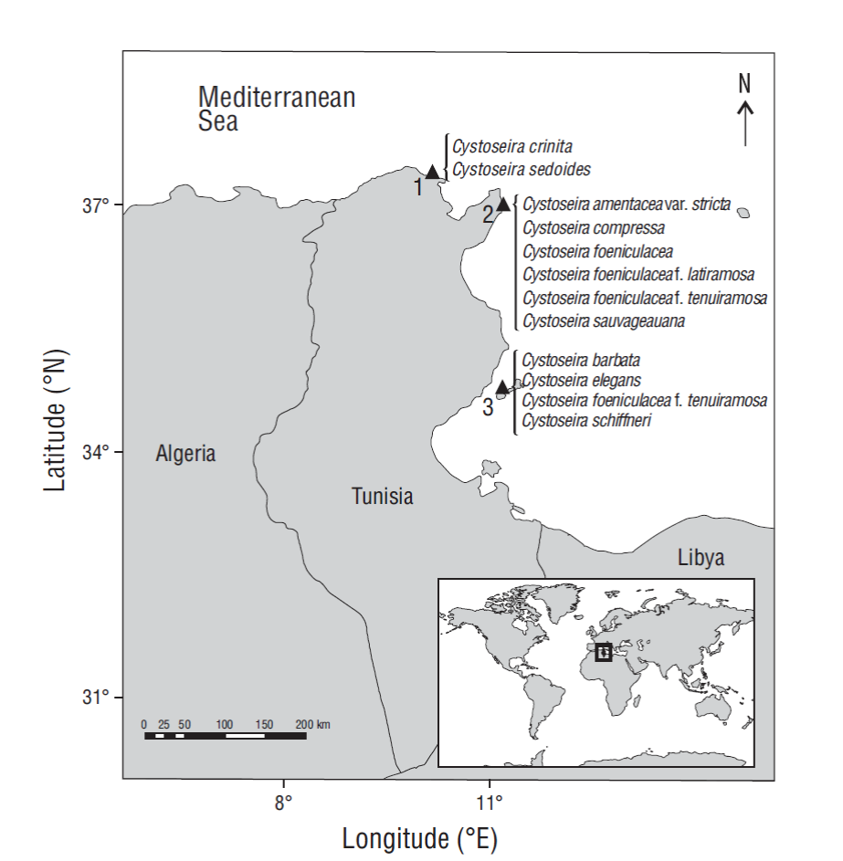
The aim of this paper is to assess the composition of FAs extracted from 11 Cystoseira species (Fig. 1) growing along the Tunisian coast (i) to evaluate the efficiency of Cystoseira FAs as chemotaxonomic markers associated with criteria performed on the morphological diversity of Tunisian Cystoseira taxa (Bouafif et al. 2014, 2016) and (ii) to estimate the nutritional characteristics of one of the most abundantly available brown macroalgae in the Mediterranean. To the best of our knowledge, this is the first report on the FA profile of some endemic Mediterranean species of Cystoseira.
Materials and methods
Fieldwork and algal samples
Eleven species and infraspecific taxa of the genus Cystoseira were collected in the upper intertidal zone between 0.5 and 2.0 m depth in October 2014: Cystoseira amentacea var. stricta, Cystoseira barbata, Cystoseira compressa, Cystoseira crinita, Cystoseira elegans, Cystoseira foeniculacea, Cystoseira foeniculacea f. latiramosa, Cystoseira foeniculacea f. tenuiramosa (from 2 localities), Cystoseira schiffneri, Cystoseira sauvageauana, and Cystoseira sedoides (Figs. 1, 2). Specimens were transported to the laboratory in an icebox. Species were identified by morphological examination according to specialized literature on the genus Cystoseira (Hamel 1931-1939, Ercegovič 1952, Gómez-Garreta et al. 2001) after careful cleaning that removed epiphytes and sand. A representative specimen of each species was deposited in the Herbarium of the Faculty of Sciences of Tunis (Tunisia) in the Department of Biology. Samples of each freshly collected and identified species were rinsed with distilled water, dried in an oven at 30 ºC for 72 h, then stored in darkness and kept in the dry at room temperature until further use. Each sample consisted of a mixture of 2 to 4 infertile adult thalli. The same stage of development of thalli was considered to pass up unpredictability linked to the physiological state of individuals, and entire algal thalli were considered when determining total lipids and FAs to avoid any intra-thallus variation among the species (Connan et al. 2007, Abdala-Díaz et al. 2014).

Figure 2 Cystoseira species collected along the Tunisian coast (scale bar = 1 cm). 1, Cystoseira amentacea var. stricta; 2, Cystoseira barbata; 3, Cystoseira compressa; 4, Cystoseira crinita; 5, Cystoseira elegans; 6, Cystoseira foeniculacea; 7, Cystoseira foeniculacea f. latiramosa; 8, Cystoseira foeniculacea f. tenuiramosa (North); 9, Cystoseira foeniculacea f. tenuiramosa (South); 10, Cystoseira schiffneri; 11, Cystoseira sauvageauana; 12, Cystoseira sedoides.
Extraction and estimation of total lipids
Five grams of each sample were pulverized in liquid nitrogen with a mortar and pestle. Extraction was done by the chloroform-methanol method according to Nomura et al. (2013). The obtained organic phase was subjected to total lipid and FA methyl esters (FAME) analysis. Total lipid content was determined gravimetrically and expressed as milligrams per gram of dry weight (DW) after evaporation of the solvent on a rotary vacuum evaporator at 30 ºC. All analyses were performed in triplicate.
Preparation of fatty acid methyl esters
FAs were transformed to their FAME by transmethylation in accordance with Prevot and Mordret (1976), with slight modifications. Briefly, 1.0 mL of hexane was added to 10 ± 1 mg of total lipid aliquot and 0.2 mL 2 N NaOH in MeOH, vortexed for 10 s, then heated at 55 ºC for 30 s. Then 0.2 mL 2 N HCl in MeOH was added and vortexed for 60 s. The mixture was centrifuged (Sorvall RC5C instruments, Fiberlite F21-8x50y rotor, Thermofisher, USA) at 1,000 × g for 5 min. The upper hexane layer containing FAME was subjected to gas chromatography coupled with mass spectrometry analysis.
Gas chromatography coupled with mass spectrometry analysis
Gas-chromatography coupled with mass spectrometry (GC-MS) was used to establish the FA profile of the lipid fraction. GC-MS analysis was done on an Agilent 7890A chromatograph equipped with an Agilent 5975C inert Mass Selective Detector and a HP-5 MS fused-silica capillary column (30.0 m × 0.25 mm, 0.25 μm film thickness). The column temperature regime was 150 ºC for 2 min, followed by 230 ºC at 4 ºC·min-1 and 230 ºC for 5 min. Helium was used as a carrier gas at a rate flow of 0.8 mL·min-1. Mass scan range was 50-550 amu at 70 eV and scan velocity was 2.86 scans per second. One microliter of each sample was injected. Identification of FAME was carried out by comparing their retention times with those of FAME standards and by comparing their mass spectra with those recorded in the NIST08 and W8N08 mass spectral libraries.
Determining lipid quality indices
For the unsaturation index (UI), the molar proportion (percentage) of each FA was multiplied by the number of double bonds after addition:
To assess the nutritional and lipid quality of Cystoseira species, health lipid indices (indices of atherogenicity [IA] and thrombogenicity [IT]) were estimated in accordance with the Ulbricht and Southgate (1991) method based on FA composition: where MUFA indicates monounsaturated FA and PUFA indicates polyunsaturated FA.
Statistical analysis
The results were expressed as mean values ± standard deviation of the 3 replicates. Mean total lipid and FA values were assessed using one-way analysis of variance (ANOVA) at P < 0.05, using the Statgraphics Centurion 16.1.11 software for Windows. Multivariate analyses (Sokal and Michener 1958) were carried out to evaluate the grouping of the studied species according to their total lipid and FA composition: (i) the cluster analysis was performed using the unweighted pair group method with arithmetic mean (UPGMA) and the Euclidean distance (DE), the UPGMA being authentic for ecological classification tools especially in species composition (Legendre and Legendre 1998); (ii) the principal component analysis (PCA) was carried out to differentiate between species and forms. Multivariate analyses were done using the MultiVariate Statistical Package (MVSP) software for Windows v3.1, following Kovach (2007).
Results
Total lipid content
The total lipid content of the 11 Cystoseira species showed wide fluctuations (Table 1). The yield for the total lipid fraction extracted varied in the range between 1.98% (C. schiffneri) and 6.82% DW (C. sauvageauana) (Table 1).
Table 1 Lipid extracts of Cystoseira species collected along the Tunisian coast. DW, dry weight; FW, fresh weight; N, northern coast of Tunisia; and S, southeastern coast of Tunisia.
| Species | Total lipids | |
| (mg·g-1 DW)* | Yield* (FW% w/w) | |
| Cystoseira amentacea var. stricta (N) | 39.60 ± 3.60cd | 5.28 ± 2.31cd |
| Cystoseira barbata (S) | 30.80 ± 2.80ef | 4.11 ± 1.80ef |
| Cystoseira compressa (N) | 46.20 ± 4.20bc | 6.16 ± 2.70bc |
| Cystoseira crinita (N) | 66.00 ± 6.00a | 8.80 ± 3.86a |
| Cystoseira elegans (S) | 47.50 ± 4.50b | 6.33 ± 2.78b |
| Cystoseira foeniculacea (N) | 52.80 ± 4.80b | 7.04 ± 3.09b |
| Cystoseira foeniculacea f. latiramosa (N) | 24.20 ± 2.20fg | 3.23 ± 1.41fg |
| Cystoseira foeniculacea f. tenuiramosa (N) | 31.20 ± 2.40e | 4.16 ± 1.82e |
| Cystoseira foeniculacea f. tenuiramosa (S) | 33.00 ± 3.00de | 4.40 ± 1.93de |
| Cystoseira sauvageauana (N) | 68.20 ± 6.20a | 9.09 ± 3.99a |
| Cystoseira schiffneri (S) | 19.80 ± 1.80g | 2.64 ± 1.16g |
| Cystoseira sedoides (N) | 33.00 ± 3.00de | 4.40 ± 1.93de |
*Data are expressed as mean ± SD, where n = 3; values in a column without a common superscript are significantly different (P < 0.05).
Fatty acid profile of lipid extract
Fourteen major FAs were identified in the lipid fraction of the 11 Cystoseira species (Table 2). The FA content of Cystoseira lipids varied widely between species. The FA profile of lipid extracts was characterized by high amounts of saturated FAs (SFAs) ranging from 40.51% (C. amentacea var. stricta) to 57.91% (C. compressa) of total FAs. Palmitic acid (C16:0) was the predominant lipid fraction, with the highest rate being found in C. foeniculacea f. tenuiramosa (44.70%). Cystoseira barbata was found to be rich in myristic acid (C14:0, 7.45%), while stearic acid (C18:0) characterized C. compressa (15.27%) (Table 2).
Table 2 Fatty acid composition and nutritional indices (expressed as percent of total fatty acids) for Cystoseira species collected along the Tunisian coast. N, north; S, south; ∑SFAs, sum of saturated fatty acids; ∑MUFAs, sum of monounsaturated fatty acids; ∑PUFAs, sum of polyunsaturated fatty acids; AA, arachidonic acid; EPA, eicosapentanoic acid; Tr., trace fatty acids that were present at <0.1%; n.d., fatty acid not detected; UI, unsaturation index; IA, index of atherogenicity; IT, index of thrombogenicity.
| Common name | Cystoseira amentacea var. stricta | Cystoseira barbata | Cystoseiracom presa | Cystoseira crinita | Cystoseira elegans | Cystoseira foeniculacea | Cystoseira foeniculacea f. latiramosa | Cystoseira foeniculacea f. tenuiramosa (N) | Cystoseira foeniculacea f. tenuiramosa (S) | Cystoseira sauvageauan α | Cystoseira schiffneri | Cystoseira sedoides |
| C14:0 | 5.82 ± 0.13abcd | 7.45 ± 0.69a | 4.39 ± 0.44def | 6.91 ± 0.13ab | 5.60 ± 2.33bcd | 5.17 ± 0.39cdef | 3.87 ± 0.02ef | 5.24 ± 1.39bcde | 5.10 ± 0.22cdef | 6.38 ± 1.82abc | 5.08 ± 0.37cdef | 3.54 ± 0.65f |
| C15:0 | n.d. | n.d. | 0.20 ± 0.17bc | 0.13 ± 0.22c | 0.23 ± 0.23bc | n.d. | 0.75 ± 0.37a | n.d. | n.d. | Tr. | 0.48 ± 0.04c | 0.44 ± 0.09b |
| C16:0 | 32.96 ± 1.76de | 32.05 ± 0.20de | 38.05 ± 1.83bc | 30.13 ± 0.33e | 36.18 ± 1.77bcd | 38.78 ± 1.18bc | 39.11 ± 0.64bc | 44.70 ± 6.44a | 39.88 ± 3.54b | 35.37 ± 0.25cd | 32.89 ± 3.09de | 35.19 ± 0.23cd |
| C18:0 | 1.73 ± 0.33e | 6.19 ± 2.21bcd | 15.27 ± 3.37a | 3.47 ± 1.76de | 4.29 ± 0.61cde | 4.14 ± 0.24cde | 7.82 ± 0.15b | 3.20 ± 0.67de | 2.50 ± 1.93e | 6.95 ± 3.39bc | 3.01 ± 3.07e | 1.60 ± 0.13e |
| ∑SFA | 40.51 ± 1.33f | 45.69 ± 1.33def | 57.91 ± 3.87a | 40.64 ± 1.72f | 47.38 ± 4.62cde | 48.09 ± 1.62bcd | 51.56 ± 0.14bc | 53.14 ± 8.41b | 54.91 ± 2.96cde | 49.33 ± 2.51bcd | 42.11 ± 0.13f | 41.90 ± 0.81ef |
| C16:1n-7 | 3.00 ± 0.28ef | 2.93 ± 0.38efg | 3.13 ± 0.26ef | 1.92 ± 0.05g | 3.81 ± 0.82e | 7.82 ± 0.63b | 5.46 ± 0.02d | 6.36 ± 0.63cd | 7.47 ± 0.30b | 2.20 ± 1.47fg | 7.26 ± 0.60bc | 9.91 ± 0.57a |
| C18:1n-9 | 26.88 ± 1.31a | 26.56 ± 2.05ab | 20.60 ± 0.67ef | 24.55 ± 1.60bc | 21.56 ± 0.32def | 17.67 ± 1.06g | 19.71 ± 0.55fg | 20.42 ± 0.93ef | 20.79 ± 2.16def | 22.05 ± 1.50de | 25.19 ± 0.42ab | 22.84 ± 0.86cd |
| Trans-18:1 | 2.35 ± 0.39bc | 1.78 ± 0.06bc | 1.57 ± 0.41c | 1.24 ± 0.05c | 4.58 ± 3.25a | 2.72 ± 0.23bc | 1.12 ± 0.05c | 2.62 ± 0.17bc | 1.28 ± 0.26c | 2.27 ± 0.12bc | 4.64 ± 0.30a | 3.49 ± 1.27ab |
| ∑MUFAs | 32.23 ± 1.80b | 31.27 ± 1.33bc | 25.30 ± 0.36f | 27.71± 1.58def | 29.93 ± 4.22bcd | 28.21 ± 0.91cdef | 26.30 ± 0.57f | 29.40 ± 0.80bcde | 29.51 ± 2.38bcde | 26.51 ± 1.83ef | 37.09 ± 1.18a | 36.17 ± 0.96a |
| C18:2n-6 | 4.81 ± 0.08bc | 5.69 ± 0.77ab | 3.86 ± 1.94bcd | 5.32 ± 0.29bc | 7.59 ± 2.13a | 5.87 ± 0.45ab | 5.94 ± 0.23ab | 3.40 ± 2.95cd | 5.73 ± 0.36ab | 5.56 ± 1.56abc | 4.15 ± 0.22bcd | 2.42 ± 0.31d |
| C18:3n-6 | 1.12 ± 0.34ab | 0.87 ± 0.75abc | Tr. | 0.99 ± 0.05ab | 1.67 ± 0.90a | 1.76 ± 0.54a | 0.38 ± 0.04bc | 0.68 ± 1.18bc | 0.23 ± 0.10bc | 0.61 ± 0.45bc | 0.77 ± 0.04bc | 0.40 ± 0.15bc |
| C18:4n-3 | 3.10 ± 0.23a | 1.46 ± 0.21bcd | 1.07 ± 0.82cd | 2.36 ± 2.06abc | 0.62 ± 0.11d | 2.20 ± 0.21abc | 2.71 ± 0.01ab | 2.06 ± 1.90abc | 1.27 ± 0.03bcd | 1.37 ± 0.24bcd | 1.62 ± 0.17bcd | 0.92 ± 0.10cd |
| C20:2n-6 | n.d. | n.d. | 0.31 ± 0.55b | 0.33 ± 0.25b | n.d. | 0.71 ± 0.32b | 0.34 ± 0.25b | 0.27 ± 0.19b | 0.20 ± 0.24b | n.d. | 0.37 ± 0.15b | 1.08 ± 0.21a |
| C20:3n-6 | 0.96 ± 0.03bcd | 0.51 ± 0.36cd | 0.87 ± 0.69cd | 2.07 ± 1.46a | 0.74 ± 0.05cd | 1.32 ± 0.12abc | 1.08 ± 0.05bc | 1.01 ± 0.93bc | 0.40 ± 0.39cd | Tr. | 0.60 ± 0.06cd | 1.92 ± 0.38ab |
| C20:4n-6 (AA) | 14.45 ± 0.41ab | 12.70 ± 0.56bc | 7.35 ± 2.46f | 15.98 ± 1.54a | 10.97 ± 0.67cd | 9.94 ± 0.79de | 7.91 ± 0.25ef | 8.20 ± 0.75ef | 12.74 ± 1.07bc | 14.56 ± 1.51ab | 10.47 ± 1.41d | 11.97 ± 1.19cd |
| C20:5n-3 | 1.74 ± 0.43cde | 1.43 ± 0.09def | 1.59 ± 0.31de | 3.31 ± 0.62a | 0.86 ± 0.05f | 2.01 ± 0.17cd | 3.01 ± 0.39ab | 1.58 ± 0.84de | 1.20 ± 0.12ef | 2.01 ± 0.72cd | 1.63 ± 0.24de | 2.42 ± 0.20bc |
| ∑PUFAs | 26.17 ± 0.62ab | 24.12 ± 0.81bc | 15.05 ± 4.14e | 30.35 ± 2.38a | 22.45 ± 0.50bc | 23.80 ± 2.08bc | 22.10 ± 0.36cd | 17.21 ± 7.62de | 21.77 ± 0.82bcd | 24.12 ± 0.77bc | 19.60 ± 1.26cde | 21.15 ± 0.90cd |
| PUFA/SFA | 0.65 ± 0.03a | 0.50 ± 0.02b | 0.26 ± 0.09d | 0.75 ± 0.09a | 0.49 ± 0.06b | 0.48 ± 0.06b | 0.41 ± 0.01bc | 0.34 ± 0.18cd | 0.45 ± 0.03bc | 0.50 ± 0.04b | 0.48 ± 0.03b | 0.52 ± 0.03b |
| Σn-3 | 4.83 ± 0.56ab | 2.89 ± 0.28cde | 2.66 ± 1.02cde | 5.67 ± 1.75a | 1.48 ± 0.06e | 4.20 ± 0.37abc | 5.69 ± 0.39a | 3.65 ± 2.68bcd | 2.47 ± 0.13de | 3.38 ± 0.13bcd | 3.24 ± 0.07bcd | 3.34 ± 0.147bcd |
| Σn-6 | 21.33 ± 0.66b | 19.76 ± 0.56bc | 12.39 ± 3.43g | 24.68 ± 0.79a | 20.97 ± 0.56bc | 19.09 ± 1.70bcd | 15.66 ± 0.32efg | 13.29 ± 5.13fg | 19.11 ± 0.50bcd | 20.73 ± 0.51bc | 16.35 ± 1.19def | 17.80 ± 0.76cde |
| Σn-6/Σn-3 | 4.41 ± 0.64cd | 5.75 ± 0.44bc | 4.66 ± 1.95bcd | 4.69 ± 1.63cd | 10.95 ± 3.57a | 4.67 ± 0.12cd | 2.76 ± 0.13d | 6.16 ± 5.63bc | 7.83 ± 0.19b | 6.26 ± 1.16bc | 5.04 ± 0.25bcd | 5.33 ± 0.08bcd |
| AA/EPA | 8.77 ± 2.74bc | 8.88 ± 0.17bc | 4.62 ± 1.37ef | 5.93 ± 1.52def | 12.85 ± 1.53a | 4.97 ± 0.30def | 2.66 ± 0.26f | 6.75 ± 4.46cde | 10.66 ± 0.96ab | 7.76 ± 2.28bcd | 6.47 ± 0.55cde | 4.94 ± 0.09def |
| UI | 126.95 ± 2.32ab | 110.56 ± 2.43c | 77.23 ± 12.90e | 137.41 ± 8.38a | 103.02 ± 2.92cd | 107.74 ± 5.57c | 100.0 ± 3.11cd | 90.26 ± 25.23de | 104.91 ± 2.90cd | 113.25 ± 5.96bc | 105.97 ± 4.92c | 111.68 ± 4.31bc |
| IA | 0.96 ± 0.05c | 1.15 ± 0.02abc | 1.40 ± 0.13ab | 0.99 ± 0.03c | 1.13 ± 0.31bc | 1.15 ± 0.08abc | 1.14 ± 0.02abc | 1.48 ± 0.59a | 1.18 ± 0.10abc | 1.20 ± 0.14abc | 0.94 ± 0.07c | 0.86 ± 0.07c |
| IT | 0.98 ± 0.04c | 1.33 ± 0.09bc | 2.23 ± 0.47a | 0.94 ± 0.16c | 1.55 ± 0.27bc | 1.32 ± 0.10bc | 1.32 ± 0.05bc | 1.88 ± 1.10ab | 1.49 ± 0.16bc | 1.44 ± 0.17bc | 1.12 ± 0.01c | 1.08 ± 0.05c |
Values are expressed as mean ± SD of 3 replicates at 95% confidence interval. Different superscript letters in the same row are significantly different (P < 0.05); values were rounded off to 2 decimal places and may not total 100%.
MUFAs ranged from 25.30% of total FAs in C. compressa to 37.1% in C. schiffneri. The major MUFA (>20% of total FAs) for most of the studied Cystoseira species was oleic acid (C18:1).
Total PUFAs varied between 15.05% (C. compressa) and 30.35% (C. crinita). Among the PUFAs, arachidonic acid (C20:4n-6) was the most abundant in the analyzed species, with a particular high level of 15.98% in C. crinita. The highest percentage of linoleic acid (C18:2n-6) was observed in C. elegans (7.59%), while the highest percentages of the eicosapentaenoic acid (C20:5n 3) and stearidonic acid (C18:4n-3) were observed in C. crinita (3.31%) and C. amentacea var. stricta (3.10%), respectively. Omega 3 PUFAs were detected in all studied species. Total omega 3 (∑n-3) ranged between 1.48% (C. elegans) and 5.69% (C. foeniculacea f. latiramosa). Cystoseira foeniculacea f. latiramosa and C. crinita were the richest species in n-3 PUFA (5.69% and 5.67% of total FAs, respectively). This richness is associated with the high rates of n-3 marine triglyceride (C20:5n-3), an important n-3 PUFA, that they contain. Stearidonic acid (C18:4n-3) was also detected in all studied species in rates varying between 0.62% (C. elegans) and 3.10% (C. amentacea var. stricta). Linoleic acid (C18:2n-6) was found in all studied species at rates ranging between 2.42% (C. sedoides) and 7.59% (C. elegans). Cystoseira crinita was the richest in the dihomo-γ-linolenic acid (C20:3n-6, 2.07%), an uncommon FA.
Discrimination of Cystoseira
The results of FA composition were evaluated using multivariate analyses to investigate whether the various lipid and FA patterns correlated with the taxonomic position of the brown Cystoseira algae and lead to distinguish between species and forms. Differentiating species according to their chemical composition could be valuable when examining any possible relationship between them. In this regard, a PCA was performed to consider the FA composition of each sample investigated. The 3 axes accounted for 86.89% of total variance. The first 2 dimensions explained 78.07% of the variability. Principal component (PC) 1 was defined by palmitic acid (C16:0), oleic acid (C18:1n-9), and arachidonic acid (C20:4n-6), while the most discriminant variable along the PC2 axis was stearic acid (C18:0) and explained 27.89% of total variation. PC3 was defined by elaidic acid (Trans C18:1), myristic acid (C14:0), palmitoleic acid (C16:1n-7), and linoleic acid (C18:2n-6). Plotting on axes 1 and 2 divided species into 3 major groups (Fig. 3). The first group included only C. compressa, the second comprised the C. foeniculacea group (C. foeniculacea and its forms tenuiramosa and latiramosa), and the third, the most heterogeneous one, comprised the other Cystoseira samples (Fig. 3).

Figure 3 Principal component analysis using fatty acid composition of Cystoseira taxa. The Cystoseira foeniculacea forms are denoted by squares. S, south; and N, north.
With the hierarchical clustering analysis, the dendrogram of FAs for the Cystoseira samples showed the presence of 4 clusters that were connected at DE = 14.7 (Fig. 4). Species were grouped independently of their geographic distribution. The greatest similarity was observed between C. amentacea var. stricta and C. crinita at DE = 5.2. As in the PCA model, the C. foeniculacea forms were closely linked and grouped in a single cluster at DE = 11.4 (Fig. 4).

Nutritional quality assessment
The PUFA/SFA ratio is commonly used to evaluate the nutritional quality of the lipid fraction of foods. Except for C. compressa and C. foeniculacea f. tenuiramosa, the Cystoseira species analyzed in the present study exhibited PUFA/SFA ratios exceeding 0.4 (Table 2). The n-3 and n-6 PUFAs represent a significant part of the Cystoseira lipids. The n-3 PUFA ranged between 1.48% (C. elegans) and 5.69% (C. foeniculacea f. latiramosa), while the n-6 PUFA varied between 12.39% (C. compressa) and 24.68% (C. crinita) of total FA. The 11 Cystoseira species exhibited high unsaturation indices, varying from 77.23 (C. compressa) to 137.41 (C. crinita). The highest IA and IT values were, respectively, 1.48 (C. foeniculacea f. tenuiramosa) and 2.23 (C. compressa) (Table 2).
Discussion
Total lipid content in the 11 Cystoseira species analyzed in this study was close to that reported for C. brachycarpa (Ragonese et al. 2014) off Italy and C. osmundacea (Khotimchenko et al. 2002) in the Sea of Japan. However, total lipid rates for Tunisian Cystoseira were higher than those for Moroccan Cystoseira from the Atlantic (between 1.3 and 16 mg·g-1 DW, Valls et al. 1993) and those for Indian Cystoseira (between 6.1 and 6.7 mg·g-1 fresh weight [FW], Kumari et al. 2013). The species investigated here are poorly studied in the world as regards their total lipid content. This study shows that Tunisian C. barbata (4.11 g·100 g-1 FW) and C. crinita (8.8 g·100 g-1 FW) are richer in total lipids than species from the Black Sea harvested during the same season (0.84 g·100 g-1 FW for C. barbata [Ivanova et al. 2012] and 0.72 g·100 g-1 FW for C. crinita [Ivanova et al. 2013]), and these differences are related to environmental conditions. In fact, it is proven that in algae, total lipid content depends on temperature, light, and salinity (Kim et al. 1996), factors that are very different between Tunisian and Black Sea waters. Intraspecific variability in total lipid content could also be dependent on the physiological state of the species, as suggested by the higher rate observed in this study for C. barbata (3.08%), compared to that reported by Frikha et al. (2011) for the same species in almost the same environmental conditions (2.51%, Gulf of Gabès, Tunisian southern coast).
In General, C. compressa was the richest in SFA, whereas C. amentacea var. stricta and C. schiffneri stood out for their high content of PUFA and MUFA. Palmitic acid (C16:0) was the main FA of the Cystoseira lipids, followed by oleic acid. These results confirm data previously reported in the literature for the profile commonly seen in brown algae, especially in Cystoseira species (Khotimchenko et al. 2002; Ivanova et al. 2012, 2013; Kumari et al. 2013; Ragonese et al. 2014; Vizetto-Duarte et al. 2015). For a given taxon, the differences between the FA profile observed in Tunisia and those reported in the literature for other regions could be related to the different seasons in which the studied samples were harvested and so to the different physiological states of the concerned species and/or to the differences in environmental conditions of habitats in the different regions. Temperature seems to be the factor that most affects the percentage of SFA and PUFA; for instance, Colombo et al. (2006) proved that algae from cold water (e.g., Canada) were usually richer in PUFA, with a higher n-3/n-6 FA ratio, than algae from warm water (e.g., China), and studies carried out in cold waters (Atlantic waters) exhibited higher SFA ratios than PUFA ratios in Cystoseira species (Vizetto-Duarte et al. 2015). However, the highest PUFA ratios found in warm-water algae from the Sea of Japan (Nomura et al. 2013) suggest that the environmental factors in the species’ habitat, other than temperature, could influence the FA profiles of algae.
Species from different localities did not show clustering behavior along the 2 axes of the PCA (Fig. 3). Furthermore, the PCA score plot of species highlighted that the FA composition of a species was independent of its origin. This low geographic differentiation could be explained by intraspecific diversity, which might affect the variability in FA composition. Samples of the C. foeniculacea group were distributed mainly along axis 2 and aggregated close to each other (group 2), showing low variation in the pool of variables considered and that species displayed a similar lipid profile (Fig. 3). The C. foeniculacea group was clustered according to its forms. Cystoseira foeniculacea f. tenuiramosa from different regions (north and south) were grouped together.
It is not surprising that C. schiffneri was segregated from the C. foeniculacea group since C. schiffneri (sensu Hamel 1939) (Hamel 1931-1939), considered as a synonym of C. foeniculacea f. schiffneri (Hamel) Gómez-Garreta, Barceló, Ribera and Rull Lluch( Gómez-Garreta et al. 2001), was recently reinstated as a specific rank, a distinct entity of the Cystoseira species (Bouafif et al. 2016). This discrimination of Cystoseira based on FAs supports the reinstatement of C. schiffneri (Hamel) as a specific rank.
Hierarchical clustering analysis revealed that species were more related to each other by their FA composition than by their geographic distribution, highlighting the intraspecific diversity in the genus Cystoseira (Fig. 4). The clustering of the C. foeniculacea group according to the FA profile supports earlier evidence that lipid composition could be a biochemical marker for each taxonomic group.
The PUFA/SFA ratios obtained in the present study are within the nutritional guidelines recommending a PUFA/SFA ratio higher than 0.4 (Wood et al. 2004). The n-6/n-3 PUFA ratios ranging from 2.76 (C. foeniculacea f. latiramosa) to 10.95 (C. elegans) for the 11 Cystoseira species were comparable to those for Portuguese Cystoseira species (Vizetto-Duarte et al. 2015), and these high ratios are explained by the high levels of n-6 PUFAs in the lipid composition of the species. Seaweed FAs are beneficial for the prevention of cardiovascular diseases and other chronic diseases, such as diabetes, hypertension, and autoimmune disease, in humans (Dawczynski et al. 2007). Some studies suggested a beneficial n-6/n-3 ratio between 1:1 and 5:1 for human nutrition (Simopoulos 2002, 2008); the Food and Agriculture Organization of the United Nations/World Health Organization committee recommended, in turn, an n-6/n-3 ratio between 5:1 and 10:1 (FAO/WHO 1995). Regarding our results, all 11 Tunisian Cystoseira species have high nutritional values.
High IA and IT values have adverse effects on human health and are considered potent indicators of and important factors for increased risk of coronary heart disease, where the higher the values the more degraded the nutritional quality (Ulbricht and Southgate 1991). The IT values for the algae analyzed in the present study were comparable to the IT values found by Ulbricht and Southgate (1991) for meat (0.7-1.1) and milk-based products (1.32). Thus, the results obtained in the present study suggest that C. amentacea, C. crinita, and C. sedoides were more antithrombogenic and antiatherogenic than the other analyzed Cystoseira species and could be used in nutraceuticals or food.
In conclusion, all studied species of Cystoseira have the same qualitative major FA composition. Interspecific quantitative variability in FA composition is, however, pointed out. This variability could be linked to environmental, biological, and physiological factors. Clustering of C. foeniculacea and its infrataxa (the latiramosa and tenuiramosa forms) according to the FA profiles suggest a possible use of FA composition for taxonomic purposes for the genus Cystoseira. This study further supports and confirms the recent study by Bouafif et al. (2016) about the reinstatement of the endemic Mediterranean C. schiffneri, considered before as a form of C. foeniculacea. The PUFA/SFA, n-6/n-3 PUFA, IA, and IT, indicators of nutritional lipid quality in food, suggest that the Cystoseira species studied here can be an interesting source of dietary FAs with beneficial effects on human health.
To our knowledge, this work is one of the rare investigations of lipid composition and nutritional value of Mediterranean Cystoseira species, some of which are rare and endemic.

