


 text in
  English (pdf)
 Article in xml format
 Article references
 Send this article by e-mail
 Cited by SciELO
  Similars in
SciELO
Similars in
SciELO 
 Permalink
Bananas and plantains (Musa spp.) are a significant source of food for a large portion of the global population. However, their production is threatened by diseases such as Fusarium wilt caused by Fusarium oxysporum f. sp. cubense (FOC), which is one of the most destructive and economically important diseases in the genus Musa. The control of this vascular pathogen using agrochemicals is expensive and harmful to the environment (FAOSTAT, 2020). Therefore, genetic improvement for resistance is considered the only effective and safe control method in recent years (Saraswathi et al., 2016). The use of biotechnological techniques to support genetic improvement for resistance to Fusarium wilt in bananas has facilitated the introduction of improvement results much faster than conventional genetic improvement methods (García et al., 2021). Moreover, it has increased the efficiency of genetic improvement programs in cultivation (Saraswathi et al., 2016). Ventura et al. (2015) obtained a wide genetic variability in the ‘Zanzíbar’ cultivar (group AAB) using a combination of tissue culture and in vitro mutagenesis in meristematic apices, selecting 15 mutants. ‘Z 13’, ‘Z 30’, and ‘Z 30 A’ were the most promising materials for genetic improvement in the cultivation, due to their high yield, elimination of superficial corms, decreased height, and ordered suckering. This study aimed to evaluate the resistance to Fusarium wilt race 1 in new banana clones ‘Z 13’, ‘Z 30’, and ‘Z 30 A’ obtained through biotechnological techniques in Cuba.
The research was carried out at the Laboratory of Plant-Pathogen Interaction of the Bioplants Center of the University of Ciego de Avila ¨Máximo Gómez Báez¨, Cuba.
Evaluated lines of clone ‘Zanzíbar’ (group AAB). The study utilized selected lines from the ‘Zanzíbar’ clone (group AAB) - ‘Z 13’, ‘Z 30’, and ‘Z 30 A’. These lines were obtained from the Musa spp. genetic improvement program at the National Institute of Tropical Root and Tuber Crops Research in Santo Domingo, Villa Clara, Cuba (INIVIT). The program utilized ionizing radiation (gamma rays) on meristematic apices of the ‘Zanzíbar’ clone in vitro culture, resulting in the selection of 15 mutants. After three vegetative cycles under field conditions, ‘Z 13’, ‘Z 30’, and ‘Z 30 A’ lines were chosen as the most advanced materials for genetic improvement in cultivation. These lines are being proposed for registration as new genotypes in Musa spp. by the International Atomic Energy Agency (IAEA) (Ventura et al., 2015).
Plant material. Leaf samples from the ‘Z 13’, ‘Z 30’ and ‘Z 30 A’ lines, eight to nine months old, planted in the INIVIT Banana Germplasm Bank, were used.
Fungal strain. We used a FOC race 1 isolate (belonging to the vegetative compatibility group (VCG) [01210] from the culture collection of the Institute of Plant Health Research (INISAV), Havana, Cuba.
Filtration of the fungus culture (FC). To obtain a liquid culture of the fungus, mycelial discs measuring 8 mm in diameter were taken from the periphery of colonies grown on Petri dishes containing Potato Dextrose Agar culture medium. Next, 100 mL of Czapek broth culture medium with a pH of 5.5 was added to a 250 mL Erlenmeyer flask. A mycelial disc was then inoculated into the flask and incubated for 24 days at 28±2 °C, 56 µmol m-2 s-1 light intensity, and a 12-hour light/12-hour dark photoperiod. The fungal culture (FC) was harvested after 24 days and filtered through four layers of gauze and Whatman No. 1 filter paper. Following filtration, it was centrifuged at 8000 rpm for 20 minutes in a Sigma-201M centrifuge to remove mycelial and conidial debris. The supernatant was then passed through filters with 0.22 μm diameter pores (Sartorius, NG, Göttingen, Germany). The FC was then concentrated to 80% (v/v) through rotary evaporation, using a Heidolph rotary evaporator, Bioblock, Paris, France. Two treatments were utilized in the experiment: 1) concentrated FC of the fungus (Czapek broth culture medium inoculated with the fungus and incubated for 24 days); and 2) Czapek broth culture medium without the fungus and incubated for 24 days, which was used as a control treatment.
Procedure for evaluating resistance to Fusarium wilt race 1. To evaluate resistance and susceptibility, six evaluation intervals were established (every two months within the same year of the experiment). Healthy middle-aged leaves were collected from each line (‘Z 13’, ‘Z 30’, and ‘Z 30 A’) in the field. Eighteen punctures were made on each leaf using a sterile needle on the adaxial side of the leaf blade, spaced 3 cm apart and in three different positions (distal, middle, and proximal). After the damage was inflicted, 5 µL of fungus culture filtrate (FC) and 5 µL of concentrated medium without fungus (control treatment) were added. The leaves were incubated for 48 hours at 28 ± 2 °C, 56 µmol m-2 s-1 light intensity, 12-hour light/12-hour dark photoperiod, and 70% relative humidity. The elliptical lesion area (mm2) of the leaves was measured after 48 hours according to Companioni et al. (2005). Leaf fragments with elliptical lesions of 5 x 5 cm were then extracted and placed in liquid nitrogen for subsequent biochemical analyses. Biochemical indicators, including peroxidase activity, chlorophyll pigment levels (a, b, and total), free and cell wall-bound phenol content, malondialdehyde, other aldehydes, and proteins were evaluated.
Peroxidase activity was assessed using the Pascual et al. (1983) method. Leaf fragments (100.0 mg) were pulverized in liquid nitrogen and then extracted with a tris-HCl buffer solution (0.01 mol L-1, pH 7.0). The reaction mixture consisted of 0.1 mL of leaf extract, 1.0 mL of tris-HCl buffer solution (0.01 mol L-1), 0.15 mL of guajacol (100 mmol L-1), and 20 µL of hydrogen peroxide. The absorbance at 470 nm was monitored every 15 seconds for three minutes, and the average change in absorbance of the linear segment of the curve was used to estimate peroxidase activity. The reaction of the guajacol and hydrogen peroxide substrates was accounted for in each determination. Guajacol peroxidase enzymatic activity was reported as U g-1 fresh leaf weight, where 1 U represents the conversion of 1 µmol of substrate per minute. Additionally, peroxidase activity was expressed as U mg-1 of protein.
The chlorophyll pigment content was determined following Porras (1991). Leaf fragments weighing 100.0 mg were macerated in liquid nitrogen and then mixed with 0.5 mL of 80% acetone. After centrifugation at 12,100 xg for 15 minutes at 4 °C, the supernatant was collected and the absorbance was measured at 647 nm and 664 nm. Chlorophyll content was calculated as: 13.19*Absorbance664nm - 2.57*Absorbance647nm. Chlorophyll b content was calculated as 22.10*Absorbance 647 nm - 5.26*Absorbance664nm. Total chlorophyll pigment content was determined by adding 7.93*Absorbance664nm + 19.53*Absorbance647 nm.
The method of Gurr et al. (1992) was used to determine the content of free and cell wall-bound phenols. Leaf fragments weighing 100.0 mg were macerated in liquid nitrogen followed by the addition of 0.5 mL of methanol. After vortexing and centrifugation at 12,100 xg for 15 minutes, the precipitate was subjected to two additional cycles of extraction with methanol. The resulting supernatant was collected and designated as the fraction of free phenols. The precipitate was treated with sodium hydroxide (2.0 mol L-1) for 16 hours at 70 °C, followed by addition of hydrochloric acid (0.25 mL, 2.0 mol L-1) and centrifugation at 12,100 xg for 15 minutes. The resulting supernatant was considered the reservoir of cell wall-bound phenols. To quantify free and cell wall-bound phenols, 20 μL of the supernatant was mixed with 980.0 μL of distilled water, and Folin- Ciocalteau’s phenol reagent (100.0 μL) was added, followed by incubation for 5 minutes. Sodium bicarbonate (600.0 μL, saturated with sodium hydroxide (0.1 mol L-1) was then added, and after 60 minutes, the absorbance at 725 nm was measured. The content of phenols was expressed in mg of phenols g-1 fresh leaf weight and was referenced to a standard curve of chlorogenic acid.
The levels of malondialdehyde and other aldehydes were evaluated following the protocol of Heath and Packer (1968). Leaf fragments weighing 100.0 mg were macerated in liquid nitrogen and mixed with 1.4 mL of distilled water, briefly vortexed. Then, 1.5 mL of thiobarbituric acid (0.5%, w/v; in trichloroacetic acid 20%, v/v) was added, and the samples were incubated at 100 °C for 25 minutes in a water bath. After cooling the samples on ice for 15 minutes, they were centrifuged at 756 xg for 10 minutes. The supernatant’s absorbance was measured at 455 nm, 532 nm, and 600 nm. The non-specific absorbance of the reaction product was measured at 600 nm and subtracted from the maximum absorbance at 532 nm for malondialdehyde measurements and at 455 nm for other aldehydes. The concentrations of malondialdehyde were calculated using an extinction coefficient of 155 mmol-1 cm-1 at 532 nm. For other aldehydes, the extinction coefficient was 45.7 mmol-1 cm-1 (average of the extinction coefficients of propanal, butanal, hexanal, and propanal-dimethylacetal) at 532 nm.
The protein concentration was determined according to Bradford (1976). Leaf fragments weighing 100.0 mg were macerated in liquid nitrogen, and extracted with a tris-HCl buffer solution (0.01 mol L-1, pH 7.0). A 0.1 mL leaf extract was mixed with 1.0 mL of Bradford reagent, and the absorbance was measured at 595 nm. The protein content was expressed in mg of protein per g of fresh leaf mass, using a standard curve of bovine serum albumin (BSA). The protein content present in the fungal culture filtrate was subtracted from the total protein content determined in the leaves, 48 hours after the application of the fungal culture filtrate.
Each treatment included three leaves from different plants in each evaluated line (‘Z 13’, ‘Z 30’ and ‘Z 30 A’). The area of elliptical lesions resulting from 54 punctures/treatment (18 punctures/leaf) was evaluated, and three independent samples (one from each leaf) were used for each biochemical determination. Using these measurements, each plant was evaluated in the discriminant functions described by Companioni et al. (2005) to assess if they were accurately classified (Table 1).
Table 1 Discriminant functions to differentiate banana clones resistant and susceptible to Fusarium oxysporum f. sp. cubense race 1 according to Companioni et al. (2005).
| Función para clones susceptibles | S: 11.87 a + 66.73 b + 24.60 c + 13.47 d + 0.09 e + 51.60 f + 20.39 g - 16.85 h + 11.20 i - 11.13 j - 49.20 |
|---|---|
| Función para clones resistentes | R: 18.94 a + 42.84 b + 7.45 c + 19.58 d + 3.84 e + 45.72 f + 15.79 g - 5.11h - 2.31i + 0.21j - 41.55 |
| Legend (The fractions on the right correspond to the reciprocal of the larger value. These are constants in the discriminant functions): | |
| a = Lesion area of leaves treated with uninoculated culture medium (mm2) * 1/1.22 mm2 | |
| b = Lesion area of leaves treated with fungal culture filtrate (mm2) * 1/48.84 mm2 | |
| c = Free phenol content in leaves treated with uninoculated culture medium (mg g-1 fresh leaf mass) * 1/43.13 mg phenols g-1 fresh leaf mass. | |
| d = Free phenol content in leaves treated with fungus culture filtrate (mg g-1 fresh leaf mass) * 1/27.51 mg phenols g-1 fresh leaf mass. | |
| e = Content of phenols bound to cell walls in leaves treated with uninoculated culture medium (mg g-1 of fresh leaf mass) * 1/118.51 mg g-1 of fresh leaf mass. | |
| f = Content of phenols bound to cell walls in leaves treated with fungus culture filtrate (mg g-1 of fresh leaf mass) * 1/103.73 mg g-1 of fresh leaf mass. | |
| g = Aldehyde content (except malondialdehyde) in leaves treated with uninoculated culture medium (µmol g-1 fresh leaf mass) * 1/1.06 µmol g-1 fresh leaf mass. | |
| h = Aldehyde content (except malondialdehyde) in leaves treated with fungus culture filtrate (µmol g-1 of fresh leaf mass) * 1/0.83 µmol g-1 of fresh leaf mass. | |
| i = Protein content in leaves treated with uninoculated culture medium (mg protein g-1 fresh leaf mass) * 1/5.88 mg g-1 fresh leaf mass. | |
| Leyenda (Las fracciones a la derecha corresponden al recíproco del valor mayor. Estas son constantes en las funciones discriminantes): | |
| a = Área de la lesión de hojas tratadas con medio de cultivo sin inocular (mm2) * 1/1.22 mm2 | |
| b = Área de la lesión de hojas tratadas con filtrado del cultivo del hongo (mm2) * 1/48.84 mm2 | |
| c = Contenido de fenoles libres en hojas tratadas con medio de cultivo sin inocular (mg g-1 de masa fresca de la hoja) * 1/43.13 mg de fenoles g-1 de masa fresca de la hoja. | |
| d = Contenido de fenoles libres en hojas tratadas con filtrado del cultivo del hongo (mg g-1 de masa fresca de la hoja) * 1/27.51 mg de fenoles g-1 de masa fresca de la hoja. | |
| e = Contenido de fenoles ligados a las paredes celulares en hojas tratadas con medio de cultivo sin inocular (mg g-1 de masa fresca de la hoja) * 1/118.51 mg g-1 de masa fresca de la hoja. | |
| f = Contenido de fenoles ligados a las paredes celulares en hojas tratadas con filtrado del cultivo del hongo (mg g-1 de masa fresca de la hoja) * 1/103.73 mg g-1 de masa fresca de la hoja. | |
| g = Contenido de aldehídos (excepto malondialdehído) en hojas tratadas con medio de cultivo sin inocular (µmol g-1 de masa fresca de la hoja) * 1/1.06 µmol g-1 de masa fresca de la hoja. | |
| h = Contenido de aldehídos (excepto malondialdehído) en hojas tratadas con filtrado del cultivo del hongo (µmol g-1 de masa fresca de la hoja) * 1/0.83 µmol g-1 de masa fresca de la hoja. | |
| i = Contenido de proteínas en hojas tratadas con medio de cultivo sin inocular (mg de proteínas g-1 de masa fresca de la hoja) * 1/5.88 mg g-1 de masa fresca de la hoja. | |
| j = Contenido de proteínas en hojas tratadas con filtrado del cultivo del hongo (mg de proteínas g-1 de masa fresca de la hoja) * 1/4.85 mg g-1 de masa fresca de la hoja . | |
As the lesion areas were measured in three groups of 18 lesions, and each biochemical determination was repeated three times, three sets of data were available for each plant in the original matrix. The three sets of data for each plant were assessed using the discriminant function for resistant clones and the discriminant function for susceptible clones. The obtained results were compared using the parametric t-test to determine statistical differences between the results of each function. The maximum probability of making a type I error was 0.05. The Statistical Package for Social Sciences (SPSS for Windows, version 8, Copyright SPSS Inc., 1989-1997) was used for the statistical processing of the data. In addition, the evaluation procedure for Fusarium race 1 resistance in the ‘Zanzíbar’ clone lines, as described by Companioni et al. (2012), is presented below (Figure 1).
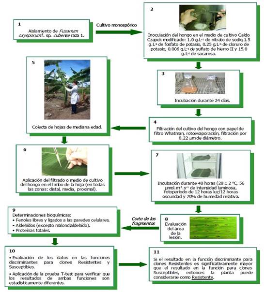
Figure 1 Method for leaf-level differentiation of resistance to Fusarium oxysporum f. sp. cubense race 1 in banana according to Companioni et al. (2012).
The study evaluated the resistance to Fusarium wilt caused by race 1 GCV [01210] in each assessed line and at every evaluation time using discriminant functions (Table 2). After applying the treatments as described and conducting biochemical analyses of the leaves from the ‘Zanzíbar’ clone lines, the discriminant functions classified 93.3% of the evaluated ‘Z 30’ mutant plants (56 out of 60) as resistant. Similarly, 91.6% of the evaluated ‘Z 13’ plants (55 out of 60) were classified as resistant, while 96.6% of the plants in line ‘Z 30A’ were classified as resistant (58 out of 60). These results suggest that the selected lines from the irradiated ‘Zanzíbar’ clone, namely ‘Z 13’, ‘Z 30’, and ‘Z 30A’, may be promising materials for genetic improvement in cultivation focused on resistance to FOC race 1 GCV [01210]. However, to confirm the resistance observed in these lines, additional field studies are necessary. The first and most crucial step when assessing a new banana clone is evaluating resistance in plants against different ‘formae specialis’ of Fusarium oxysporum (Buddenhagen, 2009).
Table 2 Classification into susceptible or resistant made by discriminant functions on selected lines of the irradiated ‘Zanzíbar’ clone.
| Momentos de evaluación | Líneas evaluadas | Número total de plantas evaluadas | Número total de plantas potencialmente resistentes según funciones discriminantes |
|---|---|---|---|
| 1 | ‘Z-30’ | 10 | 10 (100%) |
| 2 | ‘Z-30’ | 10 | 8 (80%) |
| 3 | ‘Z-30’ | 10 | 9 (90%) |
| 4 | ‘Z-30’ | 10 | 9 (90%) |
| 5 | ‘Z-30’ | 10 | 10 (100%) |
| 6 | ‘Z-30’ | 10 | 10 (100%) |
| 1 | ‘Z-13’ | 10 | 8 (80%) |
| 2 | ‘Z-13’ | 10 | 9 (90%) |
| 3 | ‘Z-13’ | 10 | 9 (90%) |
| 4 | ‘Z-13’ | 10 | 10 (100%) |
| 5 | ‘Z-13’ | 10 | 10 (100%) |
| 6 | ‘Z-13’ | 10 | 9 (90%) |
| 1 | ‘Z-30A’ | 10 | 10 (100%) |
| 2 | ‘Z-30A’ | 10 | 9 (90%) |
| 3 | ‘Z-30A’ | 10 | 9 (90%) |
| 4 | ‘Z-30A’ | 10 | 10 (100%) |
| 5 | ‘Z-30A’ | 10 | 10 (100%) |
| 6 | ‘Z-30A’ | 10 | 10 (100%) |
Even though in vitro resistance evaluations are performed, supposedly resistant plants require field studies (Dita et al., 2018). In this context, Saraswathi et al. (2016) selected banana mutants of the Rasthali cultivar (Silk, AAB) for resistance to race 1 of GCV [0124/5] of FOC through in vitro selection. To achieve this, they used toxin (fusaric acid) and the pathogen’s culture filtrate (CF) as selection agents. Individual banana mutant shoots obtained after the third or fourth subculture were transferred to multiplication medium supplemented with different concentrations of the toxin (commercial fusaric acid) (Sigma Aldrich, USA) (0.0125, 0.025, 0.0375, 0.05, and 0.0625 mM) and 3, 4, 5, 6, 7, and 8% concentrations of the pathogen’s CF, in addition to the growth regulator. After three weeks, they observed 50% survival of the explants at a concentration of 0.050 mM of the toxin and 7% with the fungal CF. At higher concentrations, there was a rapid decrease in banana mutant shoot growth. The selected mutants were transferred for pot tests under controlled conditions by inoculating the substrate with a spore suspension of 12 × 109 conidia mL-1 of the fungus for the next three months. After six months of resistance evaluation, three putatively resistant mutants to race 1 of FOC were obtained, which were massively multiplied in vitro for further interaction studies. It is important to note that determining the correct concentration of pathogen CF or toxin for the expression of differential phytotoxic activity between varieties is necessary for establishing resistance selection systems. This increases the chances of obtaining stable lines of plants with disease resistance (Portal et al., 2021).
The findings of this study demonstrate that the selected lines of the irradiated ‘Zanzíbar’ clone (‘Z 13’, ‘Z 30’, and ‘Z 30 A’) showed resistance to FOC race 1 GCV [01210] when evaluated at different selection times using the method described by Companioni et al. (2005; 2012) for differentiating susceptibility or resistance to Fusarium wilt. Additionally, the ease of evaluating resistance using the FC of the fungus and the reduced response time (48 hours) to determine plant susceptibility or resistance were also observed. Furthermore, this method facilitates the evaluation of a significant number of samples under laboratory conditions, enabling the acceleration of genetic improvement programs for this disease. These results highlight the potential of biotechnology in crop genetic improvement.

