SciELO - Scientific Electronic Library Online

 
vol.35 issue1Morphological and molecular characterization of native isolates of Trichoderma and its potential biocontrol against Phytophthora infestansPowdery mildews in agricultural crops of Sinaloa: Current status on their identification and future research lines author indexsubject indexsearch form
Home Pagealphabetic serial listing  

Services on Demand

Journal

Article

Indicators

Related links

  • Have no similar articlesSimilars in SciELO

Share


Revista mexicana de fitopatología

On-line version ISSN 2007-8080Print version ISSN 0185-3309

Rev. mex. fitopatol vol.35 n.1 Texcoco Jan. 2017

https://doi.org/10.18781/r.mex.fit.1606-9 

Review articles

Extraction and purification of large dsRNAs from virus-infected plants and fungi; applications in virus detection and identification

R. A. Valverde1  * 

R. De La Torre-Almaraz2 

1Department of Plant Pathology & Crop Physiology, Louisiana State University Agricultural Center, Baton Rouge, LA 70803, USA.

2Universidad Nacional Autónoma de México, Facultad de Estudios Superiores Iztacala, Mexico DF, Mexico.


Abstract.

Various methods for the extraction and purification of large dsRNAs are used to conduct detection and identification of viruses in plants and fungi. The most widely used protocol consists in phenol extraction and selective binding of dsRNA to cellulose. Purified viral dsRNAs from plants and fungi have been analyzed by gel electrophoresis and used as a complementary tool for virus identification. Moreover, dsRNAs have been used as reagent for reverse transcription PCR, molecular cloning, preparation of probes, and sequencing of RNA viruses. Many RNA viruses and subviral molecules infecting plants and fungi have been discovered using dsRNA extraction protocols. Recent improvements on dsRNA extraction methods have increased their efficiency, and cost effectiveness. It has been shown that viral dsRNA can be used as an initial reagent for next generation sequencing of viral genomes.

Key words: RNA viruses; plant viruses; mycoviruses; virus detection

Resumen.

Se utilizan varios métodos para realizar la extracción y purificación de ARN de doble cadena (dsRNA) largo de plantas y hongos. Los protocolos más utilizados consisten en la extracción de fenol y la unión selectiva de dsRNA a la celulosa. Los dsRNAs virales purificados de plantas y hongos se han analizado por electroforesis con gel y se han utilizado como herramienta complementaria para identificar virus. Asimismo, los dsRNAs se han utilizado como reactivos en PCR de transcriptasa reversa, clonación molecular, preparación de sondas y secuenciación de virus ARN. Se han descubierto muchos virus ARN y moléculas subvirales que infectan plantas y hongos utilizando protocolos de extracción de dsRNA. Las recientes mejoras a los métodos de extracción de dsRNA han aumentado la eficiencia y la rentabilidad. Se ha comprobado que el dsRNA viral se puede utilizar como reactivo inicial para la secuenciación de próxima generación de genomas virales.

Palabras clave: virus ARN; fitovirus; micovirus; detección de virus

Introduction

A variety of methods for the detection and identification of plant and fungal viruses are available to researchers and diagnosticians. For many years, serological methods and biological assays were commonly used in combination with viral nucleic acid detection. Today, nucleic acid-base approaches to virus detection such as polymerase chain reaction (PCR), together with sequencing, are widely used because of the high sensitivity and adaptation to the development of high throughput methods (Bonham et al., 2014). Recently, new technologies for virus detection and identification such as next-generation sequencing (NGS) have become available. NGS is a method with great potential for the detection and identification of plant viruses (Al Rwahnih et al., 2015; Bonham et al., 2014; Ho and Tzanetakis, 2013). For any nucleic acidbased method for RNA virus detection, efficient and practical RNA extraction protocols are needed. Many methods, including those of commercial kits, are currently available to extract single stranded RNA (ssRNA) from a wide range of biological tissues. Nevertheless, in some cases, the instability of ssRNA can be a factor affecting successful virus detection. The extraction, purification, and gel electrophoresis analysis of large (0.5-20.0 kb) viral double-stranded RNAs (dsRNAs) provide a complementary tool for scientists aiming to detect and identify plant viruses (Dodds et al., 1984; 1988; Morris and Dodds, 1979; Valverde et al., 1990b). Moreover, it also provides a source of RNA that can be used in other nucleic acid-based detection and identification methods for RNA viruses.

Large dsRNA has been recognized as genetic material in many plant, animal, fungal, and bacterial viruses (Libonati et al., 1980). Some biological functions of dsRNA include the capacity to induce interferon and the inhibition of protein synthesis in eukaryotes (Libonati et al., 1980). DsRNA is resistant to RNases under high ionic strength conditions. Under standard assay conditions (0.15 M NaCl, 0.015 M sodium citrate, pH 7), dsRNA is resistant to RNase A but not to RNases of the pancreatic type (Libonati and Sorrentino, 1992). Another unique property of dsRNA is that it binds to cellulose in ethanol-containing buffers. Optimally, dsRNA binds to fibrous cellulose at ethanol concentrations of about 16 % (Franklin, 1966; Morris and Dodds, 1979). The relatively high stability of dsRNA and the availability of extraction methods make the use of dsRNA practical for the detection and identification of RNA viruses.

In most plants and fungi infected with RNA viruses, dsRNAs can be found as part of: 1) genomic segments of dsRNA viruses; 2) replicative forms and replicative intermediates of single-stranded RNA viruses (ssRNA) (Buck, 1999; Horiuchi and Fukuhara, 2004; Jackson et al., 1971; Libonati et al., 1980; Nuss and Koltin, 1990; Zelcer et al., 1981); 3) subgenomic viral RNAs (sgRNAs) (Condit and Fraenkel-Conrat, 1979; Osman and Buck 1990); 4) satellite viruses (Klein and Reichman, 1970; Hillman et al., 2000; Valverde and Dodds, 1986); 5) satellite RNAs (Demler and de Zoeten, 1989; Liu et al., 2015); and 6) defective interfering RNAs (Burgyan et al., 1989; Hillman et al., 2000). Serological detection of dsRNAs from virus-infected plants and fungi has also been reported. An antiserum against polyinosinicpolycytidylic acid (polyi:c), a synthetic analog of double-stranded RNA, has been used to detect dsRNA from plants infected with Cucumber mosaic virus (CMV) and Citrus tristeza virus (CTV) and Penicillium chrysogenum infected with Penicillium chrysogenum virus using serological techniques (Aramburu et al., 1991). These techniques showed similar sensitivity for the detection of dsRNA as separation by polyacrylamide gel electrophoresis and silver staining. DsRNAs from Cowpea mosaic virus and Sugar cane mosaic virus have been detected in infected tissue extracts using serologically specific electron microscopy (Derrick et al., 1994). Monoclonal antibodies to dsRNA have been developed and used to detect Ground nut rosette virus in peanut (Schonborn et al., 1991). However, techniques based on serological methods require the development of the specific antibodies, a procedure that is more costly and lengthy than the direct visualization of dsRNA using electrophoresis or RT-PCR detection.

RNA viruses of plants and fungi

Viruses with ssRNA genomes make up more than 90 % of all known plant viruses. During replication in host cells, ssRNA viruses generate double-stranded replicative forms (RF) which consist of fully base-paired RNA molecules of the genomic RNA and replicative intermediates (Buck, 1999). They have been shown to occur in Tobacco mosaic virus (TMV) and Tobacco ringspot virus infected plants (Buck 1999; Derrick, 1978; German and de Zoeten, 1975; Jackson et al., 1971; Zelcer et al., 1981) and Oryza sativa endornavirus (Horiuchi and Fukuhara, 2004) among others. Many ssRNA viruses generate subgenomic RNAs (sgRNAs) to express internal open reading frames. These sgRNAs are synthesized either from prematurely terminated negative strands or, more commonly, by internal initiation on the negative-strand templates at specific promoter sequences (Miller and Koev, 2000). Subgenomic dsRNAs have been detected in plants infected with most RNA viruses and fungi infected with RNA mycoviruses (Condit and Fraenkel-Conrat, 1979; Kwon et al., 2007; Osman and Buck, 1990). Plant viruses with dsRNA genomes include members of the families Reoviridae, Partitiviridae, Amalgaviridae, Totiviridae, and Chrysoviridae, and their genomic dsRNA are readily detected from plant tissues (Antoniw et al., 1986; Chen et al., 2016; Karan et al., 1994; Li et al., 2013; Quito et al., 2011; Sabanadzovic et al., 2009; 2011; Wei et al., 2003). With the exception of theReoviridae, the other four families contain plant viruses that do not cause visible symptoms and have been named persistent viruses. In contrast, acute viruses cause symptoms and economically important plant diseases (Roossinck, 2010).

All major taxa of fungi have been shown to contain viruses. Although most fungal viruses described to date have dsRNA genomes, there are some with ssRNA genomes and one report of a ssDNA genome (Ghabrial et al., 2015). There are many citations in the literature of the presence of viral dsRNAs in fungi (Azzam and Gonsalves, 1999; Castillo et al., 2011; Day et al., 1977; Delye and Corio-Costet, 1998; Dickinson and Pryor, 1989; Herrero et al., 2009; Khalifa and Pearson, 2014; Kim et al., 2005; Kondo et al., 2016; Li et al., 2014; Shamoun et al., 2008); however, in most cases these viruses do not appear to affect the host’s phenotype (Enebak et al., 1994; Reyes et al., 2003). Only in some cases they have been associated with phenotypic and genotypic variations (Castanho et al., 1978; Ghabrial et al., 2015; Wei et al., 2003). The ability of some mycoviruses to induce hypovirulence has been used for biological control of their fungal hosts that are pathogenic to plants (Nuss and Koltin, 1990). The best known examples of mycovirus-induced hypovirulence are virus strains of the chestnut blight fungus Cryphonectria parasitica (Choi and Nuss, 1992; Day et al., 1977; Lee et al., 2006). Methods for the extraction and purification of dsRNA have been essential for the discovery, detection, and characterization of mycoviruses (Nuss and Koltin, 1990).

Satellite viruses, satellite RNAs, defective interfering RNAs, and viroids

Satellites are subviral agents that depend on a helper virus for their replication. Satellite viruses code for their own coat protein and their RNA is encapsidated. Satellite RNAs do not encode capsid proteins, but are packaged in a protein encoded by the helper virus. The size of most satellites associated with plant and fungal viruses range from 2.0-0.4 kb (Hillman et al., 2000; Kaper and Diaz-Ruiz, 1977; Demler and de Zoeten, 1989; Valverde and Dodds, 1987). Defective interfering RNAs (DIs) are RNAs that result from errors in virus replication (Pathak and Nagy, 2009). Satellite RNA viruses, satellite RNAs, and DI RNAs can attenuate or enhance the symptoms caused by their parent virus (Pathak and Nagy, 2009; Habili and Kaper, 1981). Some satellite viruses and RNAs associated with plant viruses accumulate relatively large quantities of dsRNA (Diaz-Ruiz and Kaper, 1977; Dodds et al., 1984; Habili and Kaper, 1981; Valverde and Dodds, 1986; 1987). In fungi, dsRNA satellites have been associated with several dsRNA viruses (Liu et al., 2015; Romanos et al., 1981), and satellite dsRNAs have been reported in Sclerotinia sclerotiorum (Liu et al., 2015). A satellite RNA of Ophiostoma novo-ulmi Mitovirus 3a in hypovirulent Isolates of Sclerotinia homoeocarpa (Deng and Boland, 2004), and a satellite and DIRNAs of Cryphonectria hypovirus 3-Grand Haven 2 (Hillman et al., 2000) have also been identified.

Viroids are some of the smallest known infectious agents. They consist of non-coding circular ssRNA molecules forming highly basepaired rod-like structures and are capable of causing economically important diseases in many crops worldwide (Kovalskaya and Hammond, 2014). Viroid derived dsRNAs have been described (Carbonell, et al., 2008).

DsRNA extraction and purification

Over the past 50 years, several methods for large dsRNA extraction from virus-infected plant, animal and fungal tissues, as well as bacteria have been reported in the literature (Akin et al., 1998; Balijja et al., 2008; Castillo et al., 2011; Delye and Corio-Costet, 1998; DePaulo and Powell, 1995; Diaz-Ruiz and Kaper, 1978; Franklin, 1966; Morris and Dodds, 1979, Morris et al., 1983; Okada et al., 2015; Tzanetakis and Martin, 2008; Valverde et al., 1990) (Table 1). Most of these methods are based on cellulose chromatography, some on enzymatic degradation or lithium chloride precipitation. Recently, Atsumi et al., (2015) developed a novel dsRNA isolation method using a recombinant dsRNA-binding protein.

Table 1. Principal Components of Some dsRNA Extraction Protocols. 

In 1979, Morris and Dodds developed a procedure for the isolation and analysis of dsRNA which has been widely used for the detection, identification, and characterization of plant viruses. It consists on the selective adsorption of dsRNA to fibrous cellulose at low ethanol concentrations and elution with ethanol-free buffer. A schematic diagram summarizing the steps involved in the method is shown in Figure 1. Although developed over 40 years ago, it is still in use today although it has been modified and improved (Khankhum et al., 2015; Valverde et al., 1990).

Figure 1. Schematic diagram of the steps involved in dsRNA extraction and purification. (Modified from Valverde et al., 1990). STE: Sodium, Tris and EDTA buffer (100 mM NaCl, 10 mM Tris-Cl, pH 8.0 and 1 mM EDTA); SDS: sodium dodecyl sulfate. 

Homogenizing by grinding fresh, infected tissue, often with mortar and pestle using liquid nitrogen, is the first step in the extraction of dsRNA. However, buffered sap extracts, fresh or lyophilized fungal mycelium, dried spores, and desiccated plant tissue (including those infected with biotrophic fungi) have also been used with successful results (Morris et al., 1983; Khankhum et al., 2015; Kousik et al., 1994; Valverde et al, 1990). Other practical and efficient approaches for disrupting the tissue include bead beaters, tissue homogenizers, etc. Desiccated tissue has broad use because this method is conventionally used for the long-term storage of virus-infected tissue. It is also the way samples are exchanged between laboratories. Furthermore, biotrophic fungi infecting plant tissues are often stored in fungaria as pressed and dried specimens from which samples can be used to test for the presence of fungal viruses (Khankhum et al., 2015). In the case of plants, most extractions are done from foliar tissue. However, bark, stem, or vein tissue are recommended for phloem-limited viruses. For fungi, mycelium or spores can be used. Negative controls are highly recommended in any dsRNA extraction. The second step involves total nucleic acid extraction using buffer-saturated phenol. With some plant tissues and particular viruses buffer may be used instead of phenol. Nevertheless, phenol extracts usually yield cleaner samples. The third step involves binding of the dsRNA to fibrous cellulose. The cellulose used by Morris and Dodds (1979) and Franklin (1966) in their original papers was Whatman CF-11 (Whatman Inc., Clifton, NJ, USA). Because this cellulose is no longer commercially available, other cellulose types such as Cellex-N1 (Bio-Rad, Hercules, CA, USA) have been used in dsRNA purifications with some success (Okada et al., 2015; Valverde et al., 1990). Khankum et al., (2015) used fibrous cellulose (Sigma-Aldrich, St. Louis, MO, USA) to extract dsRNAs from virus-infected plants and fungi. This cellulose is readily available worldwide and a good alternative to CF-11. After ethanol precipitation, dsRNA samples must be treated with DNase to eliminate potential host DNA contamination. Host ribosomal RNA may interfere with the electrophoretic detection of dsRNAs ranging from 1.0-0.5 kb. Therefore, when the target dsRNAs are within this range, digestion with S1 nuclease is recommended. After that, analysis of dsRNAs can be conducted in 6 % polyacrylamide gels, which provide high resolving power for subgenomic dsRNAs or, more simply, using 1.21.5 % agarose gels. Traditionally, ethidium bromide has been the preferred stain, although silver nitrate staining has been used as well. More recently, staining with Gel RedTM (Huang et al., 2010) has become more common. GelRed does not have the health hazards associated with ethidium bromide and has higher sensitivity. Detailed information of the methodology developed by Morris and Dodds (1979) and data showing some of its applications has been published elsewhere (Dodds et al., 1984; 1988; Valverde et al., 1986; 1990b).

Initially, the focus of the dsRNA extraction protocols was on the electrophoretic analysis and evaluation of banding patterns for virus detection and, in some cases, identification. Although analysis of viral dsRNAs by gel electrophoresis is still widely used, currently most applications are in RT-PCR, molecular cloning, sequencing, genome assembly, and virus identification.

Virus detection and identification

In many cases, particularly with ssRNA viruses infecting plants that contain high amounts of phenolic compounds and carbohydrates, diagnosticians and researchers find it difficult to obtain pure preparations of ssRNA or virus to use in identification and characterization. An alternative is to purify dsRNAs that can be used for probe preparation, RT-PCR, molecular cloning, and sequencing.

In general, different families of RNA viruses infecting plants or fungi yield characteristic electrophoretic dsRNA banding patterns that differentiate them (Dodds et al., 1984; Valverde et al., 1986). DsRNA electrophoretic profiles have been reported for members of the families Closteroviridae (Dodds and Bar-Joseph, 1983), Potyviridae (Valverde et al., 1986), Bromoviridae (Valverde and Glascock, 1991), Endornaviridae (Fukuhara et al., 2006) and the genera Comovirus (Valverde et al., 1995), Tobamovirus, Potexvirus, and Carlavirus (Valverde et al., 1986). As mentioned earlier, dsRNA profiles generally consist of one (in the case of monopartite viruses), two (bipartite) or more (multipartite) full-length genomic bands (or RFs) and several other bands corresponding to subgenomic replicative forms of RNA viruses (Condit and Fraenkel-Conrat, 1979; Osman and Buck, 1990). Figures 2 and 3 show dsRNA profiles of different plant viruses resolved in polyacrylamide and agarose gels. In the case of fungal viruses, dsRNA electrophoretic profiles have not been evaluated as extensively as a tool to differentiate between families or genera. Nevertheless, as in the case of plant RNA viruses, electrophoretic profiles vary depending upon the virus species and mixed infections are common (Figure 4).

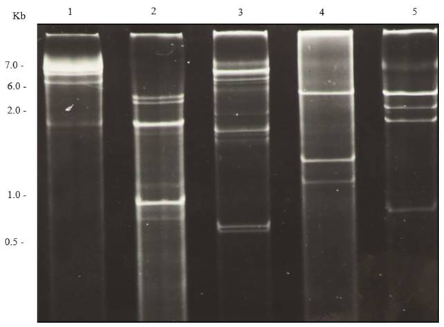
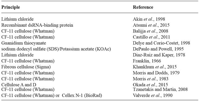
Figure 2. Polyacrylamide (6 %) gel electrophoresis of dsRNA extracted from plants infected with different viruses. Lane 1, Potato virus X; lane 2, Cucumber mosaic virus; lane 3, Tobacco mild green mosaic virus; lane 4, Tobacco necrosis virus; and lane 5, Alfalfa mosaic virus. 

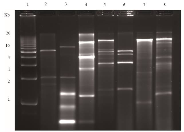
Figure 3. Agarose (1.2 %) gel electrophoresis of dsRNAs extracted from plants infected with different viruses. Lane 1, Molecular weight DNA ladder; lane 2, Tobacco necrosis virus; lane 3, Panicum mosaic virus and satellite virus; lane 4, Japanese holly fern mottle virus; lane 5, Tobacco mosaic virus, Alfalfa mosaic virus and Cucumber mosaic virus mixed infection; lane 6, Cucumber mosaic virus; lane 7,TMV; lane 8, Tomato mild green mosaic virus. 

Figure 4. Agarose (1.2 %) gel electrophoresis of dsRNAs extracted from fungi. Lane 1, Penicillium digitatum (orange); lane 2, Corn smut; lane 3, Penicillium digitatum (mandarin); lane 4, Diaporthe phaseolorum isolate B; lane 5, an unidentified totivirus from D. phaseolorum isolate C; lane 6, Penicillium chrysogenum virus; lane 7, an unidentified virus from Penicillium stoloniferum; and lane 8, 1 kb molecular weight DNA ladder. 

Extraction and electrophoretic analyses of dsRNA has been used widely to study important viruses such as Citrus tristeza virus (CTV) (Dodds and Bar-Joseph, 1983; Dodds et al., 1993) and viruses of C. parasitica, the causal agent of chestnut blight (Casthano et al., 1978; Day et al., 1977; Hansen et al., 1985; Lee et al., 2006). In the literature, there are numerous examples of the use of viral dsRNA to detect, identify, and study viruses, satellites, and DIs infecting plants (Aboughanem-Sabanadzovic et al., 2016; De la Torre, et al., 2014; 2016; Demler and Zoeten, 1989; Dodds, 1982; Falk et al., 1979; Okada et al., 2011; Valverde and Dodds, 1986; Valverde et al., 1986; Valverde et al., 1995; Ward et al, 2009) and fungi (Azzam and Gonsalves, 1999; Deng and Boland, 2004; Kousik et al., 1994; Feldman et al 2012; Pecina et al., 2000; Shamoun et al, 2008).

In plants, mixed viral infections often go undetected and can result in inadequate disease diagnoses. An advantage of dsRNAs analysis is that, unlike other virus diagnostic techniques, is nonspecific and therefore it is possible to visualize mixed viral infections (Figure 5). In fungi, mixed infections of mycoviruses are commonly detected by electrophoretic analysis of their dsRNAs (Chu et al., 2004; Herrero et al., 2012; Kousik et al., 1994). Another advantage of this technique is that in some cases, strains of the same virus can be differentiated by the mobility or the number of dsRNA fragments after gel electrophoresis. This has been shown with various strains of Tobacco mosaic virus satellite (Valverde and Dodds, 1987). Moreover, gel electrophoretic profiles of viral dsRNA have been used to differentiate strains of Tomato mosaic virus (Valverde et al., 1986), CMV (Valverde et al., 1990a), and CTV (Dodds et al., 1987). Nevertheless, in plants, members of the viral families Potyviridae and Luteoviridae generate low amounts of RF and subgenomic dsRNAs (Creamer and Falk, 1989; Dale et al., 1986; Valverde et al., 1986) making their detection by electrophoretic analysis impractical.

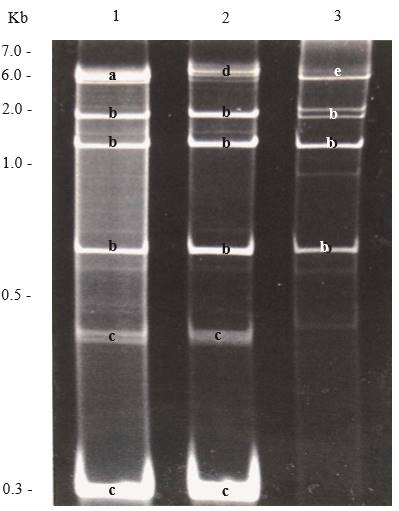
Figure 5. Polyacrylamide (6 %) gel electrophoresis showing dsRNAs extracted from plants with mixed viral infections. Lane 1, Potato virus X (PVX), Cucumber mosaic virus (CMV) and satellite RNA of CMV; lane 2, Dandelion latent virus (DLV), CMV and satellite RNA of CMV; and lane 3, Tobacco mosaic virus (TMV and CMV. Bands are labelled with letters according to the virus they belong; a, PVX; b, CMV, c, Satellite RNA of CMV; d, DLV, and e, TMV. 

Molecular probes

Radioactive and non-radioactive probes have been produced by using dsRNA to generate and label ssRNA or cDNAs (Enebak et al., 1994; Jelkmann et al., 1989; Jordan and Dodds, 1983; Rosner et al., 1983; Valverde and Dodds, 1986; Valverde et al., 1994; Valverde et al., 1990b). Jordan and Dodds used TMV dsRNA to develop P32 endlabelled ssRNA probes and used them successfully in molecular hybridization experiments. Nonradioactive probes have also been prepared by direct labelling ssRNA using dsRNA as the starting material (Valverde et al., 1994). These non-radioactive probes were used to detect Oryza sativa endornavirus, Physalis mottle virus, Pepper cryptic virus 1, and an unidentified mycovirus from the soybean stem canker pathogen (Diaporthe phaseolorum). DsRNA extracts blotted onto membranes have been used to detect Sweet potato chlorotic stunt virus (SPCSV) in (Pio-Ribeiro et al., 1996), Fiji virus (Karan et al., 1994), Satellite tobacco mosaic virus (Valverde and Dodds, 1986), and a 12 kb hypovirus from C. parasitica (Enebak et al., 1994) in northern hybridization assays.

Reverse transcription PCR, cloning, and sequencing

Viral dsRNAs have been used to clone and sequence plant and fungal RNA viruses (Aboughanem-Sabanadzovic et al., 2016; Antoniw et al., 1986; Bar-Joseph et al., 1983; Enebak et al., 1994; Herrero et al., 2009; Khalifa and Pearson, 2014; Kim et al., 2005; Okada et al. 2011; Rott and Jelkmann, 2001; Jelkmann et al., 1989; Valverde and Sabanadzovic, 2009; Valverde and Sabanadzovic; Valverde et al. 1986; 1990; 2011; Zhang and Rowhani, 2000). Virus specific primers have been developed for RT-PCR detection of most plant and fungal viruses reported to date. After heat denaturation, purified viral dsRNA has been used extensively as the template for reverse transcription PCR (RT-PCR) in plant virus testing (Rott and Jelkmann, 2001; Sabanadzovic and Valverde, 2011; Valverde and Sabanadzovic, 2009; Valverde et al., 2011; Tzanetakis et al 2005). Winter et al. (1992) and Hoyer et al. (1996) characterized Sweet potato chlorotic stunt virus using dsRNA as reagent for RT-PCR, cloning, and labeling. Zhang and Rowhani (2000) developed a rapid cDNA cloning method from dsRNA templates to partially characterized grape viruses. Random cDNA clones from CTV were obtained using denatured viral dsRNA purified from citrus bark tissue (Albiach-Marti et al., 2010). Molecular cloning of plant viruses has been accomplished from small amounts of viral dsRNA after gel purification (Jelkmann et al., 1989). Using dsRNA extracted using the method of De Paulo and Powell (1995), Kunta et al. (2007) conducted RTPCR and were able to detect, clone, and sequence full-length viroids in citrus. Similarly, De la Torre et al. (2009; 2015) detected viroids in peach and avocado using dsRNA extracted by the method of Valverde et al., (1990) followed by RT-PCR. This approach has been particularly useful, when testing plants species known to yield low quality ssRNA.

DsRNA extraction and purification has been suggested as an approach to increase viral sequences in sample preparation for NGS (Wu et al., 2015). In a metagenomics study of viruses in environmental samples, Roossinck (2012) used dsRNA-enriched samples and detected high levels of persistent viruses in plants. De novo assembly of RNAseq results has been accomplished to identify new species of endornaviruses and mycoviruses (Espach et al., 2012; Jo et al., 2015; Khalifa et al., 2016). Using viral dsRNAs, in combination with deep sequencing, researchers have been able to obtain complete genomic sequences of plant and fungal viruses (Al Rwahnih et al., 2011; Candresse et al., 2013; Coetzee et al., 2010; Deker and Parker, 2014; Espach et al., 2012; Quito-Avila et al., 2011; Magae, 2012) and to identify virus-like elements in aquatic microbial populations (Nerva et al., 2016). Deep sequencing from a single grapevine revealed a virome dominated by mycoviruses (Al Rwahnih et al., 2011).

Field surveys

Surveys of RNA viruses in plants and fungi have been conducted by extracting dsRNAs, analyzing them in agarose or polyacrylamide gels, and using them for RT-PCR and sequencing. DsRNAs, isolated from 44 vines from a diseased South African vineyard, were used in a deep sequencing analysis to build a census of the viral population (Al Rwahnih et al., 2011). In a survey for viruses from fungi associated with a parasitic plant (Cuscuta cuspidata) and its host plant (Ambrosia psilostachya) in a tallgrass prairie habitat in Oklahoma, researchers extracted dsRNA to detect fungal viruses (Feldman et al., 2012). The detected viruses were further characterized using reverse transcription-PCR and sequence analysis. A survey for endornaviruses in wild plant species of South Louisiana was conducted using a combination of dsRNA extraction and RT-PCR (Rodrigues de Souto et al., 2015). Of 140 species belonging to 58 plant families, seven were found infected with putative endornaviruses. In Mexico, Piedra et al., (2005) conducted surveys to identify viruses infecting a weed Leonotis nepetifolia (Lamiaceae) commonly present near cultivated fields. Using dsRNA extraction as a complementary tool, the authors were able to identify infections of Alfalfa mosaic virus (AMV), CMV, satellite RNA of CMV, and TMV in this plant species. In a survey for viruses infecting tree tobacco in Southern California, electrophoretic analysis of viral dsRNA was used for virus detection (Valverde and Dodds, 1986). Using dsRNA extraction and electrophoretic analysis, Herrero et al., 2009 conducted a survey of mycoviruses in a collection of 103 isolates belonging to 53 different species of endophytic fungi of grasses and detected dsRNA in 12 isolates.

Plant diseases of unknown etiology

There are several examples of economically important plant diseases of unknown etiology in which the causal agent was elucidated using dsRNA. The causal agent of pallidosis, a disease of strawberry of previously unknown etiology, was identified to be a luteovirus by using purified dsRNA (Tzanetakis et al., 2004; 2005). Similarly, a disease of unknown etiology affecting Japanese holly fern and leather leaf fern was determined to be caused by a new plant virus species from dsRNA that was used for cloning and sequencing the virus which was later named Japanese holly fern mottle virus (Valverde and Sabanadzovic, 2009). Lettuce big vein disease was first shown to be caused by a virus by detection of dsRNAs (Mirkov and Dodds, 1985). In Mexico, the causal agent of a disease of husk tomato (Physalis ixocarpa) which consists of foliar yellow mottle was unknown until 2003 when it was confirmed to be AMV by dsRNA analyses (De la Torre et al., 2003).

Persistent plant viruses

Persistent plant viruses, which are often present in asymptomatic plants, have dsRNA banding profiles that could be confused with that of disease causing acute viruses (Roossinck, 2010; Valverde and Dodds, 1986; Valverde et al., 1990). Persistent plant viruses reported to date belong to the families Amalgaviridae, Chrysoviridae, Endornaviridae, Totiviridae, and Partitiviridae, which include members that infect plants and fungi. There is no evidence that these viruses cause disease in plants. The molecular characterization of most persistent plant viruses has been accomplished by starting with genomic or RF dsRNA purification (Fukuhara, 1999; Pfeiffer, 1998; Okada, et al., 2011; 2013; Sabanadzovic and Valverde, 2011; Sabanadzovic et al., 2016; Sabanadzovic et al., 2010; Sabanadzovic et al., 2009). Using a combination of electrophoretic analysis of viral dsRNA and RT-PCR, Khankhum et al. (2015) demonstrated that two endornaviruses showed differential infection patterns between gene pools of common bean (Phaseolus vulgaris). The presence of persistent viruses in some crop cultivars and wild plant species emphasizes the importance of including samples from symptomless (assumed to be healthy) plants when using the dsRNA technique to detect and identify acute viruses. The dsRNA profiles of three persistent viruses and nucleic acid extracts from two healthy plants are shown in Figure 6.

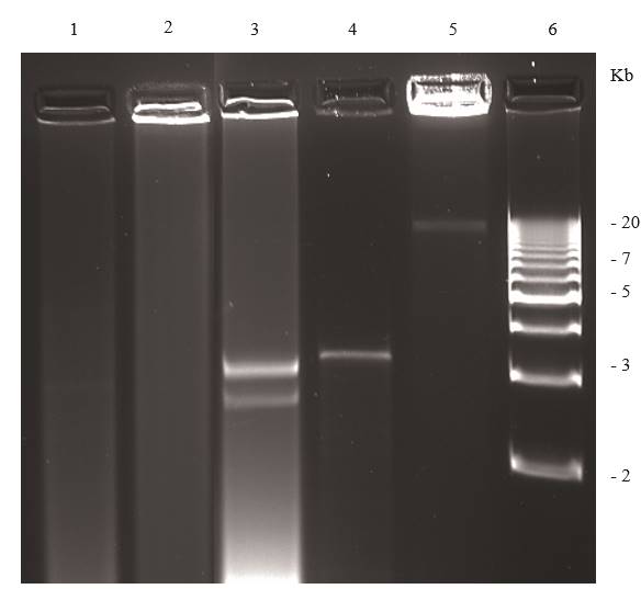
Figure 6. Agarose (1.2 %) gel electrophoresis of dsRNAs extracted from symptomless plants. Lane 1, healthy Phaseolus vulgaris; lane 2, healthy Capsicum annuum; lanes 3-5 plants infected with persistent viruses; lane 3, Persea americana infected with a putative chrysovirus; lane 4, Solanum lycopersicum infected with Southern tomato virus; lane 5 Capsicum annuum infected with Bell pepper endornavirus lane 6 DNA ladder. 

Conclusions

Although the cellulose-based protocol for the extraction and purification of viral dsRNA from plants and fungi infected with RNA viruses has been around for over 40 years, it still provides diagnosticians and researchers with a practical tool for the study, detection, and identification of viruses. The simplicity and versatility of the technique, combined with modifications that have improved dsRNA yield, quality, and lowered the overall costs, makes it practical for laboratories with limited resources. As pointed out before, the earlier use of this technique was on the electrophoretic analysis of viral dsRNAs and the use of the banding patterns (electrophoretic profiles) to detect, and in some cases, identify the viruses. However, today, the emphasis is more on the use of viral dsRNA for RT-PCR, cloning, and sequencing which leads to virus identification. It is possible that methods for the extraction of viral dsRNAs will have a place in future implementation of NGS for virus detection and identification. This has been demonstrated with the use of dsRNAs to conduct the complete sequencing of viral RNA genomes from plants and fungi and to identify virus-like elements in aquatic microbial populations using NGS (Nerva et al., 2016), deep sequencing analysis of viruses infecting grapevines (Al Rwahnih et al., 2011; Coetzee et al., 2010), and a metagenomics study of environmental samples (Roossinck, 2012).

Acknowledgments

We wish to acknowledge the partial support to RAV by research grant No. US-4725-4 F from BARD, the United States -Israel Binational Agricultural Research and Development Fund and the USDA National Institute of Food and Agriculture.

Literatura citada

Aboughanem-Sabanadzovic N, Tzanetakis IE, Lawrence A, Stephenson RC and Sabanadzovic S. 2016. A novel Ilarvirus is associated with privet necrotic ringspot disease in the Southern United States. Phytopathology 106:87-93. DOI: 10.1094/PHYTO-12-14-0387-R [ Links ]

Akin A, Wu CC and Lin TL. 1998. A comparison of two RNA isolation methods for double-stranded RNA of infectious bursal disease virus. Journal of Virological Methods 74:179-184. DOI: 10.1016/S0166-0934(98)00082-2 [ Links ]

Al Rwahnih M, Daubert S, Urbez-Torres JR, Cordero F and Rowhani A. 2011. Deep sequencing evidence from single grapevine plants reveals a virome dominated by mycoviruses. Archives of Virology 156:397-403. DOI: 10.1007/s00705-010-0869-8 [ Links ]

Al Rwahnih M, Daubert S, Golino D, Islas C and Rowhani A. 2015. Comparison of next-generation sequencing versus biological indexing for the optimal detection of viral pathogens in grapevine. Phytopathology 105:758-63. DOI: 10.1094/PHYTO-06-14-0165-R [ Links ]

Albiach-Marti MR, Robertson C, Gowda S, Tatineni S, Belliure B, Garnsey SM, Folimonova SY, Moreno P, and Dawson WO. 2010. The pathogenicity determinant of Citrus tristeza virus causing the seedling yellows syndrome maps at the 3’-terminal region of the viral genome. Molecular Plant Pathology 11:55-67. DOI: 10.1111/j.1364-3703.2009.00572.x [ Links ]

Antoniw JF, Linthorst HJM, White RF and BOL JF. 1986. Molecular cloning of the double-stranded RNA of beet cryptic viruses. Journal of General Virology 67:2047-2051. DOI: 10.1099/0022-1317-67-9-2047 [ Links ]

Aramburu J, Navas-Castillo J, Moreno P and Cambra M. 1991. Detection of double-stranded RNA by ELISA and dot immunobinding assay using an antiserum to synthetic polynucleotides. Journal of Virologial Methods 33:1-11. DOI: 10.1016/0166-0934(91)90002-H [ Links ]

Atsumi G, Sekine KT and Kobayashi K. 2015. A new method to isolate total dsRNA. Methods in Molecular Biology 1236:27-37. DOI: 10.1007/978-1-4939-1743-3_3 [ Links ]

Azzam OI and Gonsalves D. 1999. Detection of dsRNA from cleistothecia and conidia of the grape powdery mildew pathogen Uncinula necator. Plant Disease 75:964-967. DOI: 10.1094/PD-75-0964 [ Links ]

Balijja A, Kvarnheden A and Turchetti, T. 2008. A non-phenolchloroform extraction of double-stranded RNA from plant and fungal tissues. Journal of Virological Methods 152:32-37. DOI:10.1016/j.jviromet.2008.06.001 [ Links ]

Bar-Joseph M, Rosner A, Moskovitz M and Hull R. 1983. A simple procedure for the extraction of double-stranded RNA from viral infected plants. Journal of Virological Methods 6:1-8. DOI: 10.1016/0166-0934(83)90062-9 [ Links ]

Boonham N, Kreuze J, Winter S, van der Vlugt R, Bergervoet J, Tomlinson J and Mumford R. 2014. Methods in virus diagnostics: from ELISA to next generation sequencing. Virus Research 186:20-31. DOI: 10.1016/j.virusres.2013.12.007. [ Links ]

Buck KW. 1999. Replication of tobacco mosaic virus RNA. Philosophical Transactions of the Royal Society B: Biological Sciences 354:613-627. DOI: 10.1098/rstb.1999.0413 [ Links ]

Burgyan J, Grieco F and Russo M. 1989. A defective interfering RNA molecule in cymbidium ringspot virus infections. Journal of General Virology 70:235-239. DOI: 10.1099/0022-1317-70-1-235 [ Links ]

Candresse T, Marais A, Faure C and Gentit P. 2013. Association of Little cherry virus 1 (LChV1) with the Shirofugen stunt disease and characterization of the genome of a divergent LChV1 isolate. Phytopathology 103:293-308. DOI: 10.1094/PHYTO-10-12-0275-R [ Links ]

Carbonell A, Ángel-Emilio Martínez de Alba A-E, Flores R and Gago S. 2008. Double-stranded RNA interferes in a sequence-specific manner with the infection of representative members of the two viroid families. Virology 371:44-53. DOI: 10.1016/j.virol.2007.09.031 [ Links ]

Castanho B, Butler EE and Shepherd RI. 1978. The association of double-stranded RNA with Rhizoctonia decline. Phytopathology 68:1515-1519. DOI: 10.1094/Phyto-68-1515 [ Links ]

Castillo A, Cottet L, Castro M and Selpulveda F. 2011. Rapid isolation of mycoviral double-stranded RNA from Botrytis cinerea and Saccharomyces cerevisiae. Virology Journal 8:38. DOI: 10.1186/1743-422X-8-38 [ Links ]

Chen S, Cao L, Huang Q, Qian Y, and Zhou X. 2016. The complete genome sequence of a novel maize-associated totivirus. Archives of Virology 161:487. DOI: 10.1007/s00705-015-2657-y [ Links ]

Choi GH and Nuss DL. 1992. Hypovirulence of chestnut blight fungus conferred by an infectious viral cDNA. Science 257:800-803. DOI: 10.1126/science.1496400 [ Links ]

Chu Y-M, Lim WS, Yea SY, Cho JD, Lee YW and Kim KH. 2004. Complexity of dsRNA mycovirus isolated from Fusarium graminearum. Virus Genes 28:135-143. DOI: 10.1023/B:VIRU.0000012270.67302.35 [ Links ]

Coetzee B, Freeborough MJ, Maree HJ, Celton JM, Rees DJG and Burger JT. 2010. Deep sequencing analysis of viruses infecting grapevines: Virome of a vineyard. Virology 400:157-163. DOI: 10.1016/j.virol.2010.01.023 [ Links ]

Condit C and Fraenkel-Conrat H. 1979. Isolation of replicative forms of 3’ terminal subgenomic RNAs of tobacco necrosis virus. Virology 97:122-30. DOI:10.1016/0042-6822(79)90378-7 [ Links ]

Creamer R and Falk BW. 1989. Characterization of nonspecifically aphid-transmitted aphid CA-RPV isolate of barley yellow dwarf virus. Phytopathology 79:942-946. DOI: 10.1094/Phyto-79-942 [ Links ]

Dale JL, Phillips DA and Parry JN. 1986. Double-stranded RNA in banana plants with bunchy top disease. Journal of General Virology 67:371-375. DOI: 10.1099/0022-1317-67-2-371 [ Links ]

Day PR, Dodds JA, Elliston JE, Jaynes RA and Anagnostakis SI. 1977. Double-stranded RNA in Endothia parasitica. Phytopathology 67:1393-1396. DOI: 10.1094/Phyto-67-1393 [ Links ]

De la Torre AR, Pallas V and Sanchez-Navarro JA. 2016. First report of Cucumber mosaic virus (CMV) and CARNA-5 in carnation in Mexico. Plant Disease 100:1509. DOI: org/10.1094/PDIS-01-16-0110-PDN [ Links ]

De la Torre AR, Sánchez NJ, Pallás V. 2014. Detección del Prunus necrotic ringspot virus en durazno (Prunus persica (L.) en México y caracterización molecular de su componente ARN-3. Agrociencia 48:583-598. [ Links ]

De la Torre AR, Pallas V and Sanchez-Navarro JA. 2015. First report of Peach latent mosaic viroid in peach trees from Mexico. Plant Disease 99:899. DOI: org/10.1094/PDIS-01-15-0005-PDN [ Links ]

De la Torre AR, Salazar-Segura M and Valverde RA. 2003. Etiología del moteado amarillo del tomate de cáscara (Physalis ixocarpa) en México. Agrociencia 37:227-289. [ Links ]

De la Torre AR, Teliz-Ortiz D, Pallas V and Sanchez-Navarro JA. 2009. First report of Avocado sunblotch viroid in avocado from Michoacán, México. Plant Disease 93:202. DOI: org/10.1094/PDIS-93-2-0202B [ Links ]

Deker CJ and Parker R. 2014. Analysis of double-stranded RNA from microbial communities identifies doublestranded RNA virus-like elements. Cell Reports 7:898-906. DOI: doi.org/10.1016/j.celrep.2014.03.049 [ Links ]

Delye C and Corio-Costet M-F. 1998. Rapid isolation of both double-stranded RNA and PCR-suitable DNA from the obligate biotrophic phytopathogenic fungus Uncinula necator using a commercially available reagent. Journal of Virological Methods 74:149-153. DOI: 10.1016/S0166-0934(98)00079-2 [ Links ]

Demler SA and de Zoeten GA. 1989. Characterization of a satellite RNA associated with pea enation mosaic virus. Journal of General Virology 70:1075-1084. DOI: 10.1099/0022-1317-70-5-1075 [ Links ]

Deng F and Boland GJ. 2004. A satellite RNA of Ophiostoma novoulmi mitovirus 3a in hypovirulent isolates of Sclerotinia homoeocarpa. Phytopathology 94:917-923. DOI: 10.1094/PHYTO.2003.93.11.1407 [ Links ]

DePaulo JJ and Powell CA. 1995. Extraction of double-stranded RNA from plant tissues without the use of organic solvents. Plant Disease 79:246-248. [ Links ]

Derrick KS. 1978. Double-stranded RNA is present in extracts of tobacco plants infected with tobacco mosaic virus. Science 199:538-539. DOI: 10.1126/science.622553 [ Links ]

Derrick KS, French RC, Clark CA and Gabriel CJ. 1984. Detection of double-stranded RNA by serologically specific electron microscopy. Journal of Virological Methods 9:293-299. DOI: 10.1016/0166-0934(84)900557 [ Links ]

Diaz-Ruiz JR and Kaper JM. 1977. Cucumber mosaic virus-associated RNA 5. III. Little nucleotide sequence homology between CARNA 5 and helper RNA. Virology 80:204-13. DOI: 10.1016/0042-6822(77)90393-2 [ Links ]

Diaz-Ruiz JR and Kaper JM. 1978. Isolation of viral double-stranded RNAs using LiCI fractionation procedure. Preparative Biochemistry 8:1-17. DOI: 10.1080/00327487808068215 [ Links ]

Dickinson MJ and Pryor A. 1989. Isometric virus-like particles encapsidate the double-stranded RNA found in Puccinia striiformis, Puccinia recondita, and Puccinia sorghi. Canadian Journal of Botany 67:3420-3425. DOI: 10.1139/b89-417 [ Links ]

Dodds JA. 1982. Cross-protection and interference between electrophoretically distinct strains of cucumber mosaic virus in tomato. Virology 118:235-240. DOI: 10.1016/0042-6822(82)90338-5 [ Links ]

Dodds JA and Bar-Joseph M. 1983. Double-stranded RNA from plants infected with closteroviruses. Phytopathology 73:419-423. DOI: 10.1094/Phyto-73-419 [ Links ]

Dodds JA, Morris TJ and Jordan RL. 1984. Plant viral doublestranded RNA. Annual Review of Phytopathology 22:151-168. DOI: 10.1146/annurev.py.22.090184.001055 [ Links ]

Dodds JA, Jarupat T, Lee JG and Roistacher CN. 1987. Effect of host, time of harvest, and virus concentration on double-stranded RNA analysis of Citrus tristeza virus. Phytopathology 77:442-447. DOI: 10.1094/Phyto-77-442 [ Links ]

Dodds JA, Valverde RA and Matthews DM. 1988. Detection and interpretation of dsRNAs. Pp 309-326. In: Koltin Y and Leibowitz M J (Eds). Viruses of Fungi and Simple Eukaryotes Marcel Dekker, Inc. New York, N. Y. p [ Links ]

Enebak SA, Hillman BI and Macdonald WL. 1994. A hypovirulent isolate of Cryphonectria parasitica with multiple, genetically unique dsRNA segments. Molecular Plant-Microbe Interactions 7:590-595. DOI: 10.1094/MPMI-7-0590 [ Links ]

Espach Y, Maree HJ and Burger JT. 2012. Complete genome of a novel endornavirus assembled from next-generation sequence data. Journal of Virology 86:13142. DOI: 10.1128/JVI.02538-12 [ Links ]

Feldman TS, Morsy MR and Roossinck MJ. 2012. Are communities of microbial symbionts more diverse than communities of macrobial hosts? Fungal Biology 116:465-477. DOI: 10.1016/j.funbio.2012.01.005 [ Links ]

Franklin RM. 1966. Purification and properties of replicative intermediate of the RNA bacteriophage R17. Proceedings of the National Academy of Sciences USA 55:1504-1511. [ Links ]

Fukuhara T. 1999. Double-Stranded RNA in rice. Journal of Plant Research 112:131-138. DOI: 10.1007/PL00013855 [ Links ]

Fukuhara T, Koga R, Aoki N, Yuki C, Yamamoto N, Oyama N, Udagawa T, Horiuchi H, Miyazaki S, Higashi Y, Takeshita M, Ikeda K, Arakawa M, Matsumoto N and Moriyama H. 2006. The wide distribution of endornaviruses, large double-stranded RNA replicons with plasmid-like properties. Archives of Virology 151:995-1002. DOI: 10.1007/s00705-005-0688-5 [ Links ]

Ghabrial SA, Caston JR, Jiang DH, Nibert ML and Suzuki N. 2015. 50-plus years of fungal viruses. Virology 479:356-368. DOI: 10.1016/j.virol.2015.02.034 [ Links ]

German TL and de Zoeten GA. 1975. Purification and properties of replicative form and replicative intermediates of pea enation mosaic virus. Virology 66:172-184. DOI: 10.1016/0042-6822(75)90188-9 [ Links ]

Habili N and Kaper JM. 1981. Cucumber mosaic virusassociated RNA5. VII. Doublestranded form accumulation and disease attenuation in tobacco. Virology 112:250-261. DOI: 10.1016/0042-6822(81)90630-9 [ Links ]

Hansen DR, Van Alfen NK, Gillies K and Powell WA. 1985. Naked dsRNA associated with hypovirulence of Endothia parasitica is packaged in fungal vesicles. Journal of General Virology 66:2605-2614. DOI: 10.1099/0022-1317-66-12-2605 [ Links ]

Herrero N, Márquez SS and Zabalgogeazcoa I. 2009. Mycoviruses are common among different species of endophytic fungi of grasses. Archives of Virology 154:327-330. DOI: 10.1007/s00705-008-0293-5 [ Links ]

Herrero N, Dueñas E, Quesada-Moraga E, and Zabalgogeazcoa I. 2012. Prevalence and diversity of viruses in the entomopathogenic fungus Beauveria bassiana. Applied and Environmental Microbiology 78:85238530. DOI: 10.1128/AEM.01954-12. pmid:23001673 [ Links ]

Hillman BI, Foglia R and Yuan W. 2000. Satellite and defective RNAs of Cryphonectria hypovirus 3-Grand Haven 2, a virus species in the family Hypoviridae with a single open reading frame. Virology 276:181-189. DOI:10.1006/viro.2000.0548 [ Links ]

Ho T and Tzanetakis IE. 2014. Development of a virus detection and discovery pipeline using next generation sequencing. Virology 471-473:54-60. DOI: 10.1016/j.virol.2014.09.019 [ Links ]

Horiuchi H, Fukuhara T. 2004. Putative replication intermediates in Endornavirus, a novel genus of plant dsRNA viruses. Virus Genes 29:365-375. DOI: 10.1007/s11262-004-7441-0 [ Links ]

Hoyer U, Maiss E, Jelkmann W, Lesemann D-E and Vetten J. 1996. Identification of the coat protein of sweet potato sunken vein closterovirus isolate from Kenya and evidence of serological relationship among geographically diverse closterovirus isolates from sweet potato. Phytopathology 86: 744-750. DOI: 10.1094/Phyto-86-744 [ Links ]

Huang Q, Baum L and Fu WL. 2010. Simple and practical staining of DNA with GelRed in agarose gel electrophoresis. Clinical Laboratory 56:149-152. [ Links ]

Jackson AO, Mitchell DM and Siegel A. 1971. Replication of tobacco mosaic virus. I. Isolation and characterization of double-stranded forms of ribonucleic acid. Virology 45:182-191. DOI: 10.1016/0042-6822(71)90125-5 [ Links ]

Jelkmann W, Martin RR and Maiss E. 1989. Cloning of four viruses from small quantities of double-stranded RNA. Phytopathology 79:1250-1253. DOI: 10.1094/Phyto-79-1250 [ Links ]

Jo Y, Choi H and Cho WK. 2015. De novo assembly of a Bell pepper endornavirus genome sequencing RNA sequencing data. Genome Announcements 3 no. 2. DOI: 10.1128/genomeA.00061-15 [ Links ]

Jordan RL and Dodds JA. 1983. Hybridization of 5’-end labelled RNA to plant viral RNA in agarose and acrylamide gels. Plant Molecular Biology Reporter 1:33-37. [ Links ]

Kaper JM and Diaz-Ruiz JR. 1977. Molecular weights of the double-stranded RNAs of cucumber mosaic virus strain S and its associated RNA 5. Virology 80:214-17. DOI: 10.1016/0042-6822(77)90394-4 [ Links ]

Karan M, Dale JL, Bateson MF, Harding RM and Teakle DS. 1994. Detection and characterization of pangola stunt Fijivirus from Australia using cloned eDNA probes. Archives of Virology 135:397-404. DOI: 10.1007/BF01310023 [ Links ]

Khankhum S, Escalante C, Rodrigues de Souto E, De la Torre R and Valverde RA. 2015. A method for the extraction of viral double stranded RNA from relative small amounts of virus and fungus infected plant tissues. Revista Mexicana de Fitopatología 33:S 223 (Suplemento). [ Links ]

Khalifa ME and Pearson MN. 2014. Molecular characterization of an endornavirus infecting he phytopathogen Sclerotinia sclerotiorum. Virus Research 189:303-309. DOI: 10.1016/j.virusres.2014.06.010 [ Links ]

Khalifa ME, Varsani A, Ganley AR and Pearson MN. 2016. Comparison of Illumina de novo assembled and Sanger sequenced viral genomes: A case study for RNA viruses recovered from the plant pathogenic fungus Sclerotinia sclerotiorum. Virus Research 219:51-57. DOI: 10.1016/j.virusres.2015.11.001 [ Links ]

Kim Y-J, Park S-H, Yie S-W and Kim K-H. 2005. RT-PCR Detection of dsRNA Mycoviruses Infecting Pleurotus ostreatus and Agaricus blazei Murrill. The Plant Pathology Journal 21:343-348. DOI: 10.5423/PPJ.2005.21.4.343 [ Links ]

Klein A and Reichman ME. 1970. Isolation and characterization of two species of double-stranded RNA from tobacco leaves doubly infected with tobacco necrosis and satellite tobacco necrosis viruses. Virology 42:269-72. DOI: 10.1016/0042-6822(70)90268-0 [ Links ]

Kondo H, Hisano S, Chiba S, Maruyama K, Andik IB, Toyoda K, Fujimori F and Suzuki N. 2016. Sequence and phylogenetic analyses of novel totivirus-like doublestranded RNAs from field-collected powdery mildew fungi. Virus Research 213:353-364. DOI: 10.1016/j.virusres.2015.11.0150168-1702 [ Links ]

Kousik CS, Snow JP and Valverde RA. 1994. Comparison of double-stranded RNA components and virulence among isolates of Rhizoctonia solani AG-1 IA and AG-1 IB. Phytopathology 84:44-49. DOI: 10.1094/Phyto-84-44 [ Links ]

Kovalskaya N and Hammond RW. 2014. Molecular biology of viroid-host interactions and disease control strategies. Plant Science 228:48-60. DOI: 10.1016/j.plantsci.2014.05.006 [ Links ]

Kunta M, da Graça JV and Skaria M. 2007. Molecular detection and prevalence of citrus viroids in Texas. HortScience 42:600-604. [ Links ]

Kwon S-J, Lim W-S, Park S-H, Park M-R and Kim K-H. 2007. Molecular characterization of a dsRNA mycovirus, Fusarium graminearum virus-DK21, which is phylogenetically related to Hypoviruses but has a genome organization and gene expression strategy resembling those of plant potex-like viruses. Molecules and Cells 23:304-315. DOI: 10.1007/s10059-009-0112-1 [ Links ]

Lee SH, Moon BJ and Lee JK. 2006. Characteristics of hypovirulentstrainsofchestnutblightfungus, Cryphonectria parasitica, isolated in Korea. Mycobiology 34:61-66. DOI: 10.4489/MYCO.2006.34.2.061 [ Links ]

Li W, Zhang T, Sun H, Deng Y, Zhang A, Chen H and Wang K. 2014. Complete genome sequence of a novel endornavirus in the wheat sharp eyespot pathogen Rhizoctonia cerealis. Archives of Virology 159:1213-1216. DOI: 10.1007/s00705-013-1893-2 [ Links ]

Libonati M and Sorrentino S. 1992. Revisiting the action of bovine ribonuclease A and pancreatic-type ribonucleases on double-stranded RNA. Molecular and Cellular Biochemistry 117:139-151. DOI: 10.1007/BF00230753 [ Links ]

Libonati M, Casarna N and Furia A. 1980. Double-stranded RNA. Molecular and Cellular Biochemistry 31:147-164. DOI: 10.1007/BF00225848 [ Links ]

Liu L, Wang Q, Cheng J, Fu Y, Jiang D and Xie J. 2015. Molecular characterization of a bipartite double-stranded RNA virus and its satellite-like RNA co-infecting the phytopathogenic fungus Sclerotinia sclerotiorum. Frontiers in Microbiology 6:406. DOI: 10.3389/fmicb.2015.00406 [ Links ]

Li L, Liu J, Xu A, Wang T, Chen J and Zhu X. 2013. Molecular characterization of a trisegmented chrysovirus isolated from the radish Raphanus sativus. Virus Research 176:169-78. DOI: 10.1016/j.virusres.2013.06.004 [ Links ]

Magae Y. 2012. Molecular characterization of a novel mycovirus in the cultivated mushroom, Lentinula edodes. Virology Journal 9:60. DOI: 10.1186/1743-422X-9-60 [ Links ]

Miller WA and Koev G. 2000. Synthesis of subgenomic RNAs by positive-strand RNA viruses. Virology 273:1-8. DOI:10.1006/viro.2000.0421 [ Links ]

Mirkov TE and Dodds JA. 1985. Association of doublestranded ribonucleic acids with lettuce big vein disease. Phytopathology 75:631-635. DOI: 10.1094/Phyto-75-631 [ Links ]

Morris TJ and Dodds JA. 1979. Isolation and analysis of double-stranded RNA from virus infected plant and fungal tissue. Phytopathology 69:854-858. DOI: 10.1094/Phyto-69-854 [ Links ]

Nerva L, Ciuffo M, Vallino M, Margaria P, Varese GC, Gnavi G and Turina M. 2016. Multiple approaches for the detection and characterization of viral and plasmid symbionts from a collection of marine fungi. Virus Research DOI: 10.1016/j.virusres.2015.10.028 [ Links ]

Nuss DL and Koltin Y. 1990. Significance of dsRNA genetic elements in plant pathogenic fungi. Annual Review of Phytopathology 28:37-58. DOI: 10.1146/annurev.py.28.090190.000345 [ Links ]

Okada R, Kiyota E, Moriyama H, Fukuhara T and Natsuaki T. 2015. A simple and rapid method to purify viral dsRNA from plant and fungal tissue. Journal of General Plant Pathology 81:103-107. DOI 10.1007/s10327-014-0575-6 [ Links ]

Okada R, Young CK, Valverde RA, Sabanadzovic S, Aoki N, Hotate S, Kiyota E, Moriyama H and Fukuhara T. 2013. Molecular characterization of two evolutionally distinct endornaviruses co-infecting common bean (Phaseolus vulgaris). Journal of General Virology 93:220-229. DOI 10.1099/vir.0.044487-0 [ Links ]

Okada R, Kiyota E, Sabanadzovic S, Moriyama H, Fukuhara T, Saha P, Roossinck MJ, Severin A and Valverde RA. 2011. Bell pepper endornavirus: molecular and biological properties and occurrence in the genus Capsicum. Journal of General Virology 92:2664-2673. DOI 10.1099/vir.0.034686-0 [ Links ]

Osman TAM and Buck KW. 1990. Double-stranded RNAs isolated from plant tissue infected with red clover necrotic mosaic virus correspond to genomic and subgenomic single-stranded RNAs. Journal of General Virology 71:945-948. DOI: 10.1099/0022-1317-71-4-945 [ Links ]

Pathak KB and Nagy PD. 2009. Defective interfering RNAs: foes of viruses and friends of virologists. Viruses 1:895-919 DOI: 10.3390/v1030895 [ Links ]

Pecina V, Alvarado MJ, Williams H, De la Torre RA and Vandemark GJ. 2000. Detection of double-stranded RNA in Macrophomina phaseolina. Mycologia 92:900-907. DOI: 10.2307/3761586 [ Links ]

Piedra IE, De la Torre AR, Zuñiga G, Xoconostle CB and Ruiz MR. 2005. Leonotis nepetifolia: An important plant virus reservoir in central Mexico. Phytoparasitica 33: 480-494. DOI: 10.1007/BF02981397 [ Links ]

Pfeiffer P. 1998. Nucleotide sequence, genetic organization, and expression strategy of the double-stranded RNA associated with the ‘447’ cytoplasmic male sterility trait in Vicia faba. Journal of General Virology 79:2349-2358. DOI: 10.1099/0022-1317-79-10-2349 [ Links ]

Quito-Avila DF, Jelkmann W, Tzanetakis I, Keller K and Martin RR. 2011. Complete sequence and genetic characterization of Raspberry latent virus, a novel member of the family Reoviridae. Virus Research 155:397-405. DOI: 10.1016/j.virusres.2010.11.008 [ Links ]

Reyes PN, Marbán MN, Delgadillo SF y De la Torre AR. 2003. Variabilidad en aislamientos de Sclerotium cepivorum BERK. y su relación con ARN de cadena doble. Agrociencia 37:495-502. [ Links ]

Rodrigues de Souto E, Escalante C and Valverde RA. 2015. Detection of plant endornaviruses in wild plants of south Louisiana. Revista Mexicana de Fitopatología 33:S 224 (Suplemento). [ Links ]

Romanos MA, Buck KW and Rawlinson CJ. 1981. A satellite double-stranded RNA in a virus from Gaeumannomyces graminis. Journal of General Virology 57:375-385. DOI: 10.1099/0022-1317-57-2-375 [ Links ]

Roossinck MJ. 2012. Plant virus metagenomics: biodiversity and ecology. Annual Review of Genetics 46, 359-369. DOI: 10.1146/annurev-genet-110711-155600 [ Links ]

Roossinck MJ. 2010. Lifestyles of plant viruses. Philosophical Transactions of the Royal Society B: Biological Sciences 365:1899-1905. DOI: 10.1098/rstb.2010.0057 [ Links ]

Rosner A, Bar-Joseph M, Moscovitz M and Mevarech M. 1983. Diagnosis of specific viral RNA sequences in plant extracts by hybridization with a polynucleotide kinase-mediated 32P,- labeled double-stranded RNA probe. Phytopathology 73:699-702. DOI: 10.1094/Phyto-73-699 [ Links ]

Rott ME and Jelkmann W. 2001. Characterization and detection of several filamentous viruses of cherry: Adaptation of an alternative cloning method (DOP-PCR), and modification of an RNA extraction protocol. European Journal of Plant Pathology 107:411-420. DOI: 10.1023/A:1011264400482 [ Links ]

Sabanadzovic S and Valverde RA. 2011. Properties of two cryptoviruses from pepper (Capsicum annuum). Virus Genes 43:307-312. DOI: 10.1007/s11262-011-0634-4 [ Links ]

Sabanadzovic S, Ghanem-Sabanadzovic A and Valverde RA. 2010. Novel monopartite dsRNA virus from rhododendron. Archives of Virology 155:1859-1863. DOI: 10.1007/s00705-010-0770-5 [ Links ]

Sabanadzovic S, Wintermantel WM, Valverde RA, McCreight JD and Aboughanem-Sabanadzovic N. 2016. Cucumis melo endornavirus: Genome organization, host range and co-divergence with the host. Virus Research 214:49-58. DOI: 10.1016/j.virusres.2016.01.001 [ Links ]

Sabanadzovic S, Valverde RA, Brown JK, Martin RR and Tzanetakis IE. 2009. Southern tomato virus: the link between the families Totiviridae and Partitiviridae. Virus Research 140:130-137. DOI: 10.1016/j.virusres.2008.11.018 [ Links ]

Shamoun SF, Varga A, Valverde RA, Ramsfield T, Sumampong G, Elliott M, Masri S and James D. 2008 Identification and molecular characterization of a new dsRNA virus infecting Chondrostereum purpureum. Canadian Journal of Plant Pathology 30:604-613. DOI: 10.1080/07060660809507561 [ Links ]

Schonborn J, Oberstra J, Breyel BE, Tittgen J, Schumacher J and Lukacs N. 1991. Monoclonal antibodies to double-stranded RNA as probes of RNA structure in crude nucleic acid extracts. Nucleic Acids Research 19:2993-3000. DOI: 10.1093/nar/19.11.2993 [ Links ]

Tzanetakis IE, Halgren GB, Keller KE, Hokanson SC, Maas JL, McCarthy PL and Martin RR. 2004. Identification and detection of a virus associated with strawberry pallidosis disease. Plant Disease 88:383-390. DOI: 10.1094/PDIS.2004.88.4.383 [ Links ]

Tzanetakis IE and Martin RR. 2008. A new method for extraction of double-stranded RNA from plants. Journal of Virological Methods 149:167-70. DOI: 10.1016/j.jviromet.2008.01.014 [ Links ]

Tzanetakis IE, Keller KE and Martin RR. 2005. The use of reverse transcriptase for efficient first-and second-strand cDNA synthesis from singleand double-stranded RNA templates. Journal of Virological Methods 124:73-77. DOI: 10.1016/j.jviromet.2004.11.006 [ Links ]

Valverde RA and Dodds JA. 1986. Evidence for a satellite RNA associated naturally with the U5 strain and experimentally with the U1 strain of tobacco mosaic virus. Journal of General Virology 67:1875-1884. DOI: 10.1099/0022-1317-67-9-1875 [ Links ]

Valverde RA and Glascock CB. 1991. Further examination of the RNA and coat protein of spring beauty latent virus. Phytopathology 81:401-404. DOI: 10.1094/Phyto-81-401 [ Links ]

Valverde RA and Sabanadzovic S. 2009. A new plant virus with unique properties infecting Japanese holly fern. Journal of General Virology 90:2542-2549. DOI: 10.1099/vir.0.012674-0 [ Links ]

Valverde RA, Arancibia RA and Can F. 1994. Nonradioactive probes by direct labelling of ssRNA from dsRNA. BioTechniques 17:70-72. [ Links ]

Valverde RA, Black LL and Dufresne DJ. 1995. A comovirus affecting Tabasco pepper in Central America. Plant Disease 79:421-423. DOI: 10.1094/PD-79-0421 [ Links ]

Valverde RA, Dodds JA and Heick JA. 1986. Double stranded RNAs from plants infected with viruses having elongated particles and undivided genomes. Phytopathology 76:459-465. DOI: 10.1094/Phyto-76-459 [ Links ]

Valverde RA, Nameth ST and Jordan RL. 1990a. Analysis of double-stranded RNA for plant virus diagnosis. Plant Disease 74:255-258. DOI: 10.1094/PD-74-0255 [ Links ]

Valverde RA, Sabanadzovic, S and Rush MC. 2011. Identification of Oryza sativa endornavirus in rice genotypes from breeding programmes in the United States. Plant Breeding 130:271-274. DOI: 10.1111/j.1439-0523.2010.01778.x [ Links ]

Valverde RA, Nameth ST, Abdallha AO, Desjardins PR and Dodds JA. 1990b. Indigenous double-stranded RNA from pepper (Capsicum annuum). Plant Science 67:195-201. DOI: 10.1016/0168-9452(90)90243-H [ Links ]

Ward LI, Delmiglio C, Hill CF and Clover GRG. 2009. First report of Tobacco ringspot virus on Sophora microphylla, a native tree of New Zealand. Plant Pathology 58:784. DOI: 10.1111/j.1365-3059.2009.02125.x [ Links ]

Wei CZ, Osaki H, Iwanami T, Matsumoto N and Ohtsu Y. 2003. Molecular characterization of dsRNA segments 2 and 5 and electron microscopy of a novel reovirus from a hypovirulent isolate, W370, of the plant pathogen Rosellinia necatrix. Journal of General Virology 84:2431-2437. DOI: 10.1099/vir.0.19098-0 [ Links ]

Winter S, Purac A, Leggett F, Frison EA, Rossel HW and Hamilton RI. 1992. Partial characterization and molecular cloning of a closterovirus from sweet potato infected with the sweet potato virus disease complex from Nigeria. Phytopathology 82:869-875. DOI: 10.1094/Phyto-82-869 [ Links ]

Wu Q, Ding S-W, Zhang Y and Zhu S. 2015. Identification of viruses and viroids by next-generation sequencing and homology dependent and homology independent algorithms. Annual Review of Phytopathology 53:425-44. DOI: 10.1146/annurev-phyto-080614-120030 [ Links ]

Zelcer A, Weaber KF, Balazs E and Zaitlin M. 1981. The detection and characterization of viral related doublestranded RNAs in tobacco mosaic virus infected plants. Virology 113:417-427. DOI: 10.1016/0042-6822(81)90171-9 [ Links ]

Zhang YP and Rowhani A. 2000. A strategy for rapid cDNA cloning from double-stranded RNA templates isolated from plants infected with RNA viruses using Taq DNA polymerase. Journal of Virological Methods 84:59-63. DOI: 10.1016/S0166-0934(99)00126-3 [ Links ]

Received: June 30, 2016; Accepted: August 09, 2016

*Autor para correspondencia: RValverde@agcenter.lsu.edu.

Creative Commons License This is an open-access article distributed under the terms of the Creative Commons Attribution License