SciELO - Scientific Electronic Library Online

 
vol.39 número especialMi nombre es Bernardino Cruces, tengo 85 años, soy productorUn análisis retrospectivo de epidemias en plantas y humanos para comprender COVID-19 índice de autoresíndice de assuntospesquisa de artigos
Home Pagelista alfabética de periódicos  

Serviços Personalizados

Journal

Artigo

Indicadores

Links relacionados

  • Não possue artigos similaresSimilares em SciELO

Compartilhar


Revista mexicana de fitopatología

versão On-line ISSN 2007-8080versão impressa ISSN 0185-3309

Rev. mex. fitopatol vol.39 no.spe Texcoco  2021  Epub 30-Nov-2022

https://doi.org/10.18781/r.mex.fit.2021-1 

COVID-19: The Virus, Disease and Epidemiology

Basic Coronavirus biology and vaccines for COVID-19

Hernan Garcia-Ruiz1  2  * 

Katherine LaTourrette1  2  3 

Mayra Teresa Garcia-Ruiz4 

1 Nebraska Center for Virology, University of Nebraska-Lincoln, Lincoln, NE, USA, 68503.

2 Department of Plant Pathology, University of Nebraska-Lincoln, Lincoln, NE, USA, 68503.

3 Complex Biosystems Interdisciplinary Life Sciences Program, University of Nebraska-Lincoln, Lincoln, NE, USA, 68503.

4Universidad Autónoma Chapingo, México, CP 56230.


Abstract.

Severe acute respiratory syndrome coronavirus 2 (SARS-CoV-2) is the causal agent of the COVID-19 pandemic. Two mRNA vaccines based on the spike protein S have been authorized by the Food and Drug Administration. Antibody-based diagnostic test detect antibodies developed against protein S. Mutations in the genome of SARS-CoV-2 might compromise the precision of diagnostic tests and the efficacy of vaccines and antiviral drugs. We recently profiled genomic variation in human coronaviruses SARS-CoV, SARS-CoV-2, and Middle East respiratory syndrome coronavirus (MERS-CoV). As in all species of the genus Betacoronavirus, the genome is hyper variable, and mutations are not random. The most variable cistron codes for the spike S protein. Hyper variation in protein S has the potential to affect the efficacy of vaccines, the reliability of antibody-based diagnostic test, and predicts potential for repeated SARS-CoV-2 infections. Here we review the basics of coronavirus biology and genomic variation, and link them to diagnostic tests, vaccines, and antiviral drugs.

Key words: Antiviral; Coronavirus; COVID-19; MERS-CoV; mRNA vaccine; protein S; spike protein.

Resumen.

El coronavirus de tipo 2 causante del síndrome respiratorio agudo severo (SARS-CoV-2) es el agente causal de la pandemia de COVID-19. Dos vacunas de ARNm basadas en la proteína espicular S han sido autorizadas por la Administración de Alimentos y Fármacos de Estados Unidos (FDA, por sus siglas en inglés). La prueba de diagnóstico basada en anticuerpos detecta anticuerpos desarrollados contra la proteína S. Las mutaciones en el genoma de SARS-CoV-2 podrían poner en riesgo la precisión de las pruebas de diagnóstico y la eficacia de las vacunas y los fármacos antivirales. Recientemente, realizamos un perfil de la variación genómica en los coronavirus humanos SARS-CoV, SARS-CoV-2 y el Coronavirus del síndrome respiratorio de Oriente Medio (MERS-CoV). Al igual que en todas las especies del género Betacoronavirus, el genoma es hipervariable y las mutaciones no son aleatorias. El cistrón más variable codifica la proteína espicular S. La hipervariación en la proteína S tiene el potencial de afectar la eficacia de las vacunas, la confiabilidad de una prueba de diagnóstico basada en anticuerpos y predice el potencial de infecciones recurrentes de SARS-CoV-2. En este trabajo revisamos lo básico de la biología y variación genómica del coronavirus y los vinculamos a pruebas de diagnóstico, vacunas y fármacos antivirales.

Palabras clave: Antivirales; COVID-19; MERS-CoV; vacuna ARNm; proteína S; proteína espicular.

The virus

The causal agent of the COVID-19 pandemic is the Severe acute respiratory syndrome coronavirus 2 (SARS-CoV-2) first described in Wuhan, China in December of 2019 (Lu et al., 2020; Zhu et al., 2020). Two other coronaviruses are highly pathogenic to humans. The Severe acute respiratory syndrome coronavirus (SARS-CoV) was described in China in 2002, and the Middle East respiratory syndrome coronavirus (MERS-CoV) was described in South Arabia in 2012 (Cui et al., 2019). Both SARS-CoV and SARS-CoV-2 originated in bats, in China, and adapted to infect humans (Cui et al., 2019; Cagliani et al., 2020; Lu et al., 2020).

Coronaviruses form spherical virions with a membrane envelope. The genome is single-stranded RNA (Cui et al., 2019). As in all RNA viruses, in coronaviruses sources of genetic variation include nucleotide insertions, deletions, substitutions and include RNA recombination. These events occur naturally during RNA replication (Sanjuán and Domingo-Calap, 2016). Genetic variation and selection favor accumulation of mutations in parts of the genome responsible for critical processes, such as host adaptation, vector transmission, entry into the cell, and suppression of antiviral defense (Obenauer et al., 2006; Nigam and Garcia-Ruiz, 2020).

At the population level, genetic variation and selection drive the formation of new strains and species (Lauring and Andino, 2010). This model supports the emergence of SARS-CoV and SARS-CoV-2 in bats followed by adaptation to humans (Cui et al., 2019; Cagliani et al., 2020; Lu et al., 2020). SARS-CoV never reached pandemic level. One of the differences is that SARS-CoV-2 is more readily transmissible than SARS-CoV. The genetic difference in transmissibility and pathogenicity maps to the spike protein S (Zhou et al., 2020).

The spike protein decorates the coronavirus virion and mediates entry into the cell to initiate infection (Li, 2016; Wrapp et al., 2020a). SARS-CoV-2 entry is mediated by the specific interaction between the spike protein S and cellular receptor angiotensin-converting enzyme 2 (ACE2) (Cai et al., 2020). Infected people develop neutralizing antibodies against the entire protein S and non-neutralizing antibodies against fractions or a subunit of protein S (Brochot et al., 2020; Cai et al., 2020). Accordingly, antibodies against the S protein are used as markers in diagnostic assays (Zhu et al., 2007; Li et al., 2008; Brochot et al., 2020). Other, coronavirus diagnostic protocols are based on the detection of viral RNA by RT-PCR, viral proteins, or antibodies developed against viral proteins (Brochot et al., 2020; Phan, 2020b; Zhu et al., 2020).

Vaccines against SARS-CoV-2 are being developed using multiple approaches, including attenuated or inactivated viruses, DNA, adenovirus-based and mRNA vaccines (Amanat and Krammer, 2020; Dearlove et al., 2020). In the United States of America, two mRNA vaccines based on protein S have been authorized for use by the Food and Drug Administration. The end goal of these vaccines is to block virus entry into the cell by activating the formation of antibodies against protein S. We recently showed that the cistron coding for protein S is the most variable in the genome of SARS-CoV-2 and in all species in the genus Betacoronavirus (LaTourrette et al., 2021), which includes SARS-CoV and MERS-CoV. The wide genetic diversity of their host has selected Betacoronavirus for hyper variation in protein S (LaTourrette et al., 2021).

The hyper variable nature of protein S has several biological functions. One is to maintain functionality and recognize a genetically diverse group of potential hosts, such as humans or bats (Zhai et al., 2020). Another is to trigger the formation of non-neutralizing antibodies that serve as decoys. Protein S variation may also escape neutralizing antibodies formed by natural infection or triggered by vaccines (Long et al., 2020; Walls et al., 2020). Accordingly, efficacy of vaccines, and reliability of antibody-based diagnostic test has potential to be affected by variation in protein S. Protein S variation also explains the occurrence of repeated SARS-CoV-2 infections (Tillett et al., 2020). Fundamental understanding of the coronavirus biology and genomic variation establish the basis for designing and deploying diagnostic tests, vaccines, and antiviral drugs.

Coronavirus taxonomy

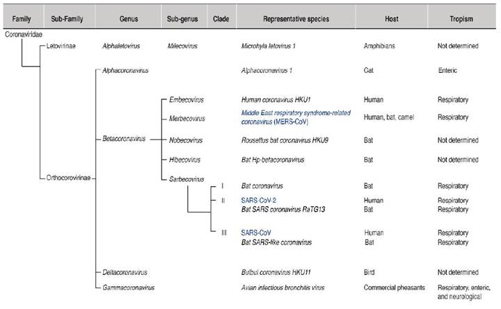
Coronaviruses belong to the order Nidovirales, the family Coronaviridae, the sub-family Orthcoronavirinae, and four genera (Alphacoronavirus, Betacoronavirus, Gammacoronavirus, and Deltacoronavirus) (Lu et al., 2020; Zhu et al., 2020) (Figure 1). Alphacoronaviruses infect mammals. Betacoronaviruses mainly infect bats and humans. Gammacoronaviruses and Deltacoronaviruses infect birds, and some species infect mammals. The genus Betacoronavirus is divided into five sub-genera (Figure 1): Embevorirus, Merbecovirus, Nobecovirus, Hibecovirus, and Sarbecovirus (Lu et al., 2020; Zhu et al., 2020). The sub-genus Sarbecovirus contains species that infect only bats or humans and includes SARS-CoV and SARS-CoV-2. Another human coronavirus, MERS-CoV, belongs to the sub-genus Merbecovirus. The sub-genera Nobecovirus, Hibecovirus are integrated by species that infect bats (Figure 1).

Figure 1 Taxonomic organization of coronaviruses. Representative species are indicated for every genus and sub-genus. Their hosts and tropism are indicated. Important human pathogens are highlighted in blue. 

Coronavirus genome organization

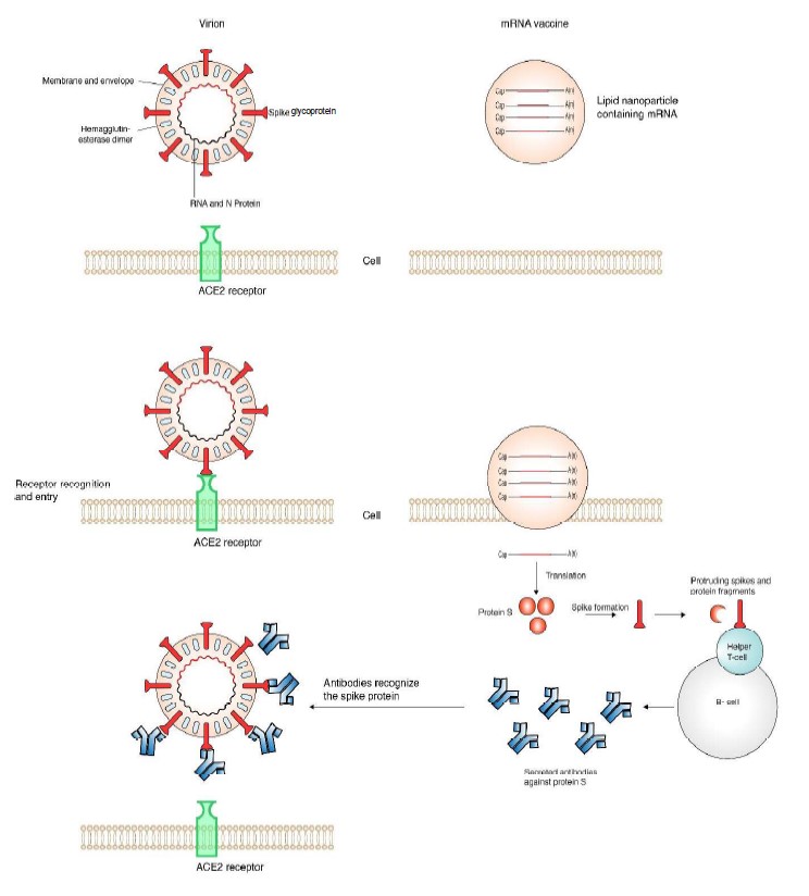
In betacoronaviruses the genome consists of a single RNA, linear, of positive polarity and is approximately 30,000 nt long. The virion is spherical, enveloped, and is 120 nm in diameter (Brian and Baric, 2005; Cui et al., 2019). The genomic RNA is protected by nucleoprotein N in a nucleocapsid. The envelope is formed by the membrane (M) protein and the small membrane protein E. A distinctive feature of the coronavirus virion is the presence of spikes formed by the glycoprotein S (protein S) ( Figure 2) (Lan et al., 2020; Walls et al., 2020; Wrapp et al., 2020a).

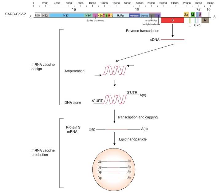
The coronavirus genomic RNA (Figure 3) is capped, polyadenylated and encodes multiple cistrons in open reading frames 1 (ORF1a) and 1b (ORF1b) joined by a ribosomal frameshift. Polyproteins 1a and 1ab are processed by papain-like proteinase NSP3 and 3C-like proteinase NSP5 to form the viral RNA-dependent RNA polymerase and several non-structural proteins necessary for RNA replication. M, E, S and other structural proteins are expressed from subgenomic RNAs co-terminal with the 3’ end, and contain a 5’ leader that is 65 to 89 nt long (Brian and Baric, 2005).

Entry into the cell

Entry into the cell is mediated by protein S spikes on the virion surface that interact with cellular receptors. The process is facilitated by entry cofactors (Gallagher and Buchmeier, 2001; Li, 2013; Cantuti-Castelvetri et al., 2020; Yan et al., 2020). Protein S is divided into S1 and S2 subunits cleaved by cellular proteases and cofactors (Millet and Whittaker, 2015; Cantuti-Castelvetri et al., 2020; Coutard et al., 2020; Xia et al., 2020). The receptor binding domain is located at the tip of the S1 head, and mediates recognition and binding to the surface of the receptor ACE2 (Cai et al., 2020). Interactions between protein S and de cellular receptor are critical for cell entry, and highly specific (Cai et al., 2020). Thus, protein S is a determinant of coronavirus host range (Gallagher and Buchmeier, 2001; Zhai et al., 2020).

antibodies against protein S triggered by natural infection or an mRNA vaccine. 

Figure 3 SARS-CoV-2 genome organization and design of a mRNA vaccine. Coordinates are based on the reference isolate Wuhan-Hu-1 (NC_045512.2). 

The mRNA vaccine

For its critical role in cell entry, the spike S protein is the common target for neutralizing antibodies and vaccines (Brochot et al., 2020; Cai et al., 2020). In people infected with coronavirus, neutralizing antibodies are formed against the entire protein S. However, non-neutralizing antibodies are also developed against the S2 subunit (Brochot et al., 2020; Cai et al., 2020).

Vaccines trigger the formation of neutralizing antibodies against protein S, in the absence of infection. Two mRNA vaccines based on protein S have been authorized. Their design is similar and are based on the genome organization and gene expression of coronaviruses (Figure 3). The cistron coding for protein S was cloned using the sequence from the reference isolate Wuhan-Hu-1 (NC_045512.2). A 5’ UTR, a 3’ UTR, and a poly A tail were added to provide stability and enhance translation efficiency. To account for variation in protein S (Becerra-Flores and Cardozo, 2020) prevalent mutations were introduced, and to further enhance translation efficiency, nucleoside modification were introduced (Pardi et al., 2018). The basic design was made by integrating all basic information previously accumulated from SARS-CoV and MERS (Corbett et al., 2020). For delivery purposes, and to avoid degradation, the mRNA is enclosed in a lipid nanodrop that releases the mRNA into the cell. Ribosomes translate the mRNA into protein S that triggers the formation of neutralizing antibodies (Pardi et al., 2018).

Genome variation

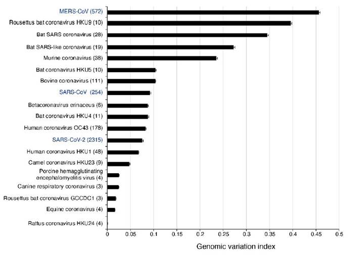
In SARS-CoV-2, mutations have been detected and are being tracked using on-line tools (Hadfield et al., 2018; Fernandes et al., 2020). We recently profiled the genomic variation in all species in the genus Betacoronavirus (LaTourrette et al., 2021). Results showed betacoronaviruses are hyper variable (Figure 4). The most diversity was observed in Rousettus bat coronavirus HKU9, other species infecting bats, and MERS-CoV. In these species, more than 25% of the nucleotides in the genome are polymorphic (Figure 4). The genome of betacoronaviruses consists of 11 to 14 cistrons. The most variable cistron codes for the spike protein S. The lowest variation was detected the cistrons that code for proteins that mediate virus replication: RNA-dependent RNA polymerase, RNA helicase, exonuclease, endo RNAse and methyltransferase, and that are located in open reading frame 1b (LaTourrette et al., 2021).

Figure 4 Genomic variation in betacoronaviruses. A genomic variation index indicates the proportion of single nucleotide et al. (2021). 

Protein S variation

Mutations in the genome of SARS-CoV-2 have the potential to affect the precision of diagnostic tests and the efficacy of vaccines. In a recent genome-wide analysis, we showed that hyper variation in protein S is a general feature of betacoronaviruses (LaTourrette et al., 2021). Hyper variation in protein S is evident in betacoronavirus highly pathogenic to humans: MERS-CoV (Figure 5A), SARS-CoV (Figure 5B), and SARS-CoV-2 (Figure 5C). The pattern is also clear in species that infect bats (Figure 5B). Specifically, in SARS-CoV-2 several regions in protein S are hyper variable, including the ACE2 receptor binding domain and the fusion peptide proximal region (Figure 5D).

Betacoronaviruses mainly infect bats and humans (Figure 1). Given the large genetic diversity of bats, and possibly humans, the cellular receptors, proteases, and entry cofactors are likely diverse (Kuo et al., 2000; Cantuti-Castelvetri et al., 2020). Accordingly, protein S hyper variation may provide an evolutionary advantage. Mechanisms driving diversifying selection in protein S may include diversity in cellular receptors, cellular proteases that process the S1/S2 cleavage site, entry cellular cofactors, and antibodies.

Several domains in protein S are intrinsically disordered (LaTourrette et al., 2021): the receptor binding domain and the C-terminal domain 2 in S1, and the fusion peptide proximal region in S2 (Figure 5D). This observation is important because intrinsically disordered proteins mediate functional diversity and interactions with genetically diverse partners such as cellular receptors and entry cofactor in bats and humans (Hebrard et al., 2009; Rantalainen et al., 2011; Charon et al., 2018). Selection for hyper variation and disorder in protein S are consistent with the bat origin of SARS-CoV and SARS-CoV-2 followed by adaptation to humans (Cui et al., 2019; Lu et al., 2020).

In betacoronaviruses, protein S is hyper variable, disordered, mutationally robust (LaTourrette et al., 2021), and is a determinant of host adaptation and host range (Kuo et al., 2000; Muth et al., 2018; Zhai et al., 2020). The emerging model is that in protein S hyper variation provides an evolutionary advantage and is an intrinsic property of betacoronaviruses (LaTourrette et al., 2021).

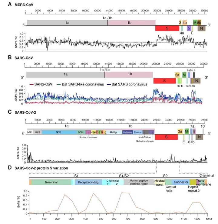
Figure 5 Distribution of genomic variation in human betacoronaviruses. Single nucleotide polymorphisms were plotted with respect to the genome. A 99% confidence interval is indicated as a horizontal line. A) MERS-CoV. B) SARS-CoV. Two species infecting bats are included for comparison. C) SARS-CoV-2. D) Single amino acid polymorphism in SARS-CoV-2 protein S. Domains are annotated and color coded. Modified from LaTourrette et al. (2021). 

Antibodies againts protein S

In infected cells, neutralizing antibodies are developed against protein S (Brochot et al., 2020; Cai et al., 2020). The receptor-binding domain is a critical antigen (Noy-Porat et al., 2020). Additionally, non-neutralizing antibodies against protein S fragments of subunit two are also present (Brochot et al., 2020; Cai et al., 2020). Non-neutralizing antibodies may serve as decoys the reduce biogenesis and targeting efficiency of neutralizing antibodies (Cai et al., 2020). Thus, the hyper variation of protein S might be a mechanism for betacoronaviruses to escape the immune system.

Variation in protein S and implications for vaccine use

Vaccines against SARS-CoV-2 induce neutralizing antibodies against protein S (Figure 2) (Cai et al., 2020; Wrapp et al.; Yuan et al., 2020). Hyper variation in protein S has potential to reduce efficacy of vaccines by multiple mechanisms. In an infected individual, new virus variants might be generated and have been detected (Jary et al., 2020) with potential to escape neutralizing antibodies. Furthermore, antibodies developed after vaccination are a selection agents with potential to favor virus variants that can escape neutralizing antibodies (Baum et al., 2020).

Under this scenario, if SARS-CoV-2 remains genetically stable, vaccines will be efficient (Dearlove et al., 2020), antibody-based diagnostic test highly reliable, and infected people who develop antibodies will likely acquire immunity to SARS-CoV-2. However, if SARS-CoV-2 differentiates into strains, vaccines will be efficient only against closely related strains, ineffective against diverse strains, and people might be repeatedly infected by SARS-CoV-2.

Re-infection in humans has been confirmed (Tillett et al., 2020), and hyper variation in protein S is a general feature of betacoronaviruses (LaTourrette et al., 2021). These observations predict that adjustments to vaccine design and antibody-based diagnostic tests will be needed. Vaccines administered to people may consist of a cocktail of protein S variants (Baum et al., 2020; Cai et al., 2020). Alternatively, or in addition, the vaccines may need to be re-designed based on SARS-CoV-2 population dynamics, structure and their geographic distribution (Korber et al., 2020; Taboada et al., 2020; LaTourrette et al., 2021).

It is likely that SARS-CoV-2 will accumulate mutations for efficient replication and differentiate into biological strains as the virus faces selection pressure from genetically different human populations (LaTourrette et al., 2021). Multiple lines of evidence support this model. Despite not reaching pandemic levels, protein S accumulated large numbers of mutations in MERS-CoV and SARS-CoV (Figure 5). SARS-CoV-2 variants infecting the same individual have been detected (Tillett et al., 2020; Jary et al., 2020), and recurrent mutations in open reading frame 1ab, and cistrons coding for NSP6 and protein S have been identified (van Dorp et al., 2020). Additionally, contrasting mutations have also be described. In Mexico, SARS-CoV-2 population were grouped into clades (Taboada et al., 2020), and in Arizona, a 27-amino acid deletion was detected in protein 7 (Holland et al., 2020).

The first genome of the SARS-CoV-2 came from the strain initially described in China, (Wuhan-Hu-1 NC_045512.2). The cistron coding for protein S contains residues that are compatible, but not optimal, for binding human receptor ACE2 (Wan et al., 2020). Thus, there is potential for protein S to accumulate mutations for more efficient entry into human cells. Consistent with this model, a D614G mutation makes the virus more transmissible, more pathogenic to humans (Becerra-Flores and Cardozo, 2020), and has replaced the initial strain (Long et al., 2020; Volz et al., 2020). The D614G mutation and others in the receptor binding domain reduce affinity to monoclonal antibody CR3022 (Long et al., 2020). This is consistent with a role for protein S variation in escaping from neutralizing antibodies.

Future challenges

Multiple lines of evidence support the model that SARS-CoV-2 is mutating (Forster et al., 2020; Korber et al., 2020; Phan, 2020a), and that as a group betacoronaviruses are hypervariable and variation mainly accumulates in protein S (LaTourrette et al., 2021). This variation has the potential to affect the both the efficacy of vaccines and the reliability of antibody-based diagnostic test. Collectively this information predicts that vaccine design and deployment will be based on fundamental understanding and characterization of proteins S, and other genes, in the SARS-CoV-2 genome in a combination of factors such as human population genetics, age groups, health underlying conditions, geographical and regional boundaries. Characterizing the genetic structure of SARS-CoV-2 at fine scale, and translating this variation into the design and deployment of SARS-CoV-2 vaccines is one of the main challenges. To answer this challenge, it will be essential profile the genetic structure of the virus in different parts of the world, in human populations of different genetic backgrounds, and before and after administration of the SARS-CoV-2 vaccines

Acknowledgments

This research was supported by NIH grant R01GM120108 to HG-R and by the Nebraska Agricultural Experiment Station with funding from the Hatch Act (Accession Number 1007272) through the USDA National Institute of Food and Agriculture.

Literature cited

Amanat F and Krammer F. 2020. SARS-CoV-2 Vaccines: Status Report. Immunity 52(4): 583-589. https://doi.org/10.1016/j.immuni.2020.03.007 [ Links ]

Baum A, Fulton BO, Wloga E, Copin R, Pascal KE, Russo V, Giordano S, Lanza K, Negron N, Ni M, Wei Y, Atwal GS, Murphy AJ, Stahl N, Yancopoulos GD and Kyratsous CA. 2020. Antibody cocktail to SARS-CoV-2 spike protein prevents rapid mutational escape seen with individual antibodies. Science 369(6506): 1014-1018. https://doi.org/10.1126/science.abd0831 [ Links ]

Becerra-Flores M and Cardozo T. 2020. SARS-CoV-2 viral spike G614 mutation exhibits higher case fatality rate. International Journal of Clinical Practice 74(8): e13525. https://doi.org/10.1111/ijcp.13525 [ Links ]

Brian DA and Baric RS. 2005. Coronavirus genome structure and replication. Current Topics and Microbiology Immunology 287: 1-30. https://doi.org/10.1007/3-540-26765-4_1 [ Links ]

Brochot E, Demey B, Touzé A, Belouzard S, Dubuisson J, Schmit J-L, Duverlie G, Francois C, Castelain S and Helle F. 2020. Anti-spike, Anti-nucleocapsid and Neutralizing Antibodies in SARS-CoV-2 Inpatients and Asymptomatic carriers. Frontiers in Microbiology. 24 p. https://doi.org/10.3389/fmicb.2020.584251 [ Links ]

Cagliani R, Forni D, Clerici M and Sironi M. 2020. Computational Inference of Selection Underlying the Evolution of the Novel Coronavirus, Severe Acute Respiratory Syndrome Coronavirus 2. Journal of Virology 94: e00411-00420. https://doi.org/10.1128/JVI.00411-20 [ Links ]

Cai Y, Zhang J, Xiao T, Peng H, Sterling SM, Walsh RM, Rawson S, Rits-Volloc, S and Chen B. 2020. Distinct conformational states of SARS-CoV-2 spike protein. Science 369(6511) 1586-1592. https://doi.org/10.1126/science.abd4251 [ Links ]

Cantuti-Castelvetri L, Ojha R, Pedro LD, Djannatian M, Franz J, Kuivanen S, van der Meer F, Kallio K, Kaya T, Anastasina M, Smura T, Levanov L, Szirovicza L, Tobi A, Kallio-Kokko H, Österlund P, Joensuu M, Meunier FA, Butcher SJ, Winkler MS, Mollenhauer B, Helenius A, Gokce O, Teesalu T, Hepojoki J, Vapalahti O, Stadelmann C, Balistreri G and Simons M. 2020. Neuropilin-1 facilitates SARS-CoV-2 cell entry and infectivity. Science 370(6518): 856-860. https://doi.org/10.1126/science.abd2985 [ Links ]

Charon J, Barra A, Walter J, Millot P, Hebrard E, Moury B and Michon T. 2018. First Experimental Assessment of Protein Intrinsic Disorder Involvement in an RNA Virus Natural Adaptive Process. Molecular Biology and Evolution 35(1): 38-49. https://doi.org/10.1093/molbev/msx249. [ Links ]

Corbett KS, Edwards DK, Leist SR, Abiona OM, Boyoglu-Barnum S, Gillespie RA, Himansu S, Schäfer A, Ziwawo CT, DiPiazza AT, Dinnon KH, Elbashir SM, Shaw CA, Woods A, Fritch EJ, Martinez DR, Bock KW, Minai M, Nagata BM, Hutchinson GB, Wu K, Henry C, Bahl K, Garcia-Dominguez D, Ma L, Renzi I, Kong W-P, Schmidt SD, Wang L, Zhang Y, Phung E, Chang LA, Loomis RJ, Altaras NE, Narayanan E, Metkar M, Presnyak V, Liu C, Louder MK, Shi W, Leung K, Yang ES, West A, Gully KL, Stevens LJ, Wang N, Wrapp D, Doria-Rose NA, Stewart-Jones G, Bennett H, Alvarado GS, Nason MC, Ruckwardt TJ, McLellan JS, Denison MR, Chappell JD, Moore IN, Morabito KM, Mascola JR, Baric RS, Carfi A and Graham BS. 2020. SARS-CoV-2 mRNA vaccine design enabled by prototype pathogen preparedness. Nature 586: 567-571. https://doi.org/10.1038/s41586-020-2622-0 [ Links ]

Coutard B, Valle C, de Lamballerie X, Canard B, Seidah NG and Decroly E. 2020. The spike glycoprotein of the new coronavirus 2019-nCoV contains a furin-like cleavage site absent in CoV of the same clade. Antiviral Research 176: 104742. https://doi.org/10.1016/j.antiviral.2020.104742 [ Links ]

Cui J, Li F and Shi ZL. 2019. Origin and evolution of pathogenic coronaviruses. Nature Reviews Microbiology 17: 181-192. https://www.nature.com/articles/s41579-018-0118-9Links ]

Dearlove B, Lewitus E, Bai H, Li Y, Reeves DB, Joyce MG, Scott PT, Amare MF, Vasan S, Michael NL, Modjarrad K and Rolland M. 2020. A SARS-CoV-2 vaccine candidate would likely match all currently circulating variants. Proceedings of the National Academy of Sciences 117(38): 23652-23662. https://doi.org/10.1073/pnas.2008281117 [ Links ]

Fernandes JD, Hinrichs AS, Clawson H, Gonzalez JN, Lee BT, Nassar LR, Raney BJ, Rosenbloom KR, Nerli S, Rao AA, Schmelter D, Fyfe A, Maulding N, Zweig AS, Lowe TM, Ares M, Corbet-Detig R, Kent WJ, Haussler D and Haeussler M. 2020. The UCSC SARS-CoV-2 Genome Browser. Nature Genetics 52: 991-998. https://www.nature.com/articles/s41588-020-0700-8Links ]

Forster P, Forster L, Renfrew C and Forster M. 2020. Phylogenetic network analysis of SARS-CoV-2 genomes. Proceedings of the National Academy of Sciences 117(17): 9241-9243. https://doi.org/10.1073/pnas.2004999117 [ Links ]

Gallagher TM and Buchmeier MJ. 2001. Coronavirus spike proteins in viral entry and pathogenesis. Virology 279(2): 371-374. https://doi.org/10.1006/viro.2000.0757 [ Links ]

Hadfield J, Megill C, Bell SM, Huddleston J, Potter B, Callender C, Sagulenko P, Bedford T and Neher RA. 2018. Nextstrain: real-time tracking of pathogen evolution. Bioinformatics 34(23): 4121-4123. https://doi.org/10.1093/bioinformatics/bty407 [ Links ]

Hebrard E, Bessin Y, Michon T, Longhi S, Uversky VN, Delalande F, Van Dorsselaer A, Romero P, Walter J, Declerck N and Fargette D. 2009. Intrinsic disorder in Viral Proteins Genome-Linked: experimental and predictive analyses. Virology Journal 6: 23. https://virologyj.biomedcentral.com/articles/10.1186/1743-422X-6-23Links ]

Holland LA, Kaelin EA, Maqsood R, Estifanos B, Wu LI, Varsani A, Halden RU, Hogue BG, Scotch M, and Lim ES. 2020. An 81-Nucleotide Deletion in SARS-CoV-2 ORF7a Identified from Sentinel Surveillance in Arizona (January to March 2020). Journal Virology 94(14): JVI.00711-00720. https://doi.org/10.1128/JVI.00711-20 [ Links ]

Jary A, Leducq V, Malet I, Marot S, Klement-Frutos E, Teyssou E, Soulié C, Abdi B, Wirden M, Pourcher V, Caumes E, Calvez V, Burrel S, Marcelin A-G and Boutolleau D. 2020. Evolution of viral quasispecies during SARS-CoV-2 infection. Clinical Microbiology and Infection 26(11):1560.e1-1560.e4. https://doi.org/10.1016/j.cmi.2020.07.032 [ Links ]

Korber B, Fischer WM, Gnanakaran S, Yoon H, Theiler J, Abfalterer W, Hengartner N, Giorgi EE, Bhattacharya T, Foley B, Hastie KM, Parker MD, Partridge DG, Evans CM, Freeman TM, de Silva TI, Angyal A, Brown RL, Carrilero L, Green LR, Groves DC, Johnson KJ, Keeley AJ, Lindsey BB, Parsons PJ, Raza M, Rowland-Jones S, Smith N, Tucker RM, Wang D, Wyles MD, McDanal C, Perez LG, Tang H, Moon-Walker A, Whelan SP, LaBranche CC, Saphire EO and Montefiori DC. 2020. Tracking Changes in SARS-CoV-2 Spike: Evidence that D614G Increases Infectivity of the COVID-19 Virus. Cell 182(4): 812-827.e819. https://doi.org/10.1016/j.cell.2020.06.043 [ Links ]

Kuo L, Godeke GJ, Raamsman MJ, Masters PS and Rottier PJ. 2000. Retargeting of coronavirus by substitution of the spike glycoprotein ectodomain: crossing the host cell species barrier. Journal Virology 74(3): 1393-1406. https://doi.org/10.1128/jvi.74.3.1393-1406.2000 [ Links ]

Lan J, Ge J, Yu J, Shan S, Zhou H, Fan S, Zhang Q, Shi X, Wang Q, Zhang L and Wang X. 2020. Structure of the SARS-CoV-2 spike receptor-binding domain bound to the ACE2 receptor. Nature 581: 215-220. https://www.nature.com/articles/s41586-020-2180-5Links ]

LaTourrette K, Holste NM, Rodriguez-Peña R, Arruda-Leme R and García-Ruiz H. 2021. Genome-wide variation in betacoronaviruses. Journal Virology Submitted. [ Links ]

Lauring AS and Andino R. 2010. Quasispecies theory and the behavior of RNA viruses. PLoS Pathog 6: e1001005. https://doi.org/10.1371/journal.ppat.1001005 [ Links ]

Li CK-f, Wu H, Yan H, Ma S, Wang L, Zhang M, Tang X, Temperton NJ, Weiss RA, Brenchley JM, Douek DC, Mongkolsapaya J, Tran B-H, Lin C-lS, Screaton GR, Hou J-l, McMichael AJ and Xu X-N. 2008. T cell responses to whole SARS coronavirus in humans. The Journal of Immunology 181(8): 5490-5500. https://doi.org/10.4049/jimmunol.181.8.5490 [ Links ]

Li F. 2013. Receptor recognition and cross-species infections of SARS coronavirus. Antiviral Research 100(1): 246-254. https://doi.org/10.1016/j.antiviral.2013.08.014 [ Links ]

Li F. 2016. Structure, Function, and Evolution of Coronavirus Spike Proteins. Annual Review of Virology 3: 237-261. https://doi.org/10.1146/annurev-virology-110615-042301 [ Links ]

Long SW, Olsen RJ, Christensen PA, Bernard DW, Davis JJ, Shukla M, Nguyen M, Saavedra MO, Yerramilli P, Pruitt L, Subedi S, Kuo H-C, Hendrickson H, Eskandari G, Nguyen HAT, Long JH, Kumaraswami M, Goike J, Boutz D, Gollihar J, McLellan JS, Chou C-W, Javanmardi K, Finkelstein IJ and Musser J. 2020. Molecular Architecture of Early Dissemination and Massive Second Wave of the SARS-CoV-2 Virus in a Major Metropolitan Area. mBio 2020.2009.2022.20199125. https://doi.org/10.1128/mBio.02707-20 [ Links ]

Lu R, Zhao X, Li J, Niu P, Yang B, Wu H, Wang W, Song H, Huang B, Zhu N, Bi Y, Ma X, Zhan F, Wang L, Hu T, Zhou H, Hu Z, Zhou W, Zhao L, Chen J, Meng Y, Wang J, Lin Y, Yuan J, Xie Z, Ma J, Liu WJ, Wang D, Xu W, Holmes EC, Gao GF, Wu G, Chen W, Shi W and Tan W. 2020. Genomic characterisation and epidemiology of 2019 novel coronavirus: implications for virus origins and receptor binding. The Lancet 395(10224): 565-574. https://doi.org/10.1016/S0140-6736(20)30251-8 [ Links ]

Millet JK, and Whittaker GR. 2015. Host cell proteases: Critical determinants of coronavirus tropism and pathogenesis. Virus Research 202: 120-134. https://doi.org/10.1016/j.virusres.2014.11.021 [ Links ]

Muth D, Corman VM, Roth H, Binger T, Dijkman R, Gottula LT, Gloza-Rausch F, Balboni A, Battilani M, Rihtaric D, Toplak I, Ameneiros RS, Pfeifer A, Thiel V, Drexler JF, Muller MA and Drosten C. 2018. Attenuation of replication by a 29 nucleotide deletion in SARS-coronavirus acquired during the early stages of human-to-human transmission. Scientific Report 8: 15177. https://doi.org/10.1038/s41598-018-33487-8 [ Links ]

Nigam D and Garcia-Ruiz H. 2020. Variation Profile of the Orthotospovirus Genome. Pathogens 9(7): 521. https://doi.org/10.3390/pathogens9070521 [ Links ]

Noy-Porat T, Makdasi E, Alcalay R, Mechaly A, Levy Y, Bercovich-Kinori A, Zauberman A, Tamir H, Yahalom-Ronen Y, Israeli Ma, Epstein E, Achdout H, Melamed S, Chitlaru T, Weiss S, Peretz E, Rosen O, Paran N, Yitzhaki S, Shapira SC, Israely T, Mazor O and Rosenfeld R. 2020. A panel of human neutralizing mAbs targeting SARS-CoV-2 spike at multiple epitopes. Nature Communications 11: 4303. https://www.nature.com/articles/s41467-020-18159-4Links ]

Obenauer JC, Denson J, Mehta PK, Su X, Mukatira S, Finkelstein DB, Xu X, Wang J, Ma J, Fan Y, Rakestraw KM, Webster RG, Hoffmann E, Krauss S, Zheng J, Zhang Z and Naeve CW. 2006. Large-Scale Sequence Analysis of Avian Influenza Isolates. Science 311(5767): 1576-1580. https://doi.org/10.1126/science.1121586 [ Links ]

Pardi N, Hogan MJ, Porter FW and Weissman D. 2018. mRNA vaccines - a new era in vaccinology. Nature Reviews Drug Discovery 17: 261-279. https://www.nature.com/articles/nrd.2017.243Links ]

Phan T. 2020a. Genetic diversity and evolution of SARS-CoV-2. Infection, Genetics and Evolution 81: 104260. https://doi.org/10.1016/j.meegid.2020.104260. [ Links ]

Phan T. 2020b. Novel coronavirus: From discovery to clinical diagnostics. Infection, Genetics and Evolution 79: 104211. https://doi.org/10.1016/j.meegid.2020.104211 [ Links ]

Rantalainen KI, Eskelin K, Tompa P, and Mäkinen K. 2011. Structural flexibility allows the functional diversity of potyvirus genome-linked protein VPg. Journal of virology 85: 2449-2457. https://jvi.asm.org/content/85/5/2449Links ]

Sanjuán R and Domingo-Calap P. 2016. Mechanisms of viral mutation. Cellular and Molecular Life Sciences 73(23): 4433-4448. https://doi.org/10.1007/s00018-016-2299-6 [ Links ]

Taboada B, Vazquez-Perez JA, Muñoz-Medina JE, Ramos-Cervantes P, Escalera-Zamudio M, Boukadida C, Sanchez-Flores A, Isa P, Mendieta-Condado E, Martínez-Orozco JA, Becerril-Vargas E, Salas-Hernández J, Grande R, González-Torres C, Gaytán-Cervantes FJ, Vazquez G, Pulido F, Araiza-Rodríguez A, Garcés-Ayala F, González-Bonilla CR, Grajales-Muñiz C, Borja-Aburto VH, Barrera-Badillo G, López S, Hernández-Rivas L, Perez-Padilla R, López-Martínez I, Ávila-Ríos S, Ruiz-Palacios G, Ramírez-González JE and Arias CF. 2020. Genomic Analysis of Early SARS-CoV-2 Variants Introduced in Mexico. Journal of Virology 94(1): e01056-01020. https://doi.org/10.1128/JVI.01056-20 [ Links ]

Tillett RL, Sevinsky JR, Hartley PD, Kerwin H, Crawford N, Gorzalski A, Laverdure C, Verma SC, Rossetto CC, Jackson D, Farrell MJ, Van Hooser S and Pandori M. 2020. Genomic evidence for reinfection with SARS-CoV-2: a case study. The Lancet Infectious Diseases 21(1): 52-58. https://doi.org/10.1016/S1473-3099(20)30764-7 [ Links ]

van Dorp L, Acman M, Richard D, Shaw LP, Ford CE, Ormond L, Owen CJ, Pang J, Tan CCS, Boshier FAT, Ortiz AT and Balloux F. 2020. Emergence of genomic diversity and recurrent mutations in SARS-CoV-2. Infection, Genetics and Evolution 83: 104351. https://doi.org/10.1016/j.meegid.2020.104351 [ Links ]

Volz EM, Hill V, McCrone JT, Price A, Jorgensen D, Toole A, Southgate JA, Johnson R, Jackson B, Nascimento FF, Rey SM, Nicholls SM, Colquhoun RM, da Silva Filipe A, Shepherd JG, Pascall DJ, Shah R, Jesudason N, Li K, Jarrett R, Pacchiarini N, Bull M, Geidelberg L, Siveroni I, Goodfellow IG, Loman NJ, Pybus O, Robertson DL, Thomson EC, Rambaut A and Connor TR. 2020. Evaluating the effects of SARS-CoV-2 Spike mutation D614G on transmissibility and pathogenicity 184(1): 64-75. https://doi.org/10.1016/j.cell.2020.11.020 [ Links ]

Walls AC, Park Y-J, Tortorici MA, Wall A, McGuire AT and Veesler D. 2020. Structure, Function, and Antigenicity of the SARS-CoV-2 Spike Glycoprotein. Cell 181(2): 281-292.e6. https://doi.org/10.1016/j.cell.2020.02.058 [ Links ]

Wan Y, Shang J, Graham R, Baric RS and Li F. 2020. Receptor Recognition by the Novel Coronavirus from Wuhan: an Analysis Based on Decade-Long Structural Studies of SARS Coronavirus. Journal of Virology 94(7): e00127-00120. https://doi.org/10.1128/JVI.00127-20 [ Links ]

Wrapp D, Wang N, Corbett KS, Goldsmith JA, Hsieh CL, Abiona O, Graham BS and McLellan JS. 2020a. Cryo-EM structure of the 2019-nCoV spike in the prefusion conformation. Science 367(6483): 1260-1263. https://doi.org/10.1126/science.abb2507 [ Links ]

Wrapp D, De Vlieger D, Corbett KS, Torres GM, Wang N, Van Breedam W, Roose K, van Schie L, Team V-CC-R, Hoffmann M, Pohlmann S, Graham BS, Callewaert N, Schepens B, Saelens X and McLellan JS. 2020b. Structural Basis for Potent Neutralization of Betacoronaviruses by Single-Domain Camelid Antibodies. Cell 181(5): 1004-1015.e15. https://doi.org/10.1016/j.cell.2020.04.031 [ Links ]

Xia S, Liu M, Wang C, Xu W, Lan Q, Feng S, Qi F, Bao L, Du L, Liu S, Qin C, Sun F, Shi Z, Zhu Y, Jiang S and Lu L. 2020. Inhibition of SARS-CoV-2 (previously 2019-nCoV) infection by a highly potent pan-coronavirus fusion inhibitor targeting its spike protein that harbors a high capacity to mediate membrane fusion. Cell Research 30: 343-355. https://www.nature.com/articles/s41422-020-0305-xLinks ]

Yan R, Zhang Y, Li Y, Xia L, Guo Y and Zhou Q. 2020. Structural basis for the recognition of SARS-CoV-2 by full-length human ACE2. Science 367(6485): 1444-1448. https://doi.org/10.1126/science.abb2762 [ Links ]

Yuan M, Wu NC, Zhu X, Lee C-CD, So RTY, Lv H, Mok CKP, and Wilson IA. 2020. A highly conserved cryptic epitope in the receptor binding domains of SARS-CoV-2 and SARS-CoV. Science 368(6491): 630-633. https://doi.org/10.1126/science.abb7269 [ Links ]

Zhai X, Sun J, Yan Z, Zhang J, Zhao J, Zhao Z, Gao Q, He W-T, Veit M and Su S. 2020. Comparison of Severe Acute Respiratory Syndrome Coronavirus 2 Spike Protein Binding to ACE2 Receptors from Human, Pets, Farm Animals, and Putative Intermediate Hosts. Journal of Virology 94: e00831-00820. https://doi.org/10.1128/JVI.00831-20 [ Links ]

Zhou H, Chen X, Hu T, Li J, Song H, Liu Y, Wang P, Liu D, Yang J, Holmes EC, Hughes AC, Bi Y and Shi W. 2020. A Novel Bat Coronavirus Closely Related to SARS-CoV-2 Contains Natural Insertions at the S1/S2 Cleavage Site of the Spike Protein. Current Biology 30(11): 2196-2203 e2193. https://doi.org/10.1016/j.cub.2020.05.023 [ Links ]

Zhu N, Zhang D, Wang W, Li X, Yang B, Song J, Zhao X, Huang B, Shi W, Lu R, Niu P, Zhan F, Ma X, Wang D, Xu W, Wu G, Gao GF, Tan W, China Novel Coronavirus I and Research T. 2020. A Novel Coronavirus from Patients with Pneumonia in China, 2019. The New England Journal of Medicine 382: 727-733. https://doi.org/10.1056/NEJMoa2001017 [ Links ]

Zhu Z, Chakraborti S, He Y, Roberts A, Sheahan T, Xiao X, Hensley LE, Prabakaran P, Rockx B, Sidorov IA, Corti D, Vogel L, Feng Y, Kim JO, Wang LF, Baric R, Lanzavecchia A, Curtis KM, Nabel GJ, Subbarao K, Jiang S and Dimitrov DS. 2007. Potent cross-reactive neutralization of SARS coronavirus isolates by human monoclonal antibodies. Proceeding of the National Academy of Sciences of United States of America 104(29): 12123-12128. https://doi.org/10.1073/pnas.0701000104 [ Links ]

Received: December 21, 2020; Accepted: March 01, 2021

*Corresponding author: hgarciaruiz2@unl.edu

Creative Commons License This is an open-access article distributed under the terms of the Creative Commons Attribution License