


Services on Demand
Journal
Article
 text in
  English (pdf)
 Article in xml format
 Article references
 Send this article by e-mail
Indicators
-
 Cited by SciELO
 -
 Access statistics
Related links
-
 Similars in
SciELO
Similars in
SciELO
Share

 Permalink
Veterinaria México OA
On-line version ISSN 2448-6760
Veterinaria México OA vol.2 n.2 Ciudad de México Apr./Jun. 2015
Original research
Stability of the B. abortus S19 vaccine strain with a eukaryotic expression plasmid encoding the G glycoprotein from the rabies virus
Nidia G. Pazos Salazara, b* 0000-0002-6451-1569, Juan C. Benítez Serranob 0000-0002-2338-9763, José L. Calderón Chamorrob 0000-0001-6766-0570, Rigoberto Hernández-Castroc 0000-0002-5656-0942, Efrén Díaz Apariciod 0000-0002-1669-1323, José A. Aguilar Setiéne 0000-0003-1339-2931
a Posgrado en Ciencias de la Salud y Producción Animal Facultad de Medicina Veterinaria y Zootecnia Universidad Nacional Autónoma de México Av. Universidad 3000, 04510, DF, México.
b Departamento de Microbiología Facultad de Ciencias Químicas Benemérita Universidad Autónoma de Puebla Av. San Claudio y 18 Sur, San Manuel, 72570 Puebla, México. *Corresponding author: Nidia G. Pazos Salazar. Tel: + 52 22-2229-5500 ext. 7379, 7390 Fax: + 52 22-2244-3106.
c Departamento de Ecología de Agentes Patógenos Hospital General "Dr. Manuel Gea González" Secretaría de Salud Tlalpan, 14080, DF, México.
d Centro Nacional de Investigación Disciplinaria en Microbiología Instituto Nacional de Investigaciones Forestales, Agrícolas y Pecuarias Carretera Federal México-Toluca km 15.5, Cuajimalpa, 05110, DF, México.
e Hospital de Pediatría Unidad de Investigación Médica en Inmunología Coordinación de Investigación Médica Instituto Mexicano del Seguro Social Centro Médico Nacional Siglo XXI 06720, DF, México.
Received: 2015-03-12.
Accepted: 2015-06-29.
Published: 2015-06-30.
Abstract
Brucella abortus S19 is an intracellular vaccine strain against bovine brucellosis. Rabies is a lethal disease in cattle. Plasmids encoding the G glycoprotein from the rabies virus induce a protective immune response in different animal species. A vector called pBBR4-CMV-Ggp-SV40+, which encodes the G gene, regulated by the cytomegalovirus eukaryotic expression promoter, and which can be used to transform the B. abortus S19 vaccine strain, was constructed. The stability of the transformant strain was tested both in vitro and in vivo. In the in vitro assays, B. abortus S19 pBBR4-CMV-Ggp-SV40+ was grown for 5 sequential passages, and for the in vivo assays, female BALB/c mice were infected. Colony-forming unit counting and plasmid identification were performed in each passage and in the spleens at 7 days post-infection. To test the plasmid stability in the strain, all parameters were determined with and without antibiotic. The bacterial concentration was lower with antibiotic than without it, but the bacterial growth was more homogeneous. The plasmid was identified in antibiotic- and non-antibiotic-treated isolated colonies under both in vitro and in vivo conditions. The plasmid construct was also transfected into BHK-21 cells, which express the G glycoprotein. The B. abortus S19 pBBR4-CMV-Ggp-SV40+ strain showed stability, representing a suitable candidate vector for developing a bivalent vaccine against brucellosis and rabies. This is the first time that a Brucella species has been transformed with a eukaryotic expression plasmid.
Keywords: G glycoprotein; Rabies virus; Brucella abortus S19; CMV promoter.
Introduction
Rabies and brucellosis are considered as major zoonotic diseases. These diseases affect cattle, constitute an animal health problem, and have repercussions for human health (Monath et al., 2013).
Bovine brucellosis (BB) is mainly caused by the intracellular bacterium Brucella abortus (Díaz, 2013). There are different vaccine strains for BB prevention. B. abortus RB51 is a rough strain that lacks the O-chain in its lipopolysaccharide (LPS), and for this reason, it does not induce detectable antibodies in diagnostic tests, which helps to differentiate vaccinated from infected animals (Schurig et al., 2002). However, this vaccine strain's level of protection is still controversial. B. abortus S19 is an attenuated smooth strain due to the presence of LPS with the O-chain, which induces antibodies that are detectable in serological assays. This feature is a major problem associated with vaccination with the S19 strain; there is also a 2-3% risk of abortion. Nevertheless, the strain forms a highly immunogenic vaccine that has proven to be the most effective vaccine against BB (Nicoletti, 1990). The vaccine's attenuation capability has been associated with a deletion in the ery operon that is detected by PCR (Sangari et al., 1994), amplifying a 361-bp fragment that distinguishes the B. abortus S19 strain from other B. abortus vaccines and field strains, in which a 1063-bp amplicon has been identified (Mukherjee et al., 2005). In Mexico, 3- to 6-month-old calves are vaccinated with the S19 vaccine at a classical dose of 1010 colony-forming units per mL (CFU/mL) of reconstituted vaccine (equivalent to 5 mL), whereas heifers older than 6 months are administered a reduced dose (3 x 10⁸ CFU/mL).
The etiologic agent of bovine paralytic rabies (BPR) is an RNA-enveloped virus of the Lyssavirus genus, which is divided into at least 11 genotypes (Rupprecht and Plotkin, 2013). Genotype 1 is distributed worldwide and includes the classical rabies virus; other genotypes are limited to specific geographic regions. On the American continent, only genotype 1 exists. The main transmitter of the virus in cattle is the hematophagous bat (Desmodus rotundus), which inhabits tropical and subtropical areas (Lee et al., 2006; Johnson et al., 2014). The rabies virus contains the G glycoprotein (Ggp), an envelope protein that has been recognized as the main viral antigen because it induces protective neutralizing antibodies (Cox et al., 1977; Ross et al., 2008). The Ggp is the only outer protein of the virus, and it contains 524 amino acids, which form several antigenic sites. Antigenic site I is formed by both linear and conformational epitopes, antigenic site II is discontinuous, and antigenic site III is a continuous conformational antigenic epitope. Another important site is referred to as G5: this linear epitope includes antigenic site IV, described as containing only one amino acid (Kuzmina et al., 2013). DNA vaccine plasmids encoding the Ggp are useful for inducing an anti-rabies immune response (Perrin et al., 2000). The pGQH plasmid contains a G gene directed by the cytomegalo-virus (CMV) eukaryotic expression promoter, which produces a high antibody titer, protects against rabies in mice and dogs (Tesoro et al., 2006), and is also efficient as post-exposure prophylaxis in rabbits and mice (Tesoro et al., 2008).
Because the main measure of control for both diseases is vaccination, alternatives to facilitate vaccine management are currently being studied. Several issues have been considered in the development of a new option:
1. Intracellular attenuated bacteria are useful as vectors to deliver DNA plasmids directly to professional antigen-presenting cells (APC), such as macrophages and dendritic cells (Dietrich et al., 2001). B. abortus is an intracellular bacterium, and virulent strains are able to survive inside macrophages. The B. abortus S19 vaccine strain is destroyed by phagocytic cells (Arenas et al., 2000; Pizarro-Cerdá et al., 1998), but it possesses the characteristics of a bacterial vector, and thus, B. abortus S19 offers the possibility of delivering a plasmid to APCs after intracellular disintegration of the strain.
2. DNA vaccine plasmids encode an antigen controlled by a eukaryotic promoter, such as the pGQH plasmid described before. B. abortus can be stably transformed with prokaryotic plasmids of the pBBR1MCS family (Elzer et al., 1995), but it has never been transformed with a eukaryotic expression plasmid.
Therefore, the objectives of this study were as follows: (1) to construct a plasmid encoding the Ggp from the rabies virus regulated by the CMV promoter and to use it to transform the B. abortus S19 vaccine strain and (2) to evaluate the in vitro and in vivo stability of the transformant strain and to assess the protein expression resulting from the plasmid construct. If these goals were achieved, the initial stage of the development of a bivalent vaccine against rabies and brucellosis would be accomplished.
Materials and Methods
Plasmids
The pGQH plasmid described by Tesoro et al. (2008) contains a G gene (1576-bp) flanked by the XbaI site. The gene used in the present study was obtained from the brain of a person who died from rabies transmitted by a hematophagous bat (isolate HQ01-IMSS). Plasmid pBBR1MCS-4 is a moderate-copy plasmid maintained as an extra-chromosomal element. This plasmid is a member of the pBBR1MCS plasmid family but differs based on marker selection: pBBR1MCS is cmr, and pB-BR1MCS-4 is ampr (Kovach et al., 1995).
Bacterial strains and culture conditions
The B. abortus S19 strain and Escherichia coli TOP-10 (F-mcrAΔ (mrr-hsdRMS-mcrBC)  80lacZΔM15 ΔlacX74 nupG recA 1 araD139Δ (ara-leu) 7697 galE15 galK16 rpsL (Str) endA1 λ-) were grown in tryptic soy broth (TSB) at 37°C with orbital shaking or in tryptic soy agar (TSA). Medium containing 100 μg/mL ampicillin (Amp) was used when needed.
Plasmid construction
Plasmid pGQH was digested with the BamHI y Bg/II restriction enzymes, with compatible site ends. The fragment containing the G gene regulated by the CMV promoter was excised and ligated into pBBR1MCS-4, which was previously digested with BamHI (Fig. 1). The insert to be cloned also included a region referred to as the SV40 enhancer/promoter. This region, which contains the origin of replication for eukaryotic cells and which induces episomal plasmid replication, was incorporated into the insert to keep the plasmid present in the host.
The resulting DNA construct was transformed in E. coli TOP-10. The new plas-mid construct was characterized by enzyme restriction: BamHI linearized it, and XbaI released the G gene. The recombinant plasmid was called pBBR4-CMV-Ggp-SV40+ and was purified using the EndoFree Plasmid Giga Kit (Qiagen, Hilden, Germany) following the manufacturer's instructions. The G gene was sequenced from the pure plasmid using the outer primers Ggp1 and Ggp2 as well as the inner primers Gli1 and Gli2. For the designed inner primers, the amplified fragment, which was 736-bp in size, included most of the glycoprotein's antigenic sites.
Transformation of B. abortus S19
Plasmid pBBR4-CMV-Ggp-SV40+ was electroporated into the B. abortus S19 strain by applying a 2.5-Kv pulse and using 3 μg of plasmid. Amp-resistant (Ampr) colonies were analyzed by PCR using pBBR primers (5'-GTAAAACGACGGCCAGT-3' and 5'-CGAGGTCGACGGTATCG-3') to amplify a 5100-bp fragment, corresponding to the insert ligated in pBBR1MCS-4. The amplification protocol was as follows: denaturation at 96°C for 1 min, 35 cycles of denaturation at 95°C for 30 sec, annealing at 64°C for 45 sec, extension at 72°C for 5 min, and a final incubation at 72°C for 10 min. The same colonies were analyzed using the ery primers described by Sangari et al. (1994) and Mukherjee et al. (2005) to identify the 361-bp amplicon of the deleted ery operon as an attenuation genetic marker. The following reaction controls were included: the B. abortus RB51 strain for the non-deleted ery operon and E. coli harboring pBBR4-CMV-Ggp-SV40+ as the negative ery control and the positive plasmid control, respectively. The transformant strain was called B. abortus S19 pBBR4-CMV-Ggp-SV40+.
In vitro stability of plasmid pBBR4-CMV-Ggp-SV40+
The stability of B. abortus S19 pBBR4-CMV-Ggp-SV40+ was evaluated following the method referenced by Elzer et al. (1995). The strain was grown in TSB-Amp for 48 h. A total of 100 μL of the culture was transferred to 5 mL of either TSB or TSB-Amp and incubated for 48 h to obtain passage 1. The process was successively repeated until passage 5. Serial dilutions of each passage were performed to determine the CFU/mL in triplicate under 3 conditions: 1) bacteria cultivated in TSB and isolated in TSA (TSB/TSA); 2) bacteria cultivated in TSB and isolated in TSA-Amp (TSB/TSA-Amp); and 3) control culture, consisting of bacteria cultivated in TSB-Amp and isolated in TSA-Amp (TSB-Amp/TSA-Amp). Randomly sampled colonies from each condition were tested by PCR to identify the 5100-bp amplicon from pBBR4-CMV-Ggp-SV40+.
Inoculation of BALB/c mice with the strain B. abortus S19 pBBR4-CMV-Ggp-SV40+
Three 6- to 8-week-old female BALB/c mice were inoculated intraperitoneally (i.p.) with 0.25 mL of PBS containing ~106 CFU of B. abortus S19 pBBR4-CMV-Ggp-SV40+ or 0.25 mL of PBS only (control group). The animals were sacrificed by cervical dislocation 7 days later. Their spleens were weighed and homogenized in 1 mL of PBS and plated on either TSA or TSA-Amp, and the CFU/spleen value was determined by serial dilution. A multiplexed PCR was applied to the spleen colonies to simultaneously identify the deleted ery operon and the G gene. In the case of the rabies protein, the following inner primers were used: Gli1, forward 5'-ACA-CAATCCGTACCCTGACT-3', and Gli2, reverse 5'-CCCGTTTACATGAGGATGAC-3'. Both amplicons of the G gene (736-bp) and of the ery operon deletion (361-bp) were obtained as follows: denaturation at 96°C for 1 min, 30 cycles of denaturation at 95°C for 30 sec, annealing at 64°C for 45 sec, extension at 72°C for 1 min, and a final incubation at 72°C for 10 min.
All experimental procedures and animal care were performed in compliance with the Guide for the care and use of laboratory animals (2011), NOM-062-ZOO-1999, which enforces the proper use and care of laboratory animals in Mexico, and with the approval of Benemérita Universidad Autónoma de Puebla (license BCB/CCUAL/118/2013).
Glycoprotein expression in eukaryotic cells
BHK-21 cells were cultured in Eagle's minimal essential medium (MEM) containing 10% fetal bovine serum (FBS) during the growth phase. Cells in the logarithmic growth phase were harvested, and 1 x 10⁶ cells were transfected by elec-troporation with 15 μg of the pBBR4-CMV-Ggp-SV40+ plasmid. A pulse of 140 V for 25 msec was applied to a 0.2-cm cell containing 0.1 mL of cellular suspension. Protein expression was monitored at 24, 48 and 72 h post-transfection; a control of non-transfected cells was also included. A polyclonal human IgG containing 150 IU/mL anti-rabies neutralizing antibodies was employed as a primary antibody at a 1:50 dilution. The protein was revealed with a 1:100 dilution of goat anti-human IgG-FITC.
Statistical analysis
The CFU data were log-transformed and after examining the residuals, the variances appeared homogeneous within the range of the treatment means. The data from the in vitro assays were analyzed by one-way ANO-VA with Dunnett's post-test, and the bacterial loads in the in vivo assays (CFU/spleen) were analyzed with Student's t-test using the software associated with GraphPad Prism 6 (GraphPad Software Inc. La Jolla, CA, USA). A value of P<0.05 was considered significant for both tests.
Results
Construction of the recombinant plasmid and transformation of the B. abortus S19 strain with plasmid pBBR4-CMV-Ggp-SV40+
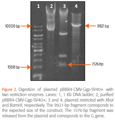
After extraction, plasmid pBBR4-CMV-Ggp-SV40+ was digested with BamHI and XbaI to verify the size of the construct and to release the G gene, respectively. Digestion with XbaI released 3 fragments of 6057-, 2283-, and 1576-bp in size. The last one corresponded to the G gene (Fig. 2). Digestion with BamHI produced a 9921-bp linearized plasmid, which indicated loss of the BglII site (Fig. 2). The complete sequence of the G gene was obtained, and preliminary results showed that Mexican isolate HQ01-IMSS maintained the amino acids considered to be invariable in all of the Ggp's antigenic sites. Figure 3 shows the deletion of the ery operon and the presence of the recombinant plasmid in the B. abortus S19 strain after electroporation.
Plasmid pBBR4-CMV-Ggp-SV40+was stable under in vitro conditions in the B. abortus S19 strain
The bacterial concentrations in each medium (Log10 CFU/mL) were as follows:
• Control culture (TSB-Amp/TSA-Amp) = 8.54
• TSB/TSA-Amp culture = 8.44
• TSB/TSA culture = 9.13.
The bacterial concentration was higher (P<0.05) in the TSB/TSA culture than under the other two culture conditions (Fig. 4a). Whereas no differences in the bacterial concentration were observed among passages in the TSB-Amp/TSA-Amp culture (P>0.05), variability was observed in both the TSB/TSA and the TSB/TSA-Amp cultures (Fig. 4b). In particular, in the TSB/TSA culture, the bacterial concentration showed a statistically significant difference (P<0.05) in passages 2, 3 and 5 relative to passage one, and in the TSB/ TSA-Amp culture, a statistically significant difference (P<0.05) was found in the bacterial concentration in passages 2 and 5 relative to passage one. In addition, plasmid pBBR4-CMV-Ggp-SV40+ was identified in all TSA-Amp colonies and in different TSA colonies (Fig. 4c).
Plasmid stability in the B. abortus S19 pBBR4-CMV-Ggp-SV40+ strain was maintained during BALB/c mouse infection
Table 1 shows the results from the spleen homogenates. No statistically significant difference was found between the bacterial concentrations in antibiotic- and non-antibiotic-complemented media. All analyzed colonies from both media were positive for the deleted ery operon. All analyzed colonies isolated in the presence of the antibiotic were positive for the G gene, and only one colony isolated in the absence of the antibiotic was positive for the G gene (Fig. 5). The plasmid was extracted from all G gene-positive colonies (data not shown).
PlasmidpBBR4-CMV-Ggp-SV40+ induced the expression of the Ggp from the rabies virus in eukaryotic cells
After BHK-21 transfection, the Ggp was detected at 24, 48 and 72 h. The glycoprotein was not detected in the control cells. Figure 6 shows protein expression at 48 h (expression at 24 and 72 h is not shown). This result indicates that the viral protein was properly expressed in eukaryotic cells with the pBBR4-CMV-Ggp-SV40+ plasmid.
Discussion
Vaccine plasmids consist of a gene of interest controlled by a strong eukaryotic expression promoter; the mRNA transcripts are stabilized by a polyadenylation sequence, and resistance genes for bacterial selection are included (Gurunathan et al, 2000). We have constructed plasmid pBBR4-CMV-Ggp-SV40+, which encodes the most immunogenic protein from the rabies virus, or the Ggp, which is ligated to the CMV promoter to strengthen gene expression in eukaryotic cells. A polyadenylation sequence and the SV40 origin of replication were also included to maintain plasmid replication in the host. The plasmid construct is based on pBBR1MCS-4, a plasmid used to transform Brucella species (Kovach et al., 1995). The G gene sequence (isolate HQ01-IMSS) is 1575-bp in size and codes for a 524 aminoacid protein containing the sequences for antigenic sites.
Employment of intracellular attenuated bacteria improves the delivery of vaccine plasmids to a host (Dietrich et al., 2001). The B. abortus S19 vaccine strain is both intracellular and attenuated, and it was transformed with the plasmid construct with the purpose of generating B. abortus S19 pBBR4-CMV-Ggp-SV40+, which maintained the marker of attenuation, or the ery operon deletion. The B. abortus S19 strain has already been transformed with plasmids encoding heterologous proteins (Comerci et al., 1998; Sabio y García et al., 2008; Sabio y García et al., 2010), but in all cases, protein expression depended on a prokaryotic promoter. This is the first report of transformation of B. abortus involving a eukaryotic expression promoter.
To evaluate the stability of the plasmid construct that encodes the G gene, in vitro and in vivo assays were performed. The strategy of culturing bacteria by successive passaging in media with or without a selection factor was previously established when evaluating the stability of plasmid pBBR1MCS in different Brucella species (Elzer et al., 1995). The in vitro stability assays in the current study showed a significantly lower concentration of B. abortus S19 pBBR4-CMV-Ggp-SV40+ in the antibiotic-complemented medium than in the medium without antibiotic, although the bacterial growth in the antibiotic-complemented cultures was homogeneous. The plasmid that we constructed is 9921-bp in size, so it might be too heavy to allow transformed bacteria to replicate. However, the variability observed in the growth of the transformant strain as well as the identification of the plasmid when these bacteria were cultured in medium without antibiotic may indicate that the plasmid confers the ability to resist adverse circumstances, although the plasmid is not essential for viability because Brucella does not naturally contain it (Crasta et al., 2008). Therefore, without selective pressure, a spontaneous loss of plasmid could occur in part of the population.
The results of the in vivo assays of the concentration of transformed bacteria recovered from the spleen homogenates were similar to those reported elsewhere (Comerci et al., 1998, Sabio y García et al., 2008). The fact that the multiplexed PCR used was able to identify the plasmid and the ery operon deletion in colonies with or without antibiotic may indicate that this is a useful diagnostic tool to differentiate between the B. abortus S19 pBBR4-CMV-Ggp-SV40+ strain and field strains and to help to differentiate infected animals from animals vaccinated with the B. abortus S19 strain (Pacheco et al., 2012). These results also demonstrate the stability of plasmid pBBR4-CMV-Ggp-SV40+ in the transformed B. abortus S19 vaccine strain in the absence of selective pressure.
Plasmid transfection in BHK-21 cells showed that the plasmid construct expressed the Ggp from the rabies virus, evidencing the functionality of pBBR4-CMV-Ggp-SV40+ in a eukaryotic model. This result is encouraging because we can now hypothesize that B. abortus S19 pBBR4-CMV-Ggp-SV40+ could deliver the plasmid to macrophages after destruction of the bacteria, and simultaneous stimulation of the anti-Brucella response inherent to this strain and of the immune response against the viral protein could occur.
Further studies are required to produce a bivalent vaccine, and we know that current vaccine strategies for both brucellosis and rabies are working properly. However, the idea of exploiting the intracellular nature of B. abortus by using the S19 strain to act as a vector that delivers a plasmid encoding the main antigen of the rabies virus directly to APCs is in itself worth pursuing. Cell culture infection assays using a macrophage line to show Ggp expression from plasmid delivered by the bacterium as well as immunization studies in mouse models will be necessary to pursue this idea, and even to perform a field study in cattle although there may be regulatory hurdles. A major benefit of a bivalent vaccine would be an increase in the number of animals immunized in official vaccination campaigns because farmers would be able to have their animals vaccinated against two diseases in one roundup. Another benefit would be a lower production cost for the vaccine because the inert media required to produce bacterial vaccines are inexpensive compared to animal tissue cultures and infrastructure necessary to produce a viral vaccine. We have shown that Brucella can accept and maintain a eukaryotic expression plasmid that encodes a viral protein; this knowledge could be used to develop vaccines for other diseases, such as viral bovine diarrhea or infectious bovine rhinotracheitis, in which the main antigens are glycoproteins (Brodersen, 2014; Suman et al., 2013).
Conclusions
The B. abortus S19 strain was successfully transformed with plasmid pBBR4-CMV-Ggp-SV40+ to induce expression of the Ggp from the rabies virus and showed in vivo and in vitro stability. In addition, evidence indicated that the plasmid constructed induces expression of the Ggp in eukaryotic cells.
Funding
This research was funded by Fondo de Investigación en Salud from Instituto Mexicano de Seguro Social, project No FIS/IMSS/PROT/1266.
Acknowledgments
Nidia Pazos Salazar had the support of a PROMEP scholarship from the Ministry of Public Education and of scholarship 366670 from the Consejo Nacional de Ciencia y Tecnología de México. We are grateful to veterinarian Carlos Escamilla Weimman from Bioterio Claude Bernard of the Benemérita Universidad Autónoma de Puebla for his guidance and excellent animal care.
Conflicts of interest
The authors declare that they have no conflicts of interest.
Author contributions
Nidia G. Pazos Salazar: conceived the study, performed all assays, and wrote the manuscript. José A. Aguilar Setién: conceived the study.
Juan C. Benítez Serrano and José L. Calderón Chamorro: helped with the construction of the plasmid.
Rigoberto Hernández-Castro and Efrén Díaz Aparicio: helped with all assays with B. abortus S19.
1) Arenas GN, Staskevich AS, Aballay A, Mayorga LS. 2000. Intracellular trafficking of Brucella abortus in J774 macrophages. Infection and Immunity, 68: 4255-4263. [ Links ]
2) Brodersen BW. 2014. Bovine viral diarrhea virus infections: manifestations of infection and recent advances in understanding pathogenesis and control. Veterinary Pathology, 51:453-464. [ Links ]
3) Comerci DJ, Pollevick GD, Vigliocco AM, Frasch ACC, Ugalde RA. 1998. Vector development for the expression of proteins in the vaccine strain Brucella abortus S19. Infection and Immunity, 66:3862-3866. [ Links ]
4) Cox JH, Dietzscold B, Schneider LG. 1977. Rabies virus glycoprotein. II. Biological and serological characterization. Infection and Immunity,16:754-9. [ Links ]
5) Crasta OR, Folkerts O, Fei Z, Mane SP, Evans C, Martino-Catt S, et al. 2008. Genome sequence of Brucella abortus vaccine strain S19 compared to virulent strains yields candidate virulence genes. PLoS ONE 3(5):e2193. doi:10.1371/ journal.pone.0002193. [ Links ]
6) Díaz AE. 2013. Epidemiology of brucellosis in domestic animals caused by Brucella melitensis, Brucella suis and Brucella abortus. Scientific and Technical Review of the Office International des Epizooties, 32:43-51. [ Links ]
7) Dietrich G, Kolb-Maürer A, Simone S, Manfred S, Werner G, Ivaylo G. 2001. Gram-positive and Gram-negative bacteria as carrier systems for DNA vaccines. Vaccine, 19:2506-2512. [ Links ]
8) Elzer PH, Kovach ME, Phillips RW, Robertson GT, Peterson KM, Roop RM. 1995. In vivo and in vitro stability of the broad-host-range cloning vector pBBR1MCS in six Brucella Species. Plasmid, 33:51-57. [ Links ]
9) National Research Council. Guide for the care and use of laboratory animals. 2011. 8th edition. Washington, DC, USA: The National Academies Press. [ Links ]
10) Gurunathan S, Klinman DM, Seder RA. 2000. DNA vaccines: immunology, application, and optimization. Annual Review of Immunology, 18:927-974. [ Links ]
11) Johnson N, Aréchiga CN, Aguilar S.A. 2014. Vampire bat rabies: ecology, epidemiology and control. Viruses, 6:1911-1928. [ Links ]
12) Kovach ME, Elzer PH, Hill DS, Robertson GT, Farris MA, Roop RM, Peterson KM. 1995. Four new derivatives of the broad-host-range cloning vector pBBR1MCS carrying different antibiotic-resistance cassettes. Gene, 166:175-176. [ Links ]
13) Kuzmina NA, Kuzmin IV, Ellison JA, Rupprecht CE. 2013. Conservation of binding epitopes for monoclonal antibodies on the rabies virus glycoprotein. Journal of Antivirals and Antiretrovirals, 5:037-043. [ Links ]
14) Lee DN, Papes, M, Van Den Bussche RA. 2012. Present and potential future distribution of common vampire bats in the Americas and the associated risk to cattle. PLoS ONE 7(8):e42466. doi:10.1371/journal.pone.0042466. [ Links ]
15) Monath TP. 2013. Vaccines against diseases transmitted from animals to humans: a one health paradigm. Vaccine, 31:5321-5338. [ Links ]
16) Mukherjee F, Jain J, Grillo MJ, Blasco JM, Nair M. 2005. Evaluation of B. abortus S19 vaccine strains by bacteriological tests, molecular analysis of ery loci and virulence in BALB/c mice. Biologicals, 33:153-160. [ Links ]
17) Nicoletti P. 1990. Vaccination. In: Nielsen K, Duncan JR (eds.) Animal brucellosis. Boca Raton, FL, USA: CRC Press. [ Links ]
18) [NOM-062] Norma Oficial Mexicana [06 dic 1999]. NOM-062-ZOO-1999. Especificaciones técnicas para la producción, cuidado y uso de los animales de laboratorio. México: DOF-Sagarpa. [ Links ]
19) Pacheco WA, Genovez ME, Pozzi CR, Silva LMP, Azevedo SS, Did CC, Piatti RM, Pinheiro ES, Castro V, Miyashiro S, Gambarini ML. 2012. Excretion of Brucella abortus vaccine B19 strain during a reproductive cycle in dairy cows. Brazilian Journal of Microbiology, 43(2):594-601. [ Links ]
20) Perrin P, Jacob Y, Aguilar-Setién A, Loza-Rubio E, Jallet C, Desmézieres E, Aubert M, Cliquet F, Tordo N. 2000. Immunization of dogs with a DNA vaccine induces protection against rabies virus. Vaccine, 18:479-486. [ Links ]
21) Pizarro-Cerdá J, Méresse S, Parton RG, Goot GVD, Sola-Landa A, López -Goñi I, Moreno E, Gorvel JP. 1998. Brucella abortus transit through the autophagic pathway and replicates in the endoplasmic reticulum of nonprofessional phagocytes. Infection and Immunity, 66:5711-5724. [ Links ]
22) Ross BA, Favi CM, Vásquez VA. 2008. Glicoproteína del virus rábico: estructura, inmunogenicidad y rol en la patogenia. Revista Chilena de Infectología, 25:14-18. [ Links ]
23) Rupprecht CE, Plotkin SA. 2013. Rabies vaccines. In: Plotkin SA, Orenstein WA, Offit PA (eds.) Vaccines. 6th ed. The University of Pensilvania, PA, USA: Elsevier Saunders. [ Links ]
24) Sabio y García JV, Bigi F, Rossetti O, Campos E. 2010. Expression of MPB83 from Mycobacterium bovis in Brucella abortus S19 induces specific cellular immune response against the recombinant antigen in BALB/c mice. Microbes and Infection, 12:1236-1243. [ Links ]
25) Sabio y García JV, Farber M, Carrica M, Cravero S, Macedo GC, Bigi F, Sergio OC, Rossetti O, Campos E. 2008. Expression of Babesia bovis rhoptry-associated protein 1 (RAP1) in B. abortus S19. Microbes and Infection, 10:635-641. [ Links ]
26) Sangari FJ, Garcia-Lobo JM, Agüero J. 1994. The Brucella abortus vaccine strain B19 carries a deletion in the erythritol catabolic genes. FEMS Microbiology Letters, 121:337-42. [ Links ]
27) Schurig G, Sriranganathan N, Corbel M. 2002. Brucellosis vaccines: past, present and future. Veterinary Microbiology, 90:479-496. [ Links ]
28) Suman B, Samiran B, Umesh D, Pabitra HP. 2013. Bovine herpesvirus-1(BHV-1)-a re-emerging concern in livestock: a revisit to its biology, epidemiology, diagnosis, and prophylaxis. Veterinary Quarterly, 33(2):68-81. [ Links ]
29) Tesoro CE, Feria RIA, López MJG, Orozco SS, Hernández GR, Blanco FF, Pérez TA, Aguilar SJA. 2008. Efficient post-exposure prophylaxis against rabies by applying a four-dose DNA vaccine intranasally. Vaccine, 26:6936-6944. [ Links ]
30) Tesoro-Cruz E, Hernández-González R, Alonso-Morales R, Aguilar-Setién A. 2006. Rabies DNA vaccination by the intranasal route in dogs. Developmental Biology, 125:221-31. [ Links ]
Note This article can be read in its full version in the following page: http://www.revistas.unam.mx/index.php/Veterinaria-Mexico  Mailing address:
Nidia G. Pazos Salazar
E-mail: nidianaye@hotmail.com

