


 text new page (beta)
 English (pdf)
 Article in xml format
 Article references
 Send this article by e-mail
 Cited by SciELO
  Similars in
SciELO
Similars in
SciELO 
 Permalink
A 39-year-old male was transferred to the hospital after having had an accident in a collision with another motorcycle. On arrival, the patient was found tachypneic, tachycardic, hypotensive, and stuporous. There were signs of head trauma, thoracic contusion, and long bone and pelvic fractures. No serious injuries were seen on a computed tomography (CT) scan of the head but a chest CT scan revealed a bilateral pneumothorax and mediastinal hematoma. Therefore, he underwent further workup to rule-out blunt cardiac injury (BCI). An ECG showed a complete right bundle branch block (Fig. 1A) and troponin I levels were found raised up to 24 ng/mL, which prompted to perform an echocardiogram that disclosed a partial rupture of the interventricular septum (PRVS) (Fig. 1B) without evidence of septal defect (VSD) on color Doppler (Fig. 1C). A second review of the chest CT scan also revealed such rupture (Fig. 1D). The patient became stable after tube thoracostomy, surgical fixation of bone fractures, and admission to the intensive care unit for mechanical ventilation. The cardiac surgeon declined any surgical intervention since there was no VSD. At 3-year follow-up, he remains free of symptoms of right ventricular failure. BCI is the cardiac damage resulting from thoracic trauma and should always be entertained as an impending condition in patients with blunt chest trauma. When both ECG and cardiac troponin I are normal at 8 h after admission, BCI is certainly excluded. 1 However, those patients with persistent hypotension, hypoxemia, heart block or arrythmias warrant an echocardiogram2 in order to rule out pericardial tamponade and valvular or myocardial injury. Although conservative management of PRVS has been previously reported,3 late rupture remains a concern.
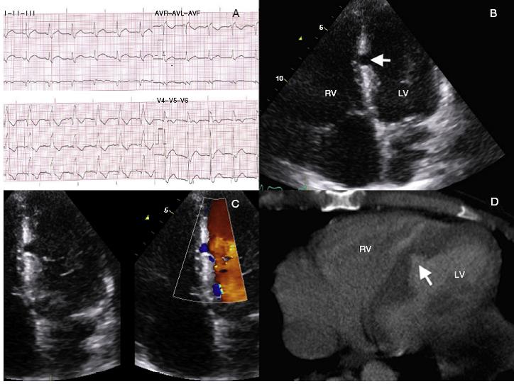
Figure 1: (A) Surface ECG showing a complete right bundle branch block. (B) Echocardiography (apical 4 chambers view) displaying an incomplete rupture of the interventricular septum (arrow) at the left side. (C) Zoomed 2D and color Doppler imaging views disclosing no shunt between the left ventricle (LV) and the right ventricle (RV). (D) Close view of the heart on chest CT scan revealing an interventricular septum tearing (arrow) noticed in a second review of the scan.
Funding
No endorsement of any kind received to conduct this study/article.
Conflict of interest
The authors declare no conflict of interest.
Ethical disclosure
Protection of people and animals. The authors declare that this research has not been conducted experiments on humans or animals. Confidentiality of data. The authors declare that they have followed the protocols of the workplace on the publication of patient data. Right to privacy and informed consent. The authors have obtained the informed consent of patients and/or subjects referred to in article consent. This document is in the possession of the corresponding author.

