




 nueva página del texto (beta)
 Inglés (pdf)
 Artículo en XML
 Referencias del artículo

 Traducción automática
 Enviar artículo por email
 Citado por SciELO
 Similares en
SciELO

 Permalink


Introduction
Genetic myopathies have gained importance nowadays due to a better understanding of the genetics underlying muscle disease; it is a rare condition in clinical practice, with a prevalence of 22.3 cases per 100,000 inhabitants.1,2 To date, more than 300 genes are associated with various inherited neuromuscular disorders, yet a significant number of patients, despite having access to these studies, remain without a definitive molecular diagnosis.3 In recent years, NGS (next-generation sequencing) of up to 301 genes for the study of neuromuscular diseases has become widespread. Given their low cost, these studies are accessible to most patients, however, due to a lack of knowledge about their use, they are not requested.4 Given the large number of genes involved in myopathies of a genetic cause, it is important to classify patients into clinical phenotypes to shorten the molecular diagnosis or to request a specific gene mutation. Currently, patients with muscular diseases are classified into one of the following phenotypes: limb-girdle muscular dystrophy myotonic dystrophy, oculopharyngeal dystrophy, facioscapulohumeral dystrophy, Emery-Dreifuss dystrophy, metabolic myopathy, channelopathy, etc.5 Certain symptoms and examination findings are particular to each phenotype; however, several clinical data are shared, namely progressive weakness, exercise intolerance, atrophy, pseudohypertrophy, and elevated creatine phosphokinase (CPK).2,6
Duchenne and Becker muscular dystrophy is the most frequent myopathy of genetic cause in the general population; they are generated by the alteration of the dystrophin protein and present an onset of symptoms at early ages.7 In the adult population, the epidemiology is different, as in some countries myotonic dystrophy type 1 is reported to be the most frequent.8,2
Despite the advance of genetics in the understanding of myopathies, there is a significant delay in diagnosis of several years. Muscle biopsy processed fresh with light microscopy continues to be a great diagnostic tool; the stains to observe the structure of muscle fibers are hematoxylin and eosin (H and E) and Gomori's modified trichrome. Other stains examine enzymatic reactions: NADH staining assesses complex II and Golgi-Cox staining assesses complex IV of the mitochondrial respiratory chain, while modified Schiff's peroxide staining (PAS) assesses glycogen deposition in the sarcoplasm. All these stains are of great help in the search for specific findings or to rule out other causes (inflammatory myopathies).2
The aim of the study is to report the frequency, clinical and preclinical characteristics, muscle biopsy findings, and results of genetic studies, performed in the different clinical phenotypes of myopathies of genetic cause in the adult population, in a reference center, as well as to review the subject.
Material and methods
Observational and cross-sectional study performed in a single center. All records of patients over 18 years of age with a clinical diagnosis of myopathy of genetic cause, seen at the neuromuscular diseases clinic of the National Institute of Neurology and Neurosurgery (INNN) from 2017 to 2020 were reviewed. Information on the following variables was obtained from the included patients: age (years) at diagnosis, time of delay of diagnosis (years), neuromuscular signs and symptoms (fatigue, exercise intolerance, weakness, winged scapula, calf hypertrophy), as well as score on the MRC (Medical Research Council) scale, that is, muscle strength grading from 0-60 points, taking into account the assessment of six muscles bilaterally (deltoids, biceps, hand extensor, iliopsoas, quadriceps and tibialis anterior). All patients were classified in some clinical phenotype of myopathy of genetic cause: myotonic dystrophy, limb-girdle dystrophy, distal myopathies, facioscapulohumeral dystrophy, metabolic myopathies, congenital myopathies, oculopharyngeal dystrophy, Emery-Dreifuss dystrophy, and axial myopathies. Information was also obtained from paraclinical studies (CPK levels, electromyography findings).
In the same way, the findings of muscle biopsies with optical microscopy were collected from the patients who had them. At the INNN, muscle biopsy processing is carried out by the Department of Neuromuscular Pathology. Muscle samples are freshly processed in 2-methyl-butane (isopentane), then cut to 8 microns thick at a temperature of -20°C and subjected to hematoxylin and eosin (H-E) techniques, modified Gomori trichrome (T-G), NADH enzymatic staining, COX enzymatic staining, and PAS staining.
At the same time, the results of the patients with a genetic study were collected. The INNN requests other institutions to perform molecular genetic studies according to the clinical diagnosis. In the case of Steinert myotonic muscular dystrophy, a molecular study is undertaken to determine the number of repeats. CTG in the DMPK (dystrophia myotonica protein kinase) gene with a determination error of +/- 2 repeats; PCR amplification is used to determine this number: an allele with 5-34 repeats is considered to be in the normal range, with 35-49 repeats it is classified in the normal-mutable range and an expanded allele with >50 repeats is classified as having full penetrance. In the case of oculopharyngeal dystrophy, it is determined by expansion of the GCN repeat in the first exon of the PABPN1 gene - both dominant (1217 repeats) and recessive inheritance (11 repeats) - by PCR amplification and Sanger nucleotide sequencing. In the case of patients with a clinical diagnosis of limb-girdle dystrophy, the NGS panel is performed for the main autosomal recessive limb-girdle dystrophies in Latin American countries (ANO5, DYSF, GAA, SGCB, SGCG, CAPN3, FKRP, SGCA, SGCD, TCAP). In some patients with more complex differential diagnosis, (congenital myopathy, muscular dystrophy, hereditary neuropathy, motor neuron disease) NGS panels and deletion/duplication analysis (MLPA, multiplex ligationdependent probe amplification) of 357 genes related to neuromuscular disorders, hereditary neuropathies, and hereditary amyotrophic lateral sclerosis panel are requested.
Results
Eighty-five records of patients with a diagnosis of myopathy of genetic cause were reviewed. The average age of the population was 37.1±16.7 years, and the female gender represented 53.5%. At diagnosis score: MRC score 45.9±8.3, modified Rankin scale score (median) 2 points (RIQ 1-2), 8 patients required a wheelchair for transfer, time of delay of diagnosis was 7 years (RIQ 4-13.5). The main neuromuscular symptoms and signs reported were: muscle weakness (100%), winged scapula (34%), myalgia (33%), exercise intolerance (29%), gastrocnemius pseudohypertrophy (11%), and muscle rippling (9%). CPK levels at diagnosis were 239 IU/dL (RIQ 90-902). Among the most frequent clinical phenotypes were found: limb-girdle muscular dystrophy (28%), myotonic dystrophy (26%), congenital myopathy (16.5%), metabolic myopathy (10.6%), oculopharyngeal dystrophy (7.1%), facioscapulohumeral dystrophy (6%), late-onset axial myopathy (2.4%), and only one patient with distal myopathy and one with Emery-Dreifuss (1.2%) (Table 1).
Regarding cardiopulmonary manifestations, 14 patients (16.47%) presented cardiac involvement. On the other hand, 22 patients (25.88%) had pulmonary involvement with the most frequent diagnosis being myotonic dystrophy type 1 and congenital myopathy (Table 1).
Table 1 Clinical characteristics of patients with genetic myopathies.
| Waist dystrophy N=24 | Myotonic dystrophy N=22 | Congenital myopathy N=14 | Metabolic myopathy N=9 | Oculopha ryngeal N=6 | Faciosca pulohumeral N=5 | Axial myopathy N=2 | Distal myopathy N=1 | Canalopathy N=2 | Emery- Dreifuss N=1 | |
|---|---|---|---|---|---|---|---|---|---|---|
| Age at diagnosis (years), mean (SD) | 22.6±15.7 | 30.4±13.5 | 12.3±12.2 | 28.2±9.5 | 53±8.6 | 42.2±17 | 69±15.5 | 29 | 30±15.5 | 33 |
| Female gender, n (%) | 12(50) | 9(41) | 10(71) | 9(100) | 3(50) | 2(40) | 0(0) | 0(0) | 0(0) | 0(0) |
| Delay in diagnosis (years), median (IQR) | 9 (5-15) | 7.5 (5-10) | 9.5 (3.7-18.2) | 4 (3-8) | 7.5 (3.7-20.5) | 4 (3-6.5) | 2.5 (2-2.5) | 5 | 12 | 20 |
| MRC score (points) median (IQR) | 41±6.5 | 46.7±7.9 | 44±9.5 | 51±5.4 | 54± 6.5 | 47±6.4 | 47±10 | 56 | 54±6 | 32 |
| Cardiac involvement, n (%) | 3(12.5) | 8(36) | 2(14) | 0(0) | 0(0) | 0(0) | 0(0) | 0(0) | 0(0) | 1(100) |
| Pulmonary involvement, n (%) | 2(8) | 14(67) | 4(29) | 0(0) | 0(0) | 1(20) | 0(0) | 0(0) | 0(0) | 1(100) |
| CPK levels (IU/dL), median (IQR). | 1019 (94-3015) | 194 (131-309) | 240 (62-1034) | 158 (47-756) | 108 (87-157) | 566 (85-8672) | 515 | 474 | 350 | 1646 |
| Muscle biopsy, n (%) | 11(46) | 1(4.5) | 12(86) | 8(89) | 2(33) | 2(40) | 1(50) | 1(100) | 2(100) | 1(100) |
| Genetic study, n (%). | 12 7-CAPN3 5-DYSF | 17 DMPK >100 repetidos CTG | 6 CAPN3, PNPLA2, RYR1,LDB3, DES,LAMA2 | 1 Negative | 5 PABN1 | 0 | 1 RYR1 | 1 ANO5 | 2 Negative, CACNA1S | 1 LAMNA |
Note: SD: standard deviation; IQR: interquartile range; CPK: creatine phosphokinase.
Forty-five patients (52.9%) had a genetic study, the main results being: 17 of 22 patients with myotonic dystrophy type 1 with >100 CTG repeats in the DMPK gene; 12 of 24 patients with limb-girdle dystrophy (7 calpain-3 and 5 dysferlin), and 5 of 6 patients with oculopharyngeal dystrophy positive for the PABPN1 gene mutation. The rest of the genes involved are shown in Table 1. Muscle biopsy was performed in 40 patients (47%) with light microscopy reading.
Discussion and review of the topic
The epidemiology of myopathies of genetic cause varies in different populations, reporting a prevalence of 16.14 to 22.3 cases per 100,000 inhabitants. The frequency of the main muscular dystrophies varies according to the age of the population studied (e.g., children, adults, or both), and between countries. A previous study in a Mexican population that included children and adult patients showed that dystrophinopathies are the most common, occurring in 61.7% of the population.1,9,10 Limb-girdle dystrophy is the most frequent in our center, which serves an adult population (≥18 years), followed by myotonic dystrophy, which differs from other reports. A population study in northern England showed that myotonic dystrophy is the most common (28.6%), followed by dystrophinopathy (22.9%), facioscapulohumeral dystrophy (10.7%), and limb-girdle dystrophy (6.2%).11 In Latin America, a retrospective study from a single center in Brazil included 1603 patients with a diagnosis of genetic myopathy who had a molecular study and/or muscle biopsy, and reported that dystrophinopathies were the most frequent (28.7%), followed by mitochondrial cytopathy (20.5%), limbgirdle dystrophies (9.7%) and myotonic dystrophy (8.6%).12 In a previous multicenter study in a Mexican population, 290 muscle biopsies were examined with immunofluorescence, detecting the following muscular dystrophies as the most frequent: dystrophinopathies (52.36%), dysferlinopathies (18.4%), sarcoglycanopathies (14.15%) and calpainopathies (11.32%).13
There is a significant delay in the diagnosis of genetically caused neuromuscular diseases worldwide. A study in Germany reported an average diagnostic delay of 4.3 years; it should be noted that the authors mention that the delay time depends on the first contact physicians: in neurologist physicians, the delay time was 3.5 years, while in nonneurologist physicians it was 5.2 years.14 Despite our case being that of a clinic specializing in neuromuscular diseases, the delay in diagnosis was longer than reported, 7 years (median). We assume that this difference is due to the fact that first-contact physicians (first or second level of care in our country) -in most cases, non-neurologists- are slow in referring these patients to our hospital (third level of care).
Limb-girdle muscular dystrophy was the most frequent, however, this data should be taken with reservation, since the population included in the study are patients who were diagnosed in adulthood. Patients with limb-girdle dystrophy present a significant delay in diagnosis, 9 years (median), longer than previously reported.14 This type of dystrophy has a clinical picture similar to that of inflammatory myopathies, i.e., involvement of proximal muscles (shoulder girdle and pelvic girdle) and elevated CPK levels, which can confuse the diagnosis, causing patients to be treated with steroids and immunosuppressants beforehand.15 The lack of clinical response to these treatments and the progressive evolution of the symptoms cause physicians to request the evaluation of other specialists (neurologists or neuromuscular specialists). Limb-girdle muscular dystrophies are divided according to the form of inheritance for their genetic study: autosomal dominant or autosomal recessive.16 In Mexico, it has been reported that the most common limb-girdle muscular dystrophies are autosomal recessive, due to a mutation in the dysferlin gene, followed by the calpain-3 gene.13 In our study, 50% of the patients had NGS molecular study, and it was found that the most frequent limb-girdle muscular dystrophy is due to mutation in the calpain-3 gene, followed by dysferlin. This result should be taken with caution because the other half of this population does not have a genetic study. A multicenter study examined the experience of the NGS study in Latin America (Brazil, Mexico, Argentina, Ecuador, Chile, and Peru), reporting the following genetic causes as the most frequent autosomal recessive causes of limb-girdle dystrophies: DYSF (dysferlinopathy), SGCA (alpha-sarcoglycanopathy), GAA (triplet for Pompe disease) and CAPN3 (calpainopathy-3).17 Eleven patients in our population underwent muscle biopsy, with the aim of finding findings that would support the diagnosis of muscular dystrophy: variability in fiber size, increased interfascicular connective tissue, degenerating fibers, and the presence of nuclear sacs (Figure 1).
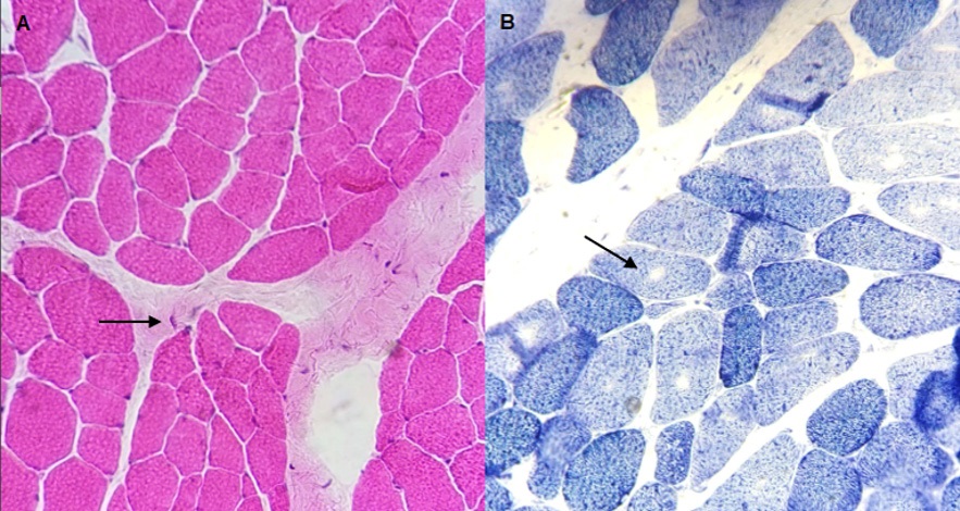
Figure 1 Image A) Deltoid muscle biopsy (left) from a 23-yearold male patient with a clinical diagnosis of limb-girdle muscular dystrophy. Hematoxylin and eosin staining shows slight to moderate variability in the shape and size of the fibers, increased interfascicular connective tissue, and the presence of nuclear sacs (arrow). Image B) Biopsy of the deltoid muscle (left) from a 17-year-old female patient with a diagnosis of congenital myopathy. NADH staining shows loss of the ¨chessboard¨ pattern and some fibers with an absence of central core reaction (arrow).
On the other hand, among the myotonic dystrophies, Steinert disease is the most common in adults, especially in Western Europe and North America (1 in 8000 inhabitants) as well as in Japan (1 in 20 000 inhabitants).18 In our population, it represents the second cause of genetic myopathy in adults. Genetically, Steinert myotonic dystrophy is autosomal dominant, and is due to increased CTG repeats (>50) in the DMPK gene on chromosome 19q; two-thirds of our patients had a genetic study and had >100 CTG repeats.18 This type of dystrophy presents a characteristic clinical phenotype: facial weakness (eyelid ptosis, horizontalization of the smile), weakness predominantly in proximal muscles in the upper extremities, weakness predominantly in distal muscles in the lower extremities, as well as myotonic phenomenon (lack of muscle relaxation after contraction). It also presents systemic alterations: androgenic alopecia, cataracts, endocrine alterations (diabetes mellitus), cardiac alterations (arrhythmias), and pulmonary alterations (obstructive sleep apnea).18,19 In the case of our population, more than half of the patients had both cardiac and pulmonary involvement.
In turn, congenital myopathies are a particular group, clinically defined as myopathy that begins in the first year of life and is characterized by non-progressive weakness and hypotonia, with normal or slightly elevated CPK levels.20 Its prevalence is estimated to be 0.6-0.9 cases per 100,000 population, and it represents 14% of the causes of hypotonia in the newborn.20,21 Patients present with myopathic facies, cleft palate, and preserved intelligence for age. Their muscle weakness is mild without some predominance in the extremities, and they may present axial muscle weakness. After adolescence, some patients may present cardiac and/or pulmonary function disorders, so it is advisable to perform spirometry and echocardiography periodically.20 Muscle biopsy with light microscopy is of great help in the suspicion of this type of myopathy; through NADH staining characteristic findings can be found, such as the presence of central-cores or multiminicores, and in the modified Gomori trichrome stain nemaline bodies can be observed (Figure 1).20 In our population, 12 patients had a biopsy, 3 of them with classification for central core congenital myopathy. This type of myopathies are of AD, AR or X-linked inheritance, the main gene involved is that of ryanodine and its receptor (RYN1), which is also related to malignant hyperthermia; only one patient in our population presented a mutation in this gene (Table 1).20,21 Recently, a study in Italy reported that congenital myopathies are mainly due to alterations causing deficiency in α-dystroglycan glycosylation (40.18%), followed by laminin-α2 deficiency (24.11%) and collagen VI deficiency (20.24%).22
Metabolic myopathies represent a diagnostic challenge since little information exists on their epidemiology; however, the prevalence of mitochondrial diseases in childhood is reported to be 1-15 cases per 10,000 population.23,24 In these cases, there are alterations in the metabolic pathways of glucose and lipids together with the respiratory chain in the mitochondria.22,24 This can lead to muscle symptoms, including poor exercise tolerance and non-severe episodes of weakness related to strenuous physical activity. On the other hand, CPK levels are generally normal and muscle atrophy is not present, and electromyography is usually normal.23,25 In muscle biopsy with light microscopy, some changes that suggest this diagnosis are: in hematoxylin and eosin staining, mild myopathic changes are present; in NADH staining, loss of the checkerboard pattern; in COX staining, presence of phantom fibers, and in PAS staining, fibers with an absence of reaction, as well as fibers with glycogen accumulation (Figure 2 and 3).24,25
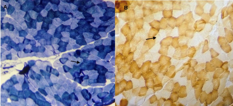
Figure 2 Biopsy of the left deltoid muscle of a 21-year-old male patient with a diagnosis of mitochondrial cytopathy. Image A) NADH staining shows myopathic changes, and loss of the "checkerboard" pattern; type I fibers tend to cluster (arrow). Image B) COX staining shows type I and type II fibers, some with an absence of reaction (COX phantom fibers, indicated with arrow).
Another less common type of genetically caused myopathy is oculopharyngeal dystrophy, which is distinguished by its late onset of symptoms, usually around the fifth decade of life.
Clinically it is characterized by palpebral ptosis without cranial nerve involvement, dysphagia, and weakness in proximal limb muscles. A significant finding in the muscle biopsy is the presence of bordered vacuoles, which is due to a mutation in the PABN1 gene.26 In our population, this phenotype represents the fifth cause of myopathy in adults. A previous study, performed in a referral center for ophthalmologic diseases in Mexico, reported a cohort of 102 Mexican-mestizo patients with oculopharyngeal dystrophy and with onset of symptoms after the age of 40 years; 91% presented dysphagia.27 Facioscapulohumeral dystrophy is rare in our population; worldwide it is estimated to have a prevalence of 7 cases per 100,000 inhabitants. It is caused by the shortening of the D4Z4 region on chromosome 4q and is autosomal dominant. Symptoms include winged scapula facial weakness in the deltoid muscle and weakness of the humeral muscles (biceps and triceps).28,29 Unfortunately, none of the patients included have had access to genetic molecular studies due to a lack of financial resources. Channelopathies are rare, with a prevalence of 1 in 100 000 inhabitants, and are mainly due to hypokalemic periodic paralysis by a mutation in the CACNA 1S gene encoding the alpha-subunit of the Cav1.1 calcium channel; they are of autosomal dominant inheritance.30 They are characterized by the presence of episodes of severe weakness, up to quadriplegia at times, with low serum potassium levels, associated with stressful events, abundant carbohydrate intake, or exercise. Vacuolar myopathy is a characteristic finding in muscle biopsy.31
In Mexico, there is currently no clinical practice guideline for the diagnostic approach and treatment of patients with suspected myopathy of genetic cause that would help health personnel not dedicated to the care of this group of patients to promptly recognize and refer them to hospital referral centers. In first-world countries such as England, despite the great progress in molecular genetic diagnosis and support of muscle biopsy in patients with suspected myopathy of genetic cause, 24.3% of patients remain without a definitive diagnosis.11 In this sense, establishing the diagnosis of probable myopathy of genetic cause, either through an adequate medical evaluation, electromyography study, CPK measurement, or through the support of muscle biopsy, is fundamental, since it will allow patients to receive an adequate rehabilitation program, avoiding treatment costs and unnecessary studies.
Conclusion
In our center, the most frequent genetic myopathy is limbgirdle dystrophy. Despite genetic advances in the study and understanding of neuromuscular diseases, there is still a significant delay in diagnosis at present. Classifying patients into clinical phenotypes helps to guide molecular diagnosis; properly processed and assessed muscle biopsy remains an excellent tool for the diagnosis of genetically caused myopathies.

