




 nueva página del texto (beta)
 Inglés (pdf)
 Artículo en XML
 Referencias del artículo

 Traducción automática
 Enviar artículo por email
 Citado por SciELO
 Similares en
SciELO

 Permalink


INTRODUCTION
Ischemic stroke is a leading cause of morbidity and mortality worldwide, but in approximately 30% of cases, the etiology remains undetermined and is termed cryptogenic stroke1, which is classified into three subgroups2: stroke with no identifiable cause after a complete diagnostic evaluation; stroke with multiple possible underlying causes; and stroke with an incomplete diagnostic evaluation. Within the subgroup with a complete diagnostic evaluation, the term embolic stroke of undetermined source (ESUS) defines a specific group of patients with non-lacunar cerebral infarction, non-stenotic carotid plaque (< 50%), and no major identifiable cardioembolic source. ESUS represents approximately 15%-20% of all ischemic strokes and is a clinically relevant entity due to its heterogeneous etiology and an annual recurrence risk of 4% to 5%1.
Non-stenotic carotid plaque (< 50%) has traditionally been considered low risk for ischemic stroke3,4. However, recent evidence suggests that high-risk plaque features such as an irregular surface, ulceration, a lipid-rich necrotic core, or intraplaque hemorrhage may cause embolism5. Other features, such as punctate calcifications6 and plaque length ≥ 10 mm7 have been considered non-high risk. Various imaging modalities have been used to evaluate the morphological characteristics of non-stenotic carotid plaque, including color Doppler ultrasound8, head and neck computed tomography angiography (CTA)5, magnetic resonance imaging (MRI)9, and positron emission tomography-computed tomography (PET-CT)10.
In patients with ESUS, the prevalence of non-stenotic carotid plaque has been reported to range from 39.1%11 to 42.1%12, and it occurs more frequently on the ipsilateral than the contralateral side. High-risk characteristics of non-stenotic carotid plaque (< 50%) have been identified by head and neck CTA5. The prevalence of morphological characteristics of non-stenotic carotid plaque on head and neck CTA in patients with ipsilateral ESUS is heterogeneous worldwide; however, information on Latin American populations is scarce11. This study describes the morphological characteristics of non-stenotic carotid plaque on head and neck CTA in Mexican patients with ipsilateral ESUS and in those without ESUS but with other neurological diagnoses.
MATERIAL AND METHODS
This retrospective cross-sectional study was conducted from March 2024 to February 2025 in the Department of Neuroimaging at the Instituto Nacional de Neurologia y Neurocirugia Manuel Velasco Suarez, in Mexico City, Mexico. Consecutive adults (≥ 18 years) with acute neurological symptoms and non-stenotic carotid plaque identified on head and neck CTA were included. Patients with non-stenotic plaques were excluded if they had an ischemic stroke of determined etiology according to TOAST criteria13, a carotid stenosis ≥ 50%, complete carotid occlusion, carotid dissection, or scans with incomplete acquisition or an inadequate arterial phase. Informed consent was waived due to the study’s retrospective design and the use of data obtained during routine clinical care. The study was approved by the Institutional Research and Ethics Committees.
Study development and variables
Clinical and imaging records were analyzed, and the following variables were collected: age, sex, smoking history, transient ischemic attack (TIA), systemic arterial hypertension, type 2 diabetes, family history of stroke in a first-degree relative, and a previous ipsilateral ischemic stroke documented by computed tomography (CT) on admission.
The morphological characteristics of high-risk non-stenotic carotid plaque, including an irregular surface, ulceration, lipid-rich necrotic core, and intraplaque hemorrhage, were recorded. Features not considered high-risk for ischemic stroke, such as punctate calcifications and plaque length ≥ 10 mm, were also recorded.
Two groups were defined: patients with ipsilateral ESUS, including those with non-stenotic carotid plaque (< 50%) in whom other etiologies were excluded according to TOAST13, and patients without ESUS but with other neurological diagnoses (aneurysm, intracranial hemorrhage, demyelinating disease, or epilepsy) who had no imaging evidence of ischemic stroke, and a non-stenotic carotid plaque as an incidental finding. In patients with ESUS, only the carotid artery on the same side as the ischemic event was evaluated. In patients without ESUS, but with other neurological diagnoses, a unilateral carotid artery with a non-stenotic plaque and greater atherosclerotic burden was assessed.
Definitions14
Irregular surface: plaque contour variability exceeding 1 mm in depth.
Ulceration: contrast extension ≥ 1 mm projecting beyond the luminal boundary into the plaque.
Lipid-rich necrotic core: a plaque component with attenuation less than 30 Hounsfield units (HU).
Intraplaque hemorrhage: subadventitial or intraplaque hyperdensity associated with adjacent fat stranding.
Punctate calcifications: hyperdense foci within the plaque > 130 HU.
Plaque length: the maximum longitudinal extent measured (mm) on multiplanar head and neck CTA reconstructions.
Image acquisition protocol
Head and neck CTA examinations were performed on a 128-slice CT scanner (Somatom Definition AS+, Siemens Healthcare, Forchheim, Germany). Automatic exposure control (CARE Dose 4D) was used with a reference tube current of 90 mAs. Tube voltage was selected based on scout acquisition (CARE kV). A total of 50 mL of non-ionic iodinated contrast medium (Iomeprol 300 mg/mL, Lomeron® 300; Bracco Imaging, Milan, Italy) was administered at 5 mL/s via a power injector (EmpowerCTA+, ACIST Medical Systems, Eden Prairie, MN, USA), followed by a 30-mL saline flush. Scanning was performed in the caudocranial direction with a pitch of 1.4 and a rotation time of 0.33 s at 120 kV. Images were reconstructed using the sinogram-affirmed iterative reconstruction (SAFIRE; Siemens Healthcare, Forchheim, Germany) algorithm with a strength level of 3. Bolus tracking was performed using a region of interest (ROI) positioned in the aortic arch; acquisition was automatically triggered when attenuation reached 70 HU.
Statistical analysis
Quantitative variables tested for normality with the Shapiro-Wilk test were reported as means ± standard deviation or as medians with interquartile ranges, as appropriate. Qualitative variables were reported as absolute and relative frequencies. Comparative analyses assessed differences in carotid plaque morphological characteristics between patients with and without ESUS using Student’s t-test or the Mann-Whitney U test for continuous variables and the chi-square test or Fisher’s exact test for categorical variables. A p value < 0.05 was considered statistically significant. Statistical analyses were performed using SPSS Statistics version 26.0 (IBM Corp., Armonk, NY, USA).
RESULTS
Four hundred ten patients with a non-stenotic carotid plaque (< 50%) were included; 175 (42.7%) in the ipsilateral ESUS group and 235 (57.3%) in the group without ESUS but with other neurological diagnoses (Table 1). Age was significantly higher in patients with ipsilateral ESUS (68.7 ± 13.0 vs 64.4 ± 13.0 years; p < 0.001). The groups were comparable by sex (n = 206, 50.3% vs n = 204, 49.7%) and smoking status (n = 80, 45.7% vs n = 104, 44.2%; p = 0.836).
Table 1 Characteristics of patients with non-stenotica carotid plaque with ipsilateral ESUS compared with patients without ESUS, but with other neurological diagnosesb assessed with head and neck CTA
| Description | Total (n = 410) | Ipsilateral ESUS group (n = 175) | Other neurological diagnoses group (n = 235) | p |
|---|---|---|---|---|
| Age, years, mean ± SD | 66.2 ± 12.6 | 68.7 ± 13.0 | 64.4 ± 13.0 | < 0.001 |
| Sex, n (%) | ||||
| Women | 206 (50.3) | 89 (50.8) | 117 (49.8) | 0.901 |
| Men | 204 (49.7) | 86 (49.1) | 118 (50.2) | 0.901 |
| Smoking, n (%) | 184 (44.9) | 80 (45.7) | 104 (44.2) | 0.836 |
| Family history of stroke in first-degree relatives, n (%) | 18 (4.4) | 12 (6.8) | 6 (2.5) | 0.068 |
| Previous ipsilateral ischemic strokec, n (%) | 144 (35.1) | 140 (80.0) | 4 (1.7) | < 0.001 |
| Presence of TIAd, n (%) | 94 (22.9) | 56 (32.0) | 38 (16.2) | < 0.001 |
| Comorbidities | ||||
| Systemic arterial hypertension, n (%) | 252 (61.5) | 127 (72.6) | 125 (53.2) | < 0.001 |
| Type 2 diabetes, n (%) | 154 (37.6) | 78 (44.6) | 76 (32.3) | < 0.05 |
aStenosis less than 50% of the arterial lumen.
bPatients with non-stenotic carotid plaque and no imaging evidence of ESUS, but with other neurological diagnoses such as aneurysm, intracranial hemorrhage, demyelinating disease, or epilepsy.
cNon-recent ipsilateral ischemic stroke, documented by CT scan on admission.
dClinical history of TIA before the current event. CTA: computed tomography angiography; ESUS: embolic stroke of undetermined source; SD: standard deviation; TIA: transient ischemic attack.
A history of ipsilateral ischemic stroke was more common in patients with ipsilateral ESUS than in the non-ESUS group (n = 140, 80.0% vs n = 4, 1.7%; p < 0.001). Similarly, TIA (n = 56, 32.0% vs n = 38, 16.2%; p < 0.001), systemic arterial hypertension (n = 127, 72.6% vs n = 125, 53.2%; p < 0.001), and type 2 diabetes (n = 78, 44.6% vs n = 76, 32.3%; p < 0.05) were significantly associated with ipsilateral ESUS.
Morphological characteristics of non-stenotic carotid plaque
In patients with non-stenotic carotid plaque and ipsilateral ESUS, the most frequent high-risk characteristics were an irregular surface (n = 168, 96.0%), followed by plaque ulceration (n = 102, 58.3%) (Table 2). Among the morphological characteristics not considered high-risk, punctate calcifications were present in 131 (74.8%) patients, followed by plaque length ≥ 10 mm (n = 111, 63.4%). Two or more morphological characteristics of non-stenotic carotid plaque were present in most patients.
Table 2 Morphological characteristics of non-stenotic carotid plaquea in patients with ipsilateral ESUS compared with patients without ESUS, but with other neurological diagnosesb assessed with head and neck CTA
| Description | Totalc (n = 410) | Ipsilateral ESUS group (n = 175) | Other neurological diagnoses groupc (n = 235) |
|---|---|---|---|
| High-risk morphological characteristics of non-stenotic carotid plaque | |||
| Irregular surface, n (%) | 386 (94.1) | 168 (96.0) | 218 (92.8) |
| Ulceration, n (%) | 233 (56.8) | 102 (58.3) | 131 (55.7) |
| Lipid-rich necrotic core, n (%) | 54 (13.2) | 20 (11.4) | 34 (14.5) |
| Intraplaque hemorrhage, n (%) | 2 (0.5) | 1 (0.6) | 1 (0.4) |
| Not considered high-risk morphological characteristics of non-stenotic carotid plaque | |||
| Punctate calcifications, n (%) | 295 (71.9) | 131 (74.8) | 164 (69.8) |
| Plaque length ≥ 10 mm, n (%) | 270 (65.8) | 111 (63.4) | 159 (67.6) |
aStenosis < 50% of the carotid arterial lumen.
bPatients with a non-stenotic carotid plaque and no imaging evidence of ESUS, but with other neurological diagnoses such as aneurysm, intracranial hemorrhage, demyelinating disease, or epilepsy.
cTwo or more morphological characteristics of non-stenotic carotid plaque were found in patients. CTA: computed tomography angiography; ESUS: embolic stroke of undetermined source.
In the group without ESUS but with other neurological diagnoses and non-stenotic carotid plaque, the most frequent high-risk characteristic was an irregular surface (n = 218, 92.8%), followed by plaque ulceration (n = 131, 55.7%). Among morphological characteristics not considered high-risk, punctate calcifications were present in 164 (69.8%) patients, followed by plaque length ≥ 10 mm (n = 159, 67.6%). Two or more morphological characteristics of non-stenotic carotid plaque were observed in most patients.
Morphological characteristics of non-stenotic carotid plaques evaluated by head and neck CTA are shown in figure 1. A head and neck CTA scan of a non-stenotic carotid plaque with high-risk morphological characteristics associated with ipsilateral ESUS is shown in figure 2. Additionally, figures 3 and 4 show high-risk morphological characteristics and other features not considered high risk in non-stenotic carotid plaque in patients without ESUS but with other neurological diagnoses, such as intracranial aneurysm and epilepsy.
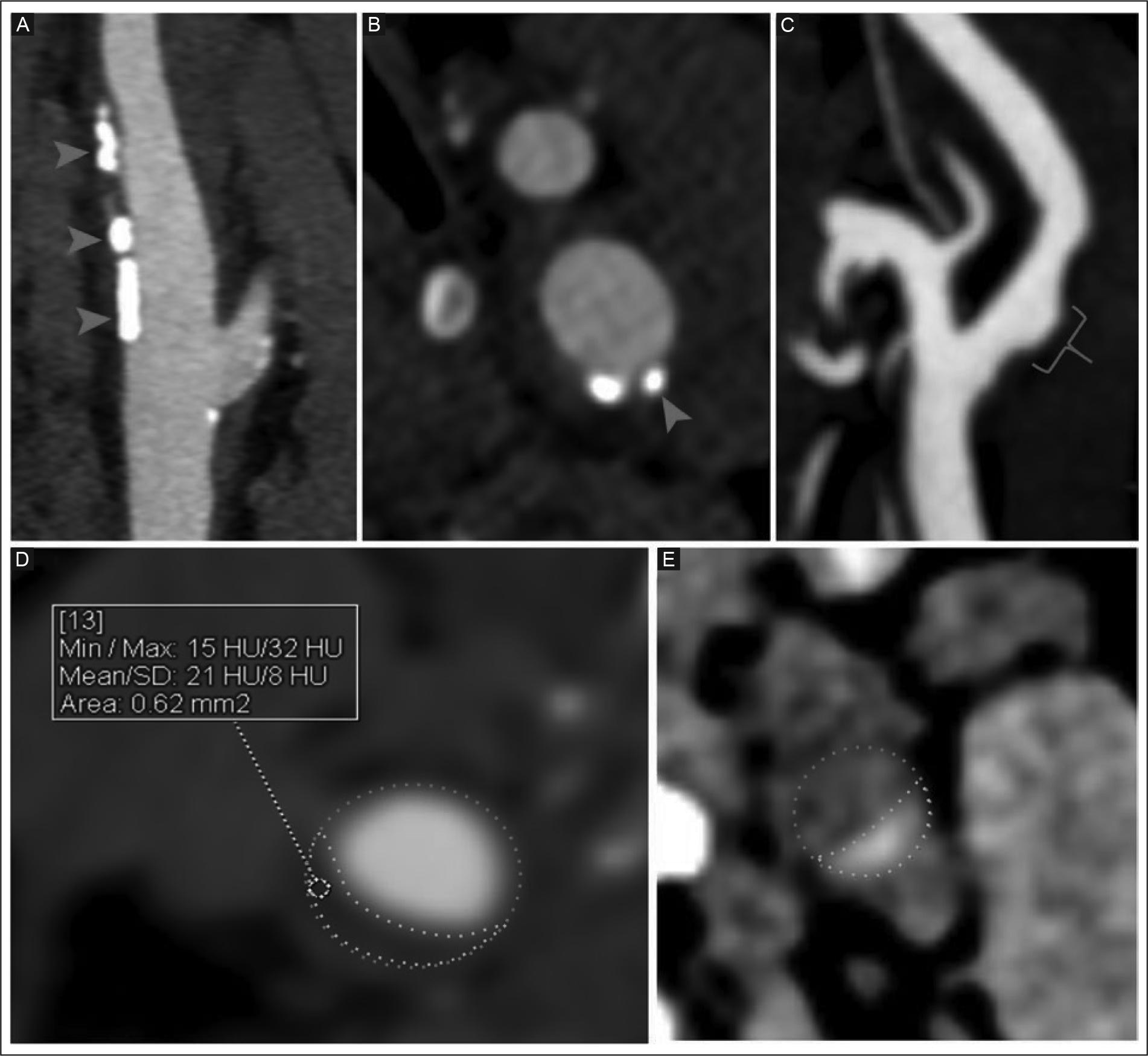
CTA: computed tomography angiography; HU: Hounsfield units; MIP: maximum intensity projection.
Figure 1 Morphological characteristics of non-stenotic carotid plaques by head and neck CTA. A: arterial phase oblique sagittal reconstruction MIP and vascular filter in the carotid bulb showing an irregular plaque surface (red arrowheads). B: arterial phase axial view of the carotid bulb showing subendothelial punctate calcifications (red arrowhead). C: arterial phase of the carotid bulb with oblique sagittal MIP reconstruction showing contrast extending > 1 mm beyond the luminal contour, indicating plaque ulceration (red curly bracket). D: arterial phase MIP of the carotid bulb, axial view. The orange dotted line marks the arterial contour, and the white dotted line marks the hypodense plaque contour, with attenuation of 21 ± 8 HU, suggesting a lipid-rich necrotic core. E: unenhanced-phase axial view of the carotid artery. The orange dotted line marks the arterial contour. The hyperdense intraplaque area, indicated by white dotted lines, suggests intraplaque hemorrhage.
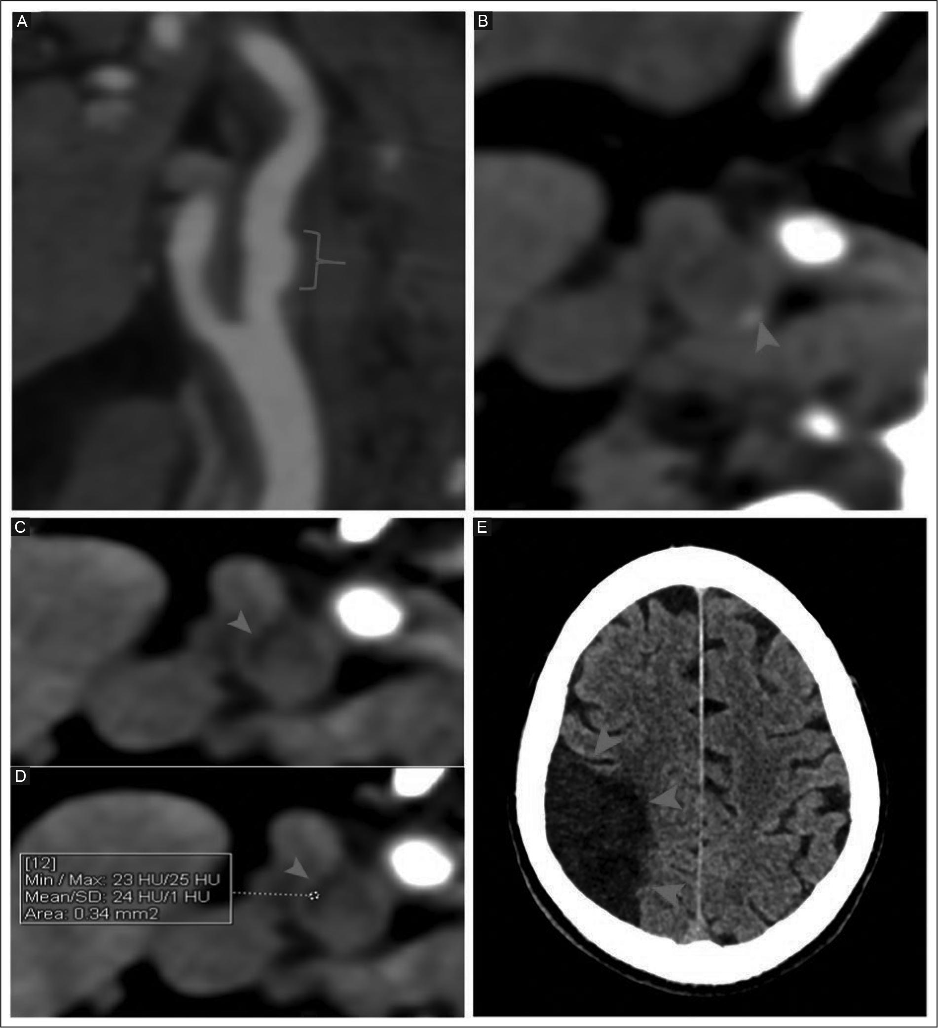
CT: computed tomography; CTA: computed tomography angiography; ESUS: embolic stroke of undetermined source; HU: Hounsfield units; MIP: maximum intensity projection; ROI: region of interest.
Figure 2 Head and neck CT of the right carotid artery shows a non-stenotic carotid plaque with high-risk morphological characteristics in a 67-year-old man presenting with sudden-onset left hemiparesis, right lip deviation, and dysarthria, without altered consciousness. The initial CT ruled out hemorrhage and demonstrated an ipsilateral cerebral infarction. A: MIP arterial phase in the oblique sagittal plane shows contrast extending > 1 mm beyond the luminal contour, indicating plaque ulceration in the right carotid bulb (red curly bracket). B: unenhanced-phase axial view showing an atherosclerotic plaque with punctate calcification (red arrowhead). C-D: unenhanced-phase views of the right carotid bulb show an intraplaque hypodense component (red arrowheads) and an ROI of 24 ± 1 HU consistent with a lipid-rich necrotic core. E: unenhanced-phase view showing a wedge-shaped hypodense area in the right cerebral hemisphere, consistent with an ipsilateral subacute cerebral infarction (red arrowheads). The diagnosis was ESUS ipsilateral to the non-stenotic carotid plaque.
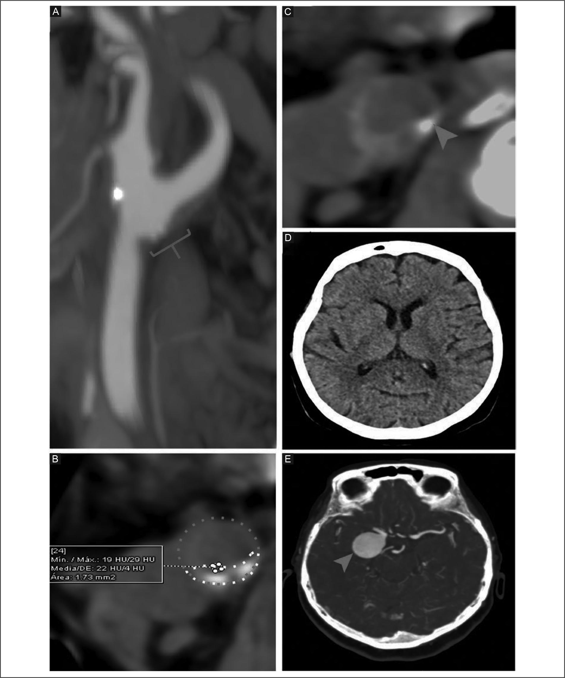
CTA: computed tomography angiography; HU: Hounsfield units; MIP: maximum intensity projection; NAS: numerical analog scale; ROI: region of interest.
Figure 3 Head and neck CTA in a 56-year-old woman with a sudden onset of right hemicranial headache (NAS 10/10), right eyelid ptosis, and retroauricular and retro-orbital pain secondary to an intracranial aneurysm. Non-stenotic carotid plaque with high-risk morphological characteristics was an incidental finding. A: arterial-phase MIP in the oblique sagittal plane of the right carotid, demonstrating contrast extending > 1 mm beyond the luminal contour, indicating plaque ulceration in the carotid bulb associated with punctate calcifications (red curly bracket). B: unenhanced-phase axial view of the right carotid bulb showing a plaque outlined by white dotted lines, with the carotid lumen indicated by orange dotted lines. The plaque contains punctate calcifications and a hypodense component with an attenuation of 22 ± 4 HU (ROI), consistent with a lipid-rich necrotic core. C: unenhanced-phase view of the right carotid bulb showing punctate subendothelial calcification (red arrowhead). D: unenhanced-phase view at the level of the basal ganglia, with no evidence of cerebral infarction. E: arterial phase axial view shows a saccular arterial dilation in the posterior communicating segment of the right internal carotid artery (red arrowhead).
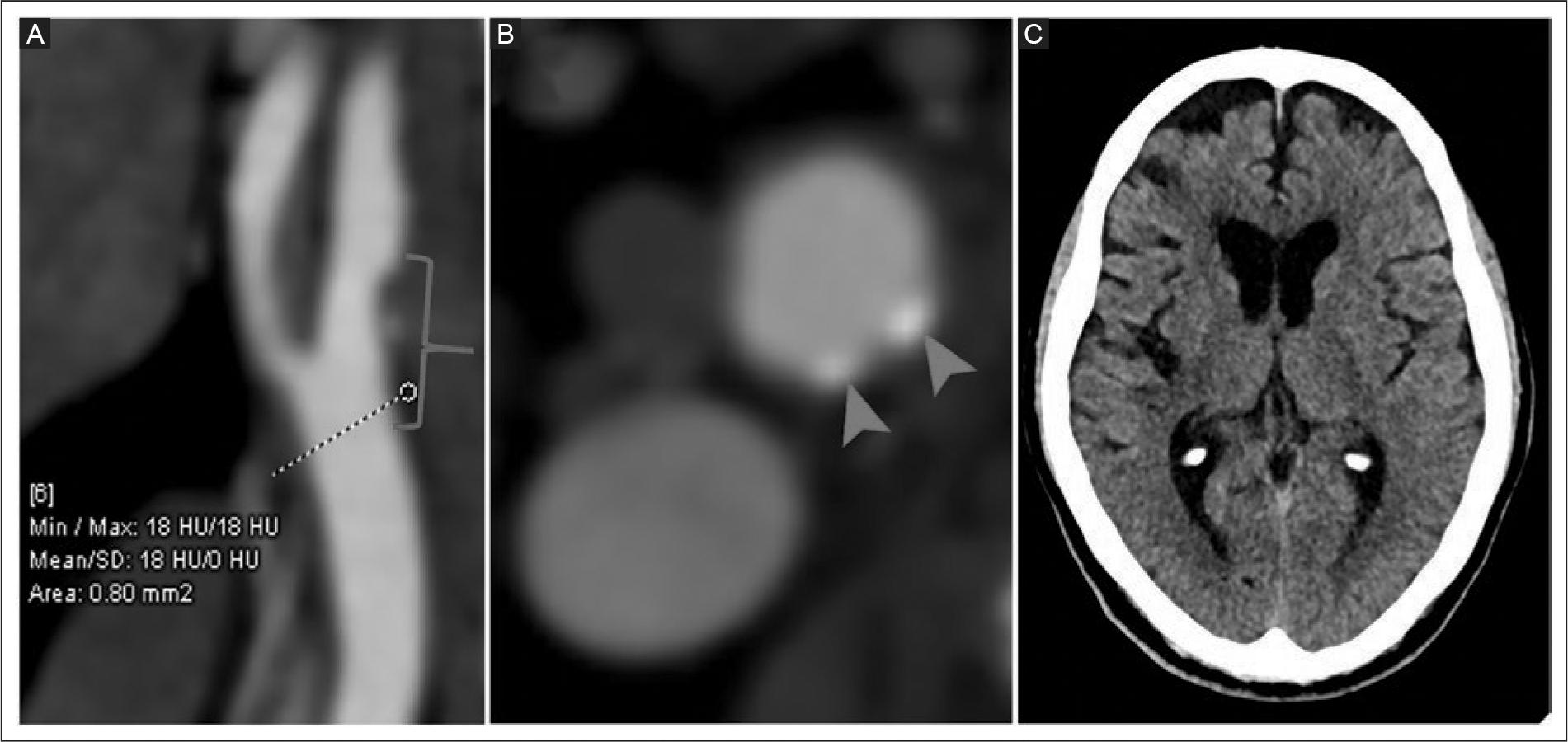
CT: computed tomography; CTA: computed tomography angiography; ESUS: embolic stroke of undetermined source; HU: Hounsfield units; MIP: maximum intensity projection; ROI: region of interest.
Figure 4 Head and neck CT of a 55-year-old woman with a history of epilepsy. The head CT showed no evidence of an acute ischemic event (not shown). A non-stenotic carotid plaque with high-risk morphologic characteristics was incidentally identified. A: oblique sagittal arterial-phase MIP shows an irregular plaque extending from the common carotid artery toward the carotid bulb and the right internal carotid artery (red curly bracket). A hypodense intraplaque area is identified with an ROI of 18 ± 0 HU, suggesting a lipid-rich necrotic core. B: arterial-phase axial view at the level of the right carotid bulb shows subendothelial punctate calcifications (red arrowheads). C: unenhanced-phase view shows decreased brain volume in the right hemisphere, with widened sulci related to atrophy.
DISCUSSION
In this study, head and neck CTA identified an irregular surface, ulceration, a lipid-rich necrotic core, and intraplaque hemorrhage as high-risk morphological characteristics of non-stenotic carotid plaque in Mexican patients with ipsilateral ESUS. Additionally, features not considered high-risk, such as punctate calcifications and plaque length ≥ 10 mm, were also commonly observed. Similar findings were observed in patients without ESUS but with other neurological diagnoses. This study is the first to describe the morphological characteristics of non-stenotic carotid plaque in Mexican patients with ipsilateral ESUS.
Non-stenotic carotid plaques can exhibit morphological features associated with instability, regardless of the degree of luminal stenosis. These features include an irregular surface, ulceration, a lipid-rich necrotic core, and intraplaque hemorrhage, which have been defined as high-risk markers of plaque vulnerability11,15,16. Demir et al.17 in a retrospective cross-sectional study using head and neck CTA evaluated 81 Turkish patients with ESUS. Non-stenotic carotid plaques were more frequent in the ipsilateral carotid artery than on the contralateral side (44/81, 54.3% vs 24/81, 29.6%; p = 0.002). Plaque thickness ≥ 3 mm (28/81, 34.6% vs 11/81, 13.6%; p = 0.003), plaque ulceration (24/81, 29.6% vs 7/81, 8.6%; p = 0.001), plaque irregularity (34/81, 42.0% vs 10/81, 12.3%; p < 0.001), noncalcified plaque (21/81, 26.0% vs 5/81, 6.2%; p = 0.001), and plaque hypodensity (20/81, 24.7% vs 5/81, 6.2%; p = 0.002) were significantly more frequent in the carotid artery on the same side as the ischemic event. The authors concluded that non-stenotic carotid plaques, particularly those with high-risk characteristics, are more prevalent on the ipsilateral side and should be considered in the clinical evaluation and treatment of ESUS. In a metaanalysis that included 10 studies with a predominance of patients with ESUS (n = 797), the prevalence of non-stenotic carotid plaques was 55% (95% CI: 42-68)11.
The authors concluded that non-stenotic carotid plaques are highly prevalent in patients with ESUS and may represent an underrecognized embolic source in ischemic stroke. Ospel et al.11 analyzed an international, multicenter cohort of 138 patients with ESUS who underwent head and neck CTA. Non-stenotic carotid plaques were more frequent in patients with ESUS than in those with non-ESUS (54/138, 39.1% vs 77/308, 25.0%; p = 0.002). Among patients with ESUS, non-stenotic carotid plaques were more frequent ipsilateral to the ischemic event than in the contralateral carotid artery (40/137, 29.2% vs 26/139, 18.7%; p = 0.041). In our study, the prevalence of high-risk and non-high-risk non-stenotic carotid plaques was elevated; however, the morphological characteristics did not show a significant association with ipsilateral ESUS compared to patients without ESUS but with other neurological diseases. This discrepancy could be due to methodological differences, particularly the lack of a contralateral carotid artery comparison and the selection of the comparison group without ESUS based on a higher atherosclerotic burden.
Non-stenotic carotid plaques with vulnerability features are associated with a higher risk of ischemic recurrence. In a meta-analysis of 2,350 patients, Rizvi et al.18 reported that intraplaque hemorrhage was associated with an annualized rate of ischemic events of 11.9%, while lipid necrotic core and rupture or thinning of the fibrous cap showed rates of 5.4% and 5.7%, respectively19. Furthermore, intraplaque hemorrhage has been identified as an independent predictor of recurrence, with ipsilateral stroke rates of 9.0% per year in patients with less than 50% carotid stenosis (odds ratio of 11.0) compared to 0.7% in the absence of intraplaque hemorrhage19. In a meta-analysis by Singh et al.20 that included 31 studies with 13,428 patients with non-stenotic carotid plaque, the incidence of associated ischemic events was evaluated using various imaging techniques-ultrasound, CTA, magnetic resonance angiography, or digital subtraction angiography. In patients with asymptomatic non-stenotic carotid plaque, the incidence of the first stroke or TIA event was 0.5 per 100 person-years; in contrast, in symptomatic non-stenotic plaques, the recurrence rate was 2.6 per 100 person-years, and in those with intraplaque hemorrhage 4.9 per 100 person-years. In our study, a high proportion of patients with a history of ipsilateral ischemic stroke related to the non-stenotic carotid plaque was identified in 140 (80.0%) patients in the ipsilateral ESUS group compared to 4 (1.7%) in the group without ipsilateral ESUS (p < 0.001), indicating a high recurrence rate in ESUS group. Likewise, a higher frequency of prior TIA was found in patients with ipsilateral ESUS (n = 56, 32.0%) compared to the group without ESUS (n = 38, 16.2%) (p < 0.001). These findings are consistent with those reported in a meta-analysis by Singh et al.20 which found that symptomatic non-stenotic carotid plaque with high-risk morphological features was associated with a higher risk of ESUS recurrence. In our study, a high frequency of history of stroke and TIA was observed in the ipsilateral ESUS group, suggesting a clinical pattern compatible with a higher risk of recurrence. Non-stenotic carotid plaque may be associated with an increased risk of recurrent ischemic events, particularly in patients with high-risk carotid plaque morphology, suggesting its potential as an embolic source.
Among the strengths of this study are the sample size, the systematic evaluation using head and neck CTA in all patients, and the characterization of morphological features of non-stenotic carotid plaque, which has traditionally been underestimated in the etiological evaluation of stroke. However, the study has limitations related to its retrospective design. Carotid plaque characterization was performed solely using head and neck CTA, without the inclusion of vascular wall MRI. Moreover, it was not possible to definitively establish a causal relationship between plaque characteristics and the ischemic event due to the possible coexistence of other causes not routinely evaluated, such as subclinical atrial fibrillation, atrial cardiomyopathy, small intracavitary thrombi, and hereditary or acquired thrombophilias. Finally, the clinical characteristics and their relationship to the morphological features of the non-stenotic carotid plaque are not described.
CONCLUSION
In our study, the most common high-risk morphological characteristics of non-stenotic carotid plaque on head and neck CTA in patients with ipsilateral ESUS were an irregular surface and ulceration. Prospective studies with larger sample sizes, including assessment of the contralateral carotid and complementary imaging techniques, are needed to enable more detailed characterization of the morphological features of non-stenotic carotid plaque in ESUS.

