Services on Demand
Journal
Article
Indicators
-
 Cited by SciELO
 -
 Access statistics
Related links
-
 Similars in
SciELO
Share
Abanico veterinario
On-line version ISSN 2448-6132Print version ISSN 2007-428X
Abanico vet vol.11 Tepic Jan./Dec. 2021 Epub Oct 11, 2021
https://doi.org/10.21929/abavet2021.28
Clinical Case
Laminitis in a Spanish pure-bred mare in Tabasco, Mexico: Case report
1División Académica de Ciencias Agropecuarias, Universidad Juárez Autónoma de Tabasco.
2Programa de Ganadería, Colegio de Postgraduados, Campus Montecillo.
3Universidad Autónoma de Chiapas, Facultad de Medicina Veterinaria y Zootecnia.
4Instituto de Investigaciones Agropecuarias y Forestales, Universidad Michoacana de San Nicolás de Hidalgo.
Laminitis is a serious, highly prevalent disease, recognized as one of the most important clinical syndromes in equines. The present study describes the case of a five-year-old Spanish pure-bred mare diagnosed with bilateral laminitis at approximately three months of age. The animal refused to walk, lying down all the time. The degree of claudication, according to the Obel scale, was degreed as 5; it also had an altered palmar digital pulse. It was prescribed 30 days of anti-inflammatory therapy with 1.1mg/kg intravenous (IV) of meglumine flunixin every 24 h, accompanied by omeprazole 2 mg/kg orally (O.V.). Additionally, hoof trimming and corrective shoeing were recommended. After eight months of treatment, the mare showed a remarkable improvement and gain in body condition, and a gestation was achieved. The anti-inflammatory treatment, hoof trimming and corrective shoeing favored the growth and hardness of the hoof, being successful in the treatment of laminitis.
Keywords: corrective shoeing; laminitis; Pure-bred Spanish Horse
La laminitis es una enfermedad grave, de alta prevalencia, reconocida como uno de los síndromes clínicos más importantes en equinos. En el presente estudio se describe el caso de una yegua Pura Raza Española de cinco años de edad, con diagnóstico de laminitis bilateral de aproximadamente tres meses. El animal se reusaba a caminar, manteniéndose echado todo el tiempo. El grado de claudicación, según la escala de Obel, se calificó de 5; asimismo, presentaba pulso palmar digital alterado. Se receto terapia de 30 días con antinflamatorios 1.1mg/kg intra venosa (IV) de flunixin de meglumine cada 24 h, acompañado de omeprazol 2 mg/kg vía oral (VO). Adicionalmente, se recomendó recorte de casco y herrajes correctivos. Después de los ocho meses de tratamiento, la yegua presentó una notable mejoría y ganancia de condición corporal, además, se logró una gestación. El tratamiento antiinflamatorio, recorte de casco y herrajes correctivos, favorecieron el crecimiento y dureza del casco, teniendo éxito en el tratamiento de laminitis.
Palabras clave: herraje correctivo; laminitis; Raza Pura Española
INTRODUCTION
Laminitis or infosura is a serious disease, of high prevalence, recognized as one of the most important clinical syndromes in equines (Paula et al., 2020). The term laminitis is used to describe a systemic pathology, which compromises the general animal condition. It is an inflammation of the sensitive hoof laminae causing degeneration, separation and necrosis of the laminar corium (Londoño-Sossa et al., 2011; Mitchell et al., 2015).
Laminitis causes acute pain to animals and it is potentially fatal, primarily affecting adult horses and ponies of any breed (Mitchell et al., 2015).
Laminitis is commonly associated with the ingestion of nonstructural carbohydrates from pasture; however, it is not only related to diet, but also to some systemic diseases (Londoño-Sossa et al., 2011; Mitchell et al., 2015; Paula et al., 2020). The clinical alterations that predispose laminitis are acute abdominal syndrome, enteritis, retention of fetal membranes, metritis, pleuropneumonia, and other pathologies. It can also occur due to improper handling of the animal, such as excessive ingestion of cold water after work or the administration of high corticosteroid levels, which decrease protein synthesis, potentiate digital vasoconstriction and induce micro-thrombosis (Londoño-Sossa et al., 2011; Mitchell et al., 2015; Paula et al., 2020). Other risk factors associated with laminitis are horse breed being more prone to draft breeds, overweight in the animal, high nutritional level and older horses with Cushing's disease (AAEP, 2020).
Laminitis can present in one or more limbs with characteristic signs, such as lameness, increased hoof temperature and increased digital pulse in the limb, pain when pressure is applied, hesitant gait, stance with forelegs extended forward to relieve pressure, in chronic cases; widening of hoof wall rings may be observed as they follow from toe to heel, bruised soles, widened white line, with appearance of seromas and/or abscesses, drooping soles, thick callus as well as concave hooves with "Aladdin's shoes" appearance (AAEP, 2020).
The Americans Association of Equine Practitioners (AAEP, 2020) established the following equine lameness scale:
Degree 1: Lameness that is difficult to observe in any situation.
Degree 2: Lameness that is difficult to observe at a walk or trot but appears in certain circumstances.
Degree 3: Permanent lameness at trot at all times.
Degree 4: Lameness is evident by nodding the head or taking a short stride.
Degree 5: Lameness is very evident and permanent with minimal weight bearing and manifests itself at rest and even more so when moving.
The knowledge and understanding of the pathophysiology of laminitis is somewhat unknown, a situation that limits efforts for its prevention and treatment. Therefore, timely diagnosis and subsequent medical treatment, supported by biomechanical methods, are of utmost importance to minimize the effects of this disease (Mitchell et al., 2015).
The objective of this study is to describe the diagnosis and treatment of a case of bilateral laminitis in a five-year-old Spanish pure-bred mare, which was treated with anti- inflammatory therapy, hoof trimming and corrective horseshoeing.
CLINICAL CASE DESCRIPTION
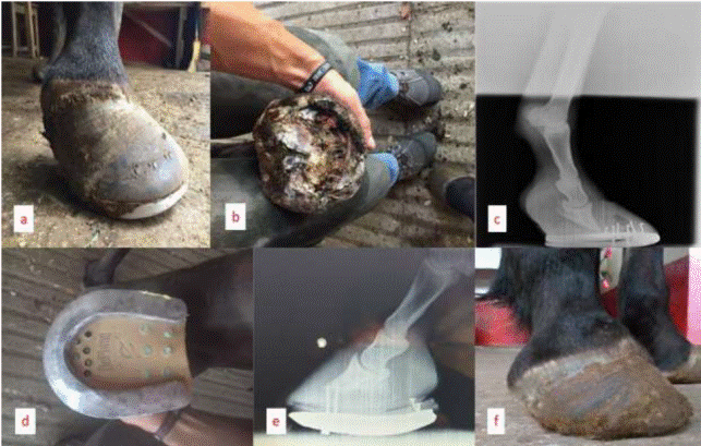
It is presented the case of a five-year-old Spanish pure-bred mare (Figure 1), that began with discomfort when walking and lying down most of the day. This problem had been present for about three months. The degree of claudication, considering the scale of EEAP (2020), it was rated from degree 5; she also presented increased digital palmar pulse. The mare was rested in a stall with a rubber floor and a bed of shavings with a thickness of approximately 70 cm to make her as comfortable as possible. Its feed was based on fresh Taiwan grass (Pennisetum purpureum) chopped at free access without additional concentrate.

Figure 1 Clinical process of laminitis in a Spanish Breed mare. a) Aspect of the laminitic hoof, b) Physical examination of the hoof clearly shows necrosis of the dermal laminae and cell death, c) MAD latero- medial image with evident 5° rotation about to perforate the solar part, d) therapeutic fitting of the hoof based on a glued aluminum horseshoe and impression material to support the palmar part of the hoof, e) radiographic aspect of the MAD after 12 months of treatment and f) aspect of the MAD hoof after 12 months of treatment.
First, a physical examination of the hoof (Figure 1 a,b) of the right/left forelimb was performed using palpation forceps. Subsequently, radiographs were taken using a latero- medial projection to determine the degree of rotation of both limbs (Figure 1 c). Once the radiographs were obtained, it was established that the rotation angle of the third phalanx (p3) was greater than 5º. Trimming was performed to stabilize and improve the position of P3 within the corneal case, so that the therapeutic hardware would perform its function of trimming the step. The hardware used in the treatment was a libero model (Mustad, Norway) with a rokert toe modification (Figure 1 e).
Thirty-day therapy was prescribed with anti-inflammatory drugs 1.1mg/kg intravenous meglumine flunixin every 24 h, accompanied by omeprazole 2 mg/kg orally to prevent possible gastric ulceration and prolong the use of anti-inflammatory drugs.
After 12 months of treatment with therapeutic shoeing (Figure 1 f), the mare presented a considerable improvement and gain in body condition, achieving a pregnancy.
Figure 2 shows the radiographic images of laminitis at the beginning as well as its evolution at six and 12 months after treatment.
DISCUSSION
Timely prognosis given by ethical clinicians is very important in the treatment of equine conditions such as laminitis. The most recommended parameters to estimate the prognosis in equines with laminitis are: claudication degree and the intensity of third phalanx rotation measured as the angle formed by the dorsal aspect of the podal bone and the hoof wall (Londoño-Sossa et al., 2011). The degree of claudication is commonly determined based on the classification defined by Obel (1948), but recently some authors (Meier et al., 2019) have made modifications to that scale and proposed a five-criteria, three-stage method that employs a severity scale of 0-12; however, this method is very limited given that many of the signs on which the criteria are based may not be present in the preclinical phase (with the exception of the digital pulse).
In the present clinical case, since the patient remained lying down and without any attempt to get up, a degree 5 was assigned, which is assigned when the animal refuses to move unless forced; which according to EEAP (2020), is the most severe degree. In this regard, Mitchell et al. (2015) describe that it is common for severely affected horses to take a specific posture to shift their weight onto their hind legs, due to intense pain that is often accompanied by anxiety and muscle twitching. It is vitally important to avoid prolonged periods of recumbency in animals due to the potential for pressure ulcers (Mitchell et al., 2015).
Lack of adequate pain control is a common reason why horses affected by laminitis are eventually euthanized (Bamford, 2019). Nonsteroidal anti-inflammatory drugs (NSAIDs) such as flunixin meglumine (1.1 mg/kg orally (OV) or intravenously (IV) and phenylbutazone (2-6 mg /kg OV), in low doses, are commonly used as a form of analgesia and to treat laminitis-induced inflammatory responses. For the chronic stage of laminitis cyclooxygenase type 2 (COX-2) inhibitor NSAIDs, such as firocoxib, provide safe and effective pain relief. Non-selective COX inhibitors may be better in the acute and developmental stages of laminitis, as vascular function is still present (Orsini, Wrigley, & Riley, 2010). A vasodilator such as acepromazine (0.002-0.0066 mg/kg VO or IV or intramuscular (IM), may be helpful in counteracting vascular dysfunction in the phalanx (Parks, 2009). Accepromacin increases digital and laminar blood flow in normal horses. The calming effects of aceppromazine may stimulate the horse to lie down, which may be beneficial. More recently, interest has developed in the use of other analgesic agents such as lidocaine and ketamine, administered as an intravenous infusion, or narcotics, either as a continuous, epidural or intravenous infusion (Parks, 2009).
On the other hand, many factors must be taken into account, especially if there is rotation of the third phalanx, an event that determines a poor prognosis in the vast majority of cases (Londoño-Sossa et al., 2011). Some authors consider that an angle greater than 11 is of poor prognosis for the return to athletic function (Londoño-Sossa et al., 2011). Radiographic studies to evaluate the degree of palmar / plantar rotation of the distal phalanx showed that in the patient in the present study, this angle was 5º, which confirms the diagnosis of laminitis (Sherlock and Parks, 2013). Stick et al. (1982) suggest that horses with a rotation less than 5.5° have a better prognosis for return to sporting activities than horses with a rotation greater than 11.5°. However, Paula et al. (2020) reported that horses with a rotation between 10.5° and 11.02° returned to normal activities.
The use of therapeutic horseshoes for the treatment of laminitis are the wodem shoe, banana shoes with wedge insoles, aluminum shoes with glued-on bench insoles, represent the first option in the treatment of chronic laminitis (Estrada, 2011). Tenotomy of the deep digital flexor tendon is another option in the treatment of laminitic horses, since it helps to improve biomechanics, decreasing the tension force that this tendon exerts on p3, allowing derotation, although the postoperative period and rehabilitation will require medication and time to restore the functionality of the limb (Carmona and López, 2011).
Radiographs are indicated in every suspected laminitic case because they provide valuable information about the presence, severity, relative chronicity and progressive nature of the disease (Sherlock and Parks, 2013). Interpretation of the horse with suspected laminitis is more complex than just identifying the presence or absence of dorsal rotation (Sherlock and Parks, 2013). A thorough evaluation of radiographs is essential as many abnormalities may be evident at the same time (Parks, 2007).
The most important factor in the long-term prognosis of the horse with laminitis is the extent of laminar pathology which influences the degree of instability between the distal phalanx and hoof wall however, this is very difficult to determine. Advanced diagnosis with the use of radiological imaging along with clinical signs may help in future evaluations, but currently the evaluation of a horse with laminitis is based on paying close attention to the details of the simple radiographic series, clinical evaluation and occasionally venographic studies.
Fortunately, the patient was able to recover satisfactorily, a situation that represents a successful case, considering that approximately 75% of horses treated for laminitis do not return to their respective physical activity, and in many cases are ultimately slaughtered (Londoño-Sossa et al., 2011).
REFERENCES
American Association Of Equine Practitioners. 2020. Laminitis prevention and treatment. https://aaep.org/horsehealth/laminitis-prevention-treatment [ Links ]
Bamford NJ (2019). Clinical insights: Treatment of laminitis. Equine Veterinary journal. 51:145-146. https://doi.org/10.1111/evj.13055 [ Links ]
Carmona JU, Lopez C. 2011. Tendinopatía del tendón flexor digital superficial y desmopatía del ligamento suspensorio en caballos: fisiopatología y terapias regenerativas. Archivos de Medicina Veterinaria. 43(3): 203-214. http://dx.doi.org/10.4067/S0301-732X2011000300002 [ Links ]
Estrada UM. 2011. Artículo de revisión fundamentos de podología equina: Recorte balanceado y herraje fisiológico. Revista de Ciencias Veterinarias. 29(2): 41-55. http://www.revistas.una.ac.cr/index.php/veterinaria/index [ Links ]
Londoño-Sossa J, Robledo-Salgado JC, Cruz-Amaya JM. 2011. Tratamiento quirúrgico de Laminitis crónica: reporte de un caso. Revista Lasallista de Investigación. 8(1): 96-103. http://www.scielo.org.co/pdf/rlsi/v8n1/v8n1a11.pdf [ Links ]
Meier A, de Laat M, Pollitt C, Walsh D, McGree J, Reiche DB, Salis-Soglio MV, Wells- Smith L, Mengeler U, Mesa SD, Droegemueller S, Sillence MN. 2019. A “modified Obel” method for the severity scoring of (endocrinopathic) equine laminitis. Peer J. 7:e7084. https://dx.doi.org/10.7717%2Fpeerj.7084 [ Links ]
Mitchell CF, Fugler LA, Eades SC. 2015. The management of equine acute laminitis. Veterinary Medicine: Research and Reports. 6: 39-47. http://dx.doi.org/10.2147/VMRR.S39967 [ Links ]
Obel N. 1948. Studies on the histopathology of acute laminitis. D. Phil., Thesis. Almquisst and Wiksells Boktryckeri, Uppsala. https://www.cabdirect.org/cabdirect/abstract/19522200184 [ Links ]
Parks A. 2007. Patterns of displacement of the distal phalanx and its sequelae. In: Proceedings of 46th British Equine Veterinary Association Congress, Equine Veterinary Journal, Newmarket. Pp. 204-205.. https://www.semanticscholar.org/paper/Proceedings-of-the-49th-British-Equine-Veterinary- [ Links ]
Parks A. 2009. Acute and Chronic Laminitis - An Overview. In: 2009 Proceedings of the American Association of Equine Practitioners - Focus Meeting Focus on the Foot, Columbus, Ohio, USA [online] 2009; 132-139. https://www.ivis.org/library/aaep/aaep-focus-meeting-focus-on-foot-columbus-2009/acute-and-chronic-laminitis-an-overview [ Links ]
Paula LAO, Lera KRJL, Schuh BRF, Silva FFA, Michelon do Nascimento E, Pagliosa GM. 2020. Laminite endocrinopática em equinos com síndrome metabólica: características clínicas, tratamento e evolução em três pacientes ˗ relato de caso. Arquivo Brasileiro de Medicina Veterinária e Zootecnia. 72(4): 1375-1380. https://doi.org/10.1590/1678-4162-11778 [ Links ]
Sherlock C, Parks A. 2013. Radiographic and radiological assessment of laminitis. Equine Veterinary Education. 25(10): 524-535. https://doi.org/10.1111/eve.12065 [ Links ]
Stick JA, Jann HW, Scott EA, Robinson NE. 1982. Pedal bone rotation as an indicator of laminitis in horses. Journal of the American. Veterinary Medical Association. 180: 251-253. https://www.ncbi.nlm.nih.gov/pubmed/7056672 [ Links ]
Received: October 26, 2020; Accepted: June 03, 2021


 text in


