




Servicios Personalizados
Revista
Articulo
 texto en
 Inglés (pdf)
 Artículo en XML
 Referencias del artículo

 Traducción automática
 Enviar artículo por email
Indicadores
-
 Citado por SciELO
-
 Accesos
Links relacionados
-
 Similares en
SciELO
Compartir

 Permalink

Revista odontológica mexicana
versión impresa ISSN 1870-199X
Rev. Odont. Mex vol.26 no.1 Ciudad de México ene./mar. 2022 Epub 08-Ago-2025
Original research
Evaluation of pH, setting, and compressive strength of two commercial MTA cements and a newly developed MTA
*Escuela Militar de Odontología
§Profesor de asignatura. Facultad de Estudios Superiores-Zaragoza, Universidad Nacional Autónoma de México.
¶Profesor de asignatura. Facultad de Odontología, Universidad Nacional Autónoma de México.
||Profesor de asignatura. Facultad de Estomatología. Benemérita Universidad Autónoma de Puebla.
**Investigador académico. Laboratorio de Cristalofísica y Materiales Naturales. Instituto de Física. Universidad Nacional Autónoma de México.
The aim of this study was to evaluate the pH and compressive strength in relation to the setting time of three retrograde filling cements. MTA-Angelus® (MBA) and MTA Viarden® (MV) root sealing cements, the latter of Mexican origin, were used as controls for the experimental cement (EC). The tests to be carried out were SEM-EDS, X-ray diffraction, pH, setting time, and compressive strength at different hardening times (1 day, 7 days,and28days). Theobtained results showed that the tricalcium silicate phase (Ca3SiO5) is present in all three cements, but with the absence of other phases and the presence of trace elements in MV. The highest pH values obtained 1 day after the onset of the reaction were shown by MV and EC. At 28 days, the pH of MBA and EC remained stable while MV remained below them. Something similar happened during the compressive strength test, where EC obtained the highest values at different hardening times (1 day-23.5 MPa, 7 days-36.5 MPa, and 28 days- 36.7 MPa) and the lowest was MV (1 day-14.7 MPa, 7 days-17 MPa and 28 days-19.5 MPa). Regarding setting time, EC registered the shortest time, and MV, the longest. MBA remained with intermediate values in all tests. It is hereby concluded that the low results shown by MV were due to the presence of trace elements and possibly to the low formation of portlandite and calcium silicate hydrate (CSH) in the MV. It should be noted that so far EC is a good candidate to compete with other commercial MTA cements.
Keywords: MTA; Portland cement; pH; X-ray diffraction; SEM
El objetivo de este trabajo fue evaluar pH y resistencia a la compresión con relación al tiempo de fraguado de tres cementos de obturación retrógrada. Como controles se emplearon los cementos de sellado radicular MTA-Angelus® (MBA) y MTA Viarden® (MV), este último de origen mexicano y uno experimental (CE). Las pruebas a realizar fueron SEM-EDS, difracción de rayos X, pH, tiempo de fraguado y resistencia a la compresión a distintos tiempos de endurecimiento (un día, siete días y 28 días). Obteniéndose como resultado que en los tres cementos se encuentra presente la fase de silicato tricálcico (Ca3SiO5), pero con ausencia de otras fases y presencia de elementos traza en MV. Los valores más altos en pH a un día de iniciada la reacción se mostraron por los cementos MV y CE. A los 28 días se estabilizó el pH de MBA y CE, quedando por debajo de ellos el MV. Sucediendo algo similar en la prueba de resistencia a la compresión, en donde CE obtuvo los valores más altos a los distintos tiempos de endurecimiento (un día-23.5 MPa, siete días-36.5 MPa y 28 días-36.7 MPa) y el más bajo fue MV (un día-14.7 MPa, siete días-17 MPa y 28 días-19.5 MPa). Así como también en tiempo de fraguado, en donde el tiempo más corto registrado fue el de CE y el más largo fue MV. El MBA se mantuvo con valores intermedios en todas las pruebas. Llegando a la conclusión de que los bajos resultados mostrados por MV son debido a la presencia de elementos traza ajenos a su composición principal y posiblemente a la poca formación de portlandita y silicato de calcio hidratado (CSH) en el MV. Cabe señalar que hasta el momento CE es un buen candidato para competir con otros cementos MTA comerciales.
Palabras clave: MTA; cemento Portland; pH; difracción de rayos X; SEM
INTRODUCTION
The wide range of dental materials that exist in the market offers certain particularities according to the situation in which they can be used nowadays; thus, there is a constant search for new biomaterials that offer greater advantages to patients. An example of this is MTA, which is a retrograde filling material that shows dimensional stability, adhesion to the dental structure, and bactericidal properties among other attributes.1 It was developed in 1990 at the University of Loma Linda. Its main ingredients are tricalcium silicate (C3S) and dicalcium silicate (C2S), tricalcium alumínate (C3A), and calcium sulfate.2-4 The bestknown applications of these cements are pulp capping and pulpotomy.5 MTA is also considered the material of choice for the nonsurgical treatment of strip or furcation perforations.5-10
An MTA that has been used for a long time in Mexico for retrograde fillings has been the MTAAngelus™. Its composition is 80% Portland cement and 20% bismuth oxide in powder and distilled water as a catalyst.11 Among its advantages are the aforementioned applications. The negative attributes are handling difficulty7,12 as well as a long setting time.13,14 Recently, a new MTA cement has been introduced to the market by Viarden™, a Mexican company. However, so far there are no reports regarding its properties.
The aim of this study was to evaluate the pH and compressive strength in relation to the setting time of three retrograde filling cements (two commercial cements and one experimental cement).
MATERIAL AND METHODS
For the synthesis of MTA, a chemical equilibrium was performed and SiO2 (SIGMA Aldrich), Al2O3 (SIGMA Aldrich), andCaCO3 (J.T. Baker) reactants were used to obtain 10 g of clinker. The components were mixed and brought, in a platinum crucible, to a temperature above 1200 oC for several hours. After the clinker was removed from the kiln, it was ground in an agate mortar and sieved with a 300 Mesh to homogenize the particle size.
Three cements were analyzed, two control groups which were MTA-AngelusTM (Angelus, Londrina, PR, Brazil) (MBA), MTA ViardenTM (MV), and an experimental MTA cement (EC). The analytical tests performed were Scanning Electron Microscopy/Energy Dispersive X-ray Spectroscopy (SEM-EDS) and X-ray Diffraction (XRD) for elemental and phase characterization.
The specimens were prepared under controlled temperature and humidity (23 ± 2 oC and 25% rH), in the Dental Biomaterials Laboratory of the Division of Post-graduate Studies and Research of the Faculty of Dentistry of the National Autonomous University of Mexico, according to ISO 6876:200115 and ADA standard no. 30. The cement was prepared at a 1:3 ratio (1g of powder:0.33 ml of catalyst) in the EC and according to the ratio recommended by the manufacturer for MBA and MV.
Scanning Electron Microscopy/Energy Dispersive X-Ray Spectroscopy (SEM-EDS)
Micrographs were taken on a low vacuum scanning electron microscope, JEOL JSM 5600 LV from the Central Microscopy Laboratory, IF-UNAM. The powder of each specimen was placed on aluminum barrels with carbon tape. Subsequently, gold was deposited on only half of the surface for 5 minutes using carbon tape to cover the other half. This was made in order to obtain both images and EDS of each sample.
X-Ray Diffraction (XRD)
Powder specimens were analyzed in a «Bruker AXS» X-Ray Diffractometer with a Cu-Kα X-Ray generator at the Institute of Physics, UNAM at room temperature. Data were collected over a 2θ interval from 5 to 110o at 35 kV and 25 mA operating conditions in the X-ray generator. The analysis was carried out by means of a phase identification software called «Match». The analysis was obtained from powder and pellet samples. The thickness of the pellets was 1 cm. They were kept in deionized water at a temperature of 37 ± 1 oC and 95 %rH for subsequent milling.
pH
Test tubes of 5 mm diameter ×1 mmthickness were prepared. The samples were stored in cylindrical containers with 10 mL of deionized water at 37 ± 1 oC and 95 %rH for the duration of the study (24 hours, 7, and 28 days). The procedure reported by Massi et al.16 was followed to perform the test.
Setting time
Samples of 10 mm in diameter and 1 mm in height were prepared. A Gilmore needle (weight 100 ± 0.5 g and 2 ± 0.1 mm diameter) was used. Indentations were performed every 5 minutes after the mixing time recommended by the manufacturer and until the indentation mark was no longer observed. The setting time was recorded up to this point according to ISO 6876/2001.
Compressive strength
The samples were created with the following dimensions: 6 ± 0.05 mm in height by 3 ± 0.01 mm in diameter, following the protocol established in A.D.A. standard no. 30. They were stored at 100 % humidity in deionized water for 24 hours, 7 and 28 days at 37 ± 1 oC. After this time, each sample was subjected to a compression test in the INSTRON® Universal Mechanical Testing Machine, applying the compressive load directed to the longitudinal axis of the sample at a speed of 1 mm/min.
RESULTS
SEM-EDS
Using SEM images, the particles of both MBA and MV powder appeared to be compact, with a smooth surface and irregular shapes. Specifically, we observed a coral shape in EC and regular smooth elongated particles in MBA (Figure 1).
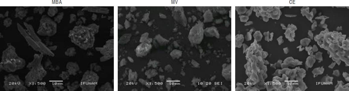
Figure 1 SEM micrographs showing compact particles, smooth surface and irregular shapes in MBA and MV. EC shows coral-shaped particles.
As for the elemental composition obtained by EDS, we were able to corroborate the presence, in the three cements of base elements such as C4+, Ca2+, O2- and Si4+ responsible for the formation of tricalcium and dicalcium silicate. The most abundant observed element after oxygenwas calcium followed by carbon, except in MV (Table 1). Magnesium and sodium were also present in MV. Sodium was only present in MBA (Table 1). Sulfur, potassium, and titanium were found only in MV (Table 1).
Table 1 Elements in weight percentage (wt%) present in MTA MBA, MV and EC.
| Elemento | MBA Wt% |
MV Wt% |
CE Wt% |
| C | 14.3 | 21.04 | 10.42 |
| O | 31.33 | 41.29 | 46.4 |
| Si | 3.13 | 13.51 | 7.52 |
| Ca | 16.95 | 13.34 | 23.79 |
| Na | 0.11 | 1.21 | - |
| Bi | 32.63 | - | - |
| Zr | - | - | 2.34 |
| Al | 1.04 | 3.29 | 0.17 |
| Ti | - | 3.57 | - |
| Mg | - | 0.32 | - |
| K | - | 1.33 | - |
Bismuth was also observed in MBA and zirconium in CE as radiopacifying agents.
XRD
The main component present in the three powder cements was tricalcium silicate. In MBA (Figure 2) and EC (Figure 3), the crystalline phase of dicalcium silicate and the radiopacifying agent were also present. These findings corroborate the results obtained by SEM-EDS with regard to the elements that were only present in MV (Figure 4), showing the rutile, magnesium oxide, and calcite phases.
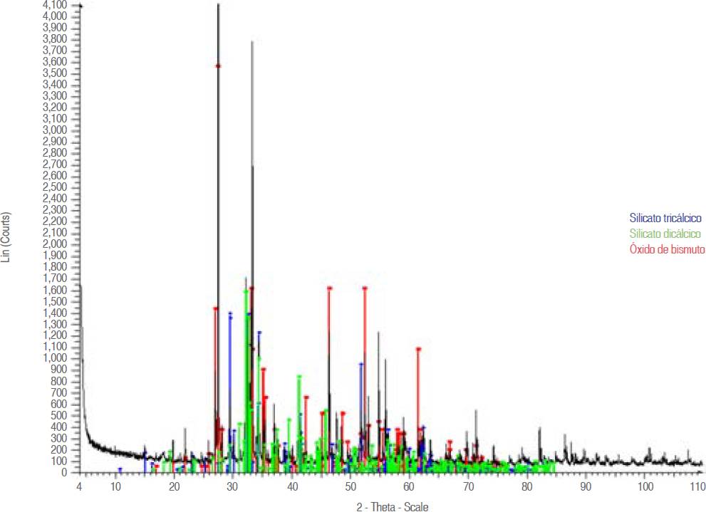
Figure 2 Diffractogram of MBA powder, showing the two main phases of the cement which are tricalcium silicate and dicalcium silicate. The radiopacifying agent may also be observed.
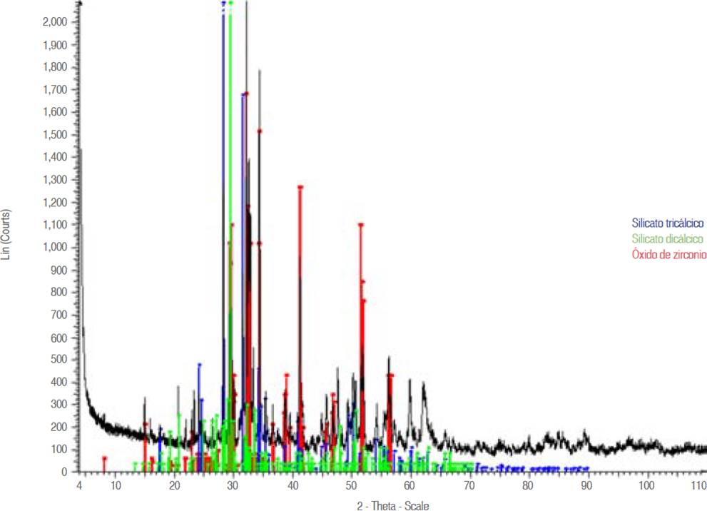
Figure 3 Powder EC diffractogram showing the main phases equal to MBA, except Bi2O3; ZrO2 is present instead.
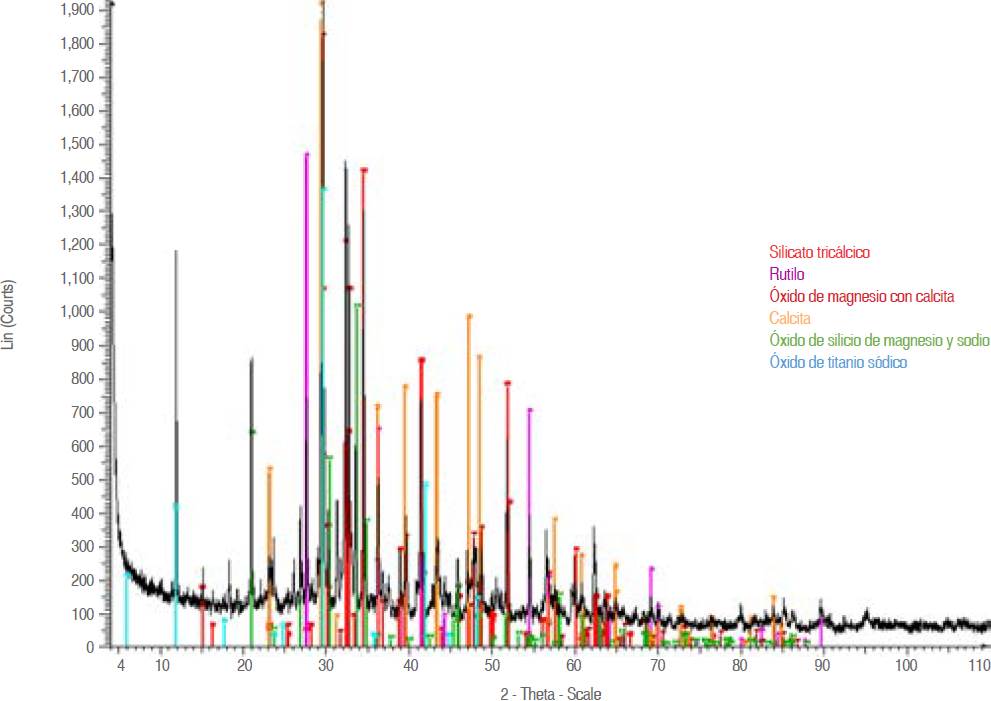
Figure 4 Powdered MV diffractogram showing the tricalcium silicate phase and other phases different from those present in the other two analyzed cements.
The results obtained in the cement 24 hours after setting showed the presence of tricalcium silicate (S) in MBA (Figure 5), MV (Figure 6), and EC (Figure 7). Bismuth oxide (B) and zirconium oxide (Z) were also found as radiopacifying agents in MBA and EC, respectively. In EC the calcium alumínate (A) and dicalcium silicate (L) phases were observed. Only in MV the phases of hatrurite (H), inyoite (I), ettringite (E), and gypsum (Y) could be observed and they were seen throughout the 28 days of hardening.
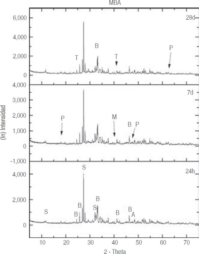
Figure 5 Diffractogram of MBA at different setting times. S tricalcium silicate, B radiopacifier (bismuth oxide), P portlandite and T C-S-H gel.
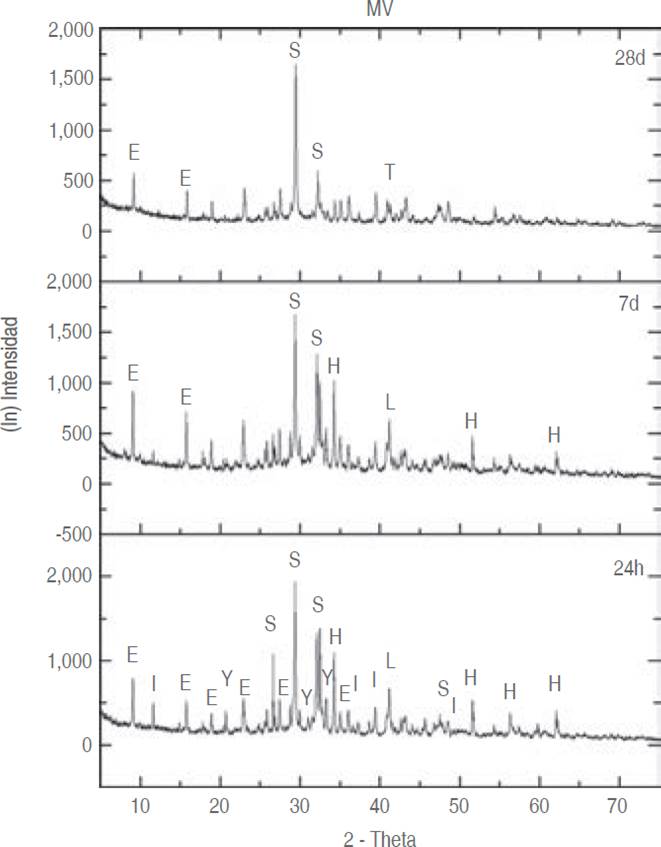
Figure 6 MV diffractogram at different setting times. S tricalcium silicate, L dicalcium silicate, E ettringite, Y gypsum, I inyoite, H hatrurite and T C-S-H gel.
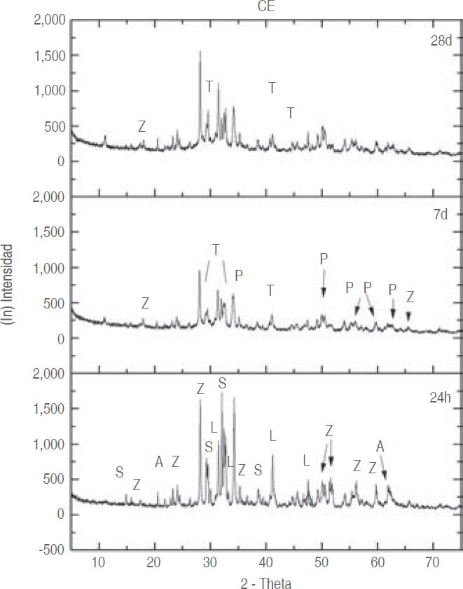
Figure 7 EC diffractogram at different setting times. S tricalcium silicate, L dicalcium silicate, A tricalcium alumínate, Z radiopacifier (zirconium oxide), P portlandite and T C-S-H gel.
At 7 and 28 days the formation of two extra phases such as calcium silicate hydrate (CSH gel) and portlandite (Ca (OH)2) could be identified in both MBA (Figure 5) and EC (Figure 7), but not in MV (Figure 6).
pH
The groups that showed statistically significant differences were as follows: at 24 hours MBA (pH 8.5) ≠ MV (pH 10) and EC (pH 10); at 7 days all showed differences between them, MBA (pH 8.5), MV (pH 7.8) and EC (pH 10.6) and at 28 days MV (pH 7.9) ≠ MBA (pH 8.2) and EC (8.2). These results are shown in Table 2.
Table 2 Means and standard deviations of pH, setting time, compressive strength and dimensional change tests.
| MBA 27 ± 5.6 |
MV 29 ± 3.6 |
CE 21.9 ± 2.3 |
|||||||
| Tiempo de fraguado [min] | 24 horas | 5.67 días | 28 días | 24 horas | 3.67 días | 28 días | 24 horas | 2.37 días | 28 días |
| pH | 8.5 ± 0.1 | 8.5 ± 0.3 | 8.2 ± 0.2 | 10 ± 0.5 | 7.8 ± 0.5 | 7.9 ± 0.1 | 10 ± 0.3 | 10.6 ± 0.5 | 8.2 ± 0.04 |
| Resistencia a la comprensión [Mpa] | 14.3 ± 5 | 23 ± 7.8 | 32 ± 11 | 14.7 ± 5 | 17 ± 3.9 | 19.5 ± 5.1 | 23.5 ± 3 | 36.3 ± 9 | 36.7 ± 6 |
Setting time
Similarly, statistically significant differences were found between EC with MBA and MV. As shown in Table 2, MBA showed 27 minutes, MV 29 minutes, and CE 22 minutes.
Compressive strength
The results of the statistical analysis showed that statistically significant differences werefound between MBA, MV, and EC. The groups that showed differences were as follows: at 24 hours and 7 days EC ≠ MBA and MV ≠ EC; 28 days MV ≠ MBA and EC. This means that the specimens that showed higher compressive strength at 24 hours, 7 days, and 28 days after the start of the setting of the material were those of EC since they showed 23.5 MPa, 36.3 MPa, and 36.7 MPa, respectively. MV showed 14.7, 17, and 19.5 MPa and MBA 14.3, 23, and 32 MPa showing a constant increase over time (Table 2).
DISCUSSION
The ideal properties of MTA cement are indispensable for its clinical application. Such attributes are mainly: easy handling, adequate setting time, dimensional stability, and low solubility in living tissues. Therefore, constant improvements of this cement are made or new cements are created to try to meet the desired requirements. In this study, an analysis of two commercial MTA cements and an experimental MTA cement was performed by means of SEM-EDS, XRD, and some physical-mechanical tests.
It was found by means of SEM and corroborated by XRD, that MV cement presented the phases corresponding to a Portland cement Type 1, including the gypsum phase, whose function is to be an excellent setting retardant agent17 without interfering in the properties of the cement, as long as the maximum percentage of content is below 4%18 because the ions could degrade the cement surface.19 Another important phase that appears in MV is ettringite, (Ca6Al2(SO4)3(OH) .26H2O), which is formed from the sulfate content of gypsum and tricalcium aluminate (C3A).17 This cement also has among its components sodium and magnesium silicon oxide, of which Mg is very commonly used for phase stabilization and Na, to lower the melting temperature.20 Na was found in both MV and MBA. An unexpected finding was that, apparently, MV showed no radiopacifier or any element responsible for it, unlike MBA and EC which contain Bi2O3 and ZrO2, respectively. Therefore, it is recommended to perform a radiopacity test in the future in order to confirm the absence of such an agent, as well as a more exhaustive chemical analysis.
Regarding the physical-mechanical tests, an interesting phenomenon was observed: the shortest setting time was exhibited by EC (22 minutes) and the longest, was that of MV (29 minutes), leaving MBA with an intermediate time of 27 minutes. These findings do not coincide with those reported by Flores21 who described a time of 18 minutes. These differences can be attributed to the fact that EC does not possess gypsum among its components. It has already been mentioned that gypsum is a retarder.17
pH is a very important factor within MTA cements since it confers them a bactericidal property since, due to its high alkalinity, it regulates the catalytic activity of some enzymes influencing the reaction speed. When the external medium is modified, the cellular activity of the bacteria may be affected.22 It has been reported that MTA has a great capacity to release OH-, increasing pH and Ca2+ improving the biocompatibility of the material,16 as well as being useful to decrease microfiltration.23 In this study, it could be seen that MV and EC show a pH of 10 within the first 24 h and MBA is slightly below them. However, as the cement hardening time progresses, the pH of MV decreases more than the other two cements, thus the three cements end with a pH close to 8. This rise and fall of pH over time may be caused by the formation of the portlandite phase (Ca(OH)2), which provides the medium with OH- and Ca2+ hence supersaturating it.24 This phase can be observed in the MBA and EC diffractograms but not in MV. This does not mean that it is not present in MV but possibly it appears in very low quantity
Another test that was performed was resistance to compression, even though it is known that this type of material, being an apical filling material, does not need to support direct pressures.25 It was decided to perform this test in order to follow up on the hardening of the cement since Portland-type cements take approximately 30 days to harden from the beginning of the mixture. The cement with the highest compressive strength at 24 hours was EC with 23.47 MPa, MV with 14.73 MPa and the lowest was MBA with 11.2 MPa. These results coincide with those reported by Flores.21 The cements finished at 28 days with 32 MPa for MBA, 19.5 MPa for MV and the highest value was for EC with 36.7 MPa. EC values at 24 hours were in agreement with those of an experimental cement reported by Grech in 2013.26
This high strength could be due to the formation of portlandite, which forms at the same time as the hydrated calcium silicate gel (C-S-H gel), a phase that is very difficult to see by diffraction because it is amorphous in nature.27 The importance of C-S-H gel relies on the fact that it is the primary bonding phase in the cement and that there are amorphous nanoporous particles that surround the cement grains,24 thus increasing its strength. The portlandite phase also helps to increase strength because it nucleates and grows within voids or between pore spaces.28 Therefore, since these phases do not appear in MV cement (by means of XRD), its low compressive strength is understood,attributingitto slightly deficient hydration. As hydration progresses, the C-S-H gel network stabilizes a few minutes after hydration has begun.24 It is worth mentioning that as the cement hardens its compressive strength increases over time. This is considered an attribute that stands out in the different studies conducted by several authors, provided that there is a minimum of humidity in the environment where it is located, since, as mentioned above, this material sets in the presence of humidity.29
CONCLUSIONS
The properties of MTA cements are strongly influenced by the presence of trace elements foreign to their main composition, hydration efficiency, as well as the formation of portlandite and C-S-H gel, as can be observed in the results obtained from MTA Viarden™.
The experimental cement so far is a good candidate to compete with commercial MTA cements, however, further tests are needed.
ACKNOWLEDGMENTS
To Diego Armando Quiterio Vargas and Manuel Aguilar Franco for the sample preparation and SEMEDS imaging performed at the Central Microscopy Laboratory of IFUNAM. To Antonio Espino for obtaining and analyzing the diffractograms at the X-Ray Laboratory of the Institute of Physics, UNAM. To Teresa Baeza Kingston for facilitating materials and equipment used at the Dental Biomaterials Laboratory of the Division of Post-graduate Studies and Research, Faculty of Dentistry, UNAM.
REFERENCES
1. Ingle J, Taintor JF. Endodoncia. 5ta ed. México: McGraw-Hill Interamericana; 2004. [ Links ]
2. Al-Hezaimi K, Al-Shalan TA, Naghshbandi J, Oglesby S, Simon JHS, Rotstein I. Antibacterial effect of two mineral trioxide aggregate (MTA) preparations against Enterococcus faecalis and Streptococcus sanguis in vitro. J Endod. 2006; 32 (11): 1053-1056. doi: 10.1016/j.joen.2006.06.004 [ Links ]
3. Torabinejad M, White DJ. Tooth filing material and method of use [online]. United States Patent US5769638A. June 23, 1998. [Consultado septiembre 2017] 8-10 p. Disponible en: Disponible en: https://patentimages.storage.googleapis.com/13/b6/a1/818b44ab8f72b0/US5769638.pdf [ Links ]
4. Camilleri J, Pitt Ford TR. Mineral trioxide aggregate: A review of the constituents and biological properties of the material. Int Endod J. 2006; 39 (10): 747-754. doi: 10.1111/j.1365-2591.2006.01135.x [ Links ]
5. Hsien H-C, Cheng YA, Lee YL, Lan WH, Lin CP. Repair of perforating internal resorption with mineral trioxide aggregate: a case report. J Endod. 2003; 29 (8): 538-539. doi: 10.1097/00004770-20030800000011 [ Links ]
6. Faraco Junior IM, Holland R. Histomorphological response of U dogs' dentalpulpcappedwith white mineral trioxide aggregate. Braz Dent J. 2004; 15 (2): 104-108. doi: 10.1590/s0103-64402004000200004. [ Links ]
7. Menezes R, Bramante CM, Letra A, Carvalho VG, Garcia RB. Histologic evaluation of pulpotomies in dog using two types of mineral trioxide aggregate and regular and white Portland cements as wound dressings. Oral Surgery Oral Med Oral Pathol Oral Radiol Endodontology. 2004; 98 (3): 376-379. doi: 10.1016/S107921040400215X. [ Links ]
8. Holland R, de Mello W, Nery MJ, de Souza V, Bernabe P, Otoboni Filho J. Healing process of dog dental pulp after pulpotomy and pulp covering with calcium hydroxide in powder or paste form. Acta Odontol Pediatr. 1981; 2 (2): 47-51. [ Links ]
9. Felippe WT, Felippe MC, Rocha MJ. The effect of mineral trioxide aggregate on the apexi cation and periapical healing of teeth with incomplete root formation. Int Endod J . 2006; 39 (2): 2-9. doi: 10.1111/j.1365-2591.2005.01037.x. [ Links ]
10. Holland R, de Souza V, Nery MJ, Otoboni Filho JA, Bernabé PFE, Dezan Júnior E. Reaction of dogs' teeth to root canal filling with mineral trioxide aggregate or a glass ionomer sealer. J Endod. 1999; 25 (11): 728-730. doi: 10.1016/s0099-2399(99)80118-6. [ Links ]
11. Bortoluzzi E, Broon N, Bramante C, Garcia R, de Moraes I, Bernardineli N. Sealing ability of MTA and radiopaque portland cement with or without calcium chloride for root-end filling. J Endod. 2006; 32 (9): 897-900. doi: 10.1016/j.joen.2006.04.006. [ Links ]
12. Granados S, Alcalde C, Guzman J, Melendez D, Torres C, Velasquez Z. Cementos a base de silicato de calcio: factor clave en el éxito del recubrimiento pulpar directo. Revisión de la literatura. RevEstomatol Herediana. 2022; 32 (1): 52-60. doi: 10.20453/reh.v32i1.4183. [ Links ]
13. Lee YL, Lee BS, Lin FH, Yun Lin A, Lan WH, Lin CP. Effects of physiological environments on the hydration behavior of mineral trioxide aggregate. Biomaterials. 2004; 25 (5): 787-793. doi: 10.1016/s0142-9612(03)00591-x. [ Links ]
14. Camilleri J, Montesin FE, Di Silvio L, Pitt Ford TR. The chemical constitution and biocompatibility of accelerated Portland cement for endodontic use. Int Endod J . 2005; 38 (11): 834-842. doi: 10.1111/j.1365-2591.2005.01028.x. [ Links ]
15. International Standards Organization. ISO 6876:2001. Dental root canal sealing materials. [Internet]. [Consultado septiembre 2017]. Disponible en: Disponible en: https://www.iso.org/standard/34965.html [ Links ]
16. Massi S, Tanomaru-Filho M, Silva GF, Duarte MA, Grizzo LT, Buzalaf MA et al. pH, calcium ion release, and setting time of an experimental mineral trioxide aggregate-based root canal sealer. J Endod. 2011; 37 (6): 844-846. doi: 10.1016/j.joen.2011.02.033 [ Links ]
17. Calleja J. Puntos de vista sobre el contenido de yeso de los cementos Portland. Mater Construcción. 1965; 15 (120): 41-57. doi: 10.3989/mc.1965.v15.i120.1725. [ Links ]
18. Crammond NJ. Quantitative X-ray diffraction analysis of ettringite, thaumasite and gypsum in concretes and mortars. Cem Concr Res. 1985; 15 (3): 431-441. doi: 10.1016/0008-8846(85)90116-4. [ Links ]
19. Silva Hermo BM, Prieto Lamas BL, Rivas Brea T, Pereira Pardo L. Origen y efectos deteriorantes del yeso en monumentos graníticos del noroeste de España. Materiales de construcción. Mater Construcción . 2010; 60 (297): 97-110. doi: 10.3989/mc.2010.46808 [ Links ]
20. Iwamoto N, Tsunawaki Y, Fuji M, Hatfori T Raman spectra of K,OSiO2 and K2OSiO2TiO2 glasses. J Non Cryst Solids. 1975; (2): 303-306. doi: 10.1016/0022-3093(75)90029-0. [ Links ]
21. Flores LA. Influencia de la Wollastonita y vidrio bioactivo en las propiedades físicas de un cemento tipo Portland. [Tesis maestría]. México: Universidad Nacional Autónoma de México; 2011. 98 pp. Disponible en: https://ru.dgb.unam.mx/handle/DGB_UNAM/TES01000680222 [ Links ]
22. Estrela C, Sydney GB, Bammann LL, Felippe Júnior O. Mechanism of action of calcium and hydroxyl ions of calcium hydroxide on tissue and bacteria. Braz Dent J. 1995; 6 (2): 85-90. [ Links ]
23. Camilleri J, Pitt Ford TR. Evaluation of the effect of tracer pH on the sealing ability of glass ionomer cement and mineral trioxide aggregate. J Mater Sci Mater Med. 2008; 19 (8): 2941-2948. doi: 10.1007/s10856-008-3429-y. [ Links ]
24. Jonsson B, Wennerstrom H, Nonat A, Cabane B. Onset of cohesion in cement paste. Langmuir. 2004; 20 (16): 6702-6709. doi: 10.1021/la0498760. [ Links ]
25. Torabinejad M, Hong CU, McDonald F, Pitt Ford TR. Physical and chemical properties of a new rootend filling material. J Endod. 1995; 21 (7): 349-353. doi: 10.1016/S0099-2399(06)80967-2 [ Links ]
26. Grech L, Mallia B, Camilleri J. Investigation of the physical properties of tricalcium silicate cementbased root-end filling materials. Dent Mater. 2013; 29 (2): e20-28. doi: 10.1016/j.dental.2012.11.007. [ Links ]
27. Kirkpatrick J, Yarger JL, McMillan PF, Yu P, Cong X. Raman spectroscopy of C-S-H, tobermorite, and jennite. Adv Cem Bas Mat. 1997; 5 (3-4): 93-99. doi: 10.1016/S1065-7355(97)00001-1. [ Links ]
28. Zhao W, Wang J, Zhai W, Wang Z, Chang J. The self-setting properties and in vitro bioactivity of tricalcium silicate. Biomaterials. 2005; 26 (31): 6113-6121. doi: 10.1016/j.biomaterials.2005.04.025 [ Links ]
29. Hong CU, Torabinejad M, Kettering JD. The effects of three retrofilling materials on selected oral bacteria. J Endod. 1993;(4): 200. doi: 10.1016/S0099-2399(06)80752-1. [ Links ]
Received: January 01, 2018; Accepted: September 01, 2018
 Este es un artículo publicado en acceso abierto bajo una licencia Creative Commons
