




 nueva página del texto (beta)
 Inglés (pdf)
 Artículo en XML
 Referencias del artículo

 Traducción automática
 Enviar artículo por email
 Citado por SciELO
 Similares en
SciELO

 Permalink


Introduction
Traumatic axonal injury (TAI) is a mechanical disruption of neuronal axon cytoskeleton resulting in proteolysis, swelling, and molecular changes secondary to traumatic blunt injury to the brain1. In this context, it may present as a single brain injury in a traumatized patient, or accompany localized brain injuries, such as hematomas, lacerations, etc.,1.
Traumatic brain injury (TBI) is a global public health problem and has the highest incidence of all common neurological disorders. The TBI population-based incidence annual rates reported by the first Lancet Neurology Commission on TBI were 811/100,000 in New Zealand, and 979/100,000 in North America; hospital discharge rates in the European Union were 287.2/100 000 people/year.2 The mortality rate is equally as worrying as the years of life lost (YLLs) and years lived with disability. In Europe, each TBI death is associated with 24 YLLs.2 Although there are no global statistics on TAI, it is known that 60% of patients with severe TBI have TAI; patients with severe TBI and TAI have a mortality rate of 16%.2
In the United States, over 1.5 million cases of TBI are reported annually, according to the Centers for Disease Control and Prevention. Glasgow coma scale (GCS), classifies TBI in mild (13-15), moderate (9-12), and severe (< 8) injury.3 TAI affects the white matter tracts in the brain, and the manifestations that clinically affected patients may present range from the minimal to being in a comatose state. Our goal was to make a review of TAI, to provide the current concepts in this topic, familiarize the physician with this pathology and analyze the type and focus of the published articles.
Materials and methods
For this review, only the search terms "Traumatic axonal injury" were used. PubMed databases were screened from January 2023 to October 2024. All articles published during the selected period were included for review, provided that at least the abstract was in English. The Preferred Reporting Items for Systematic reviews and Meta-Analyses guidelines were employed (Fig. 1). The inclusion criteria were: articles focused on TAI in humans and animals. The exclusion criteria were: focus on TBI but not TAI, and duplicated articles. The search in PubMed with these keywords provided 401 articles in the period studied. In spite of the search terms, most of these articles focused on different types of brain trauma, not specifically on TAI.
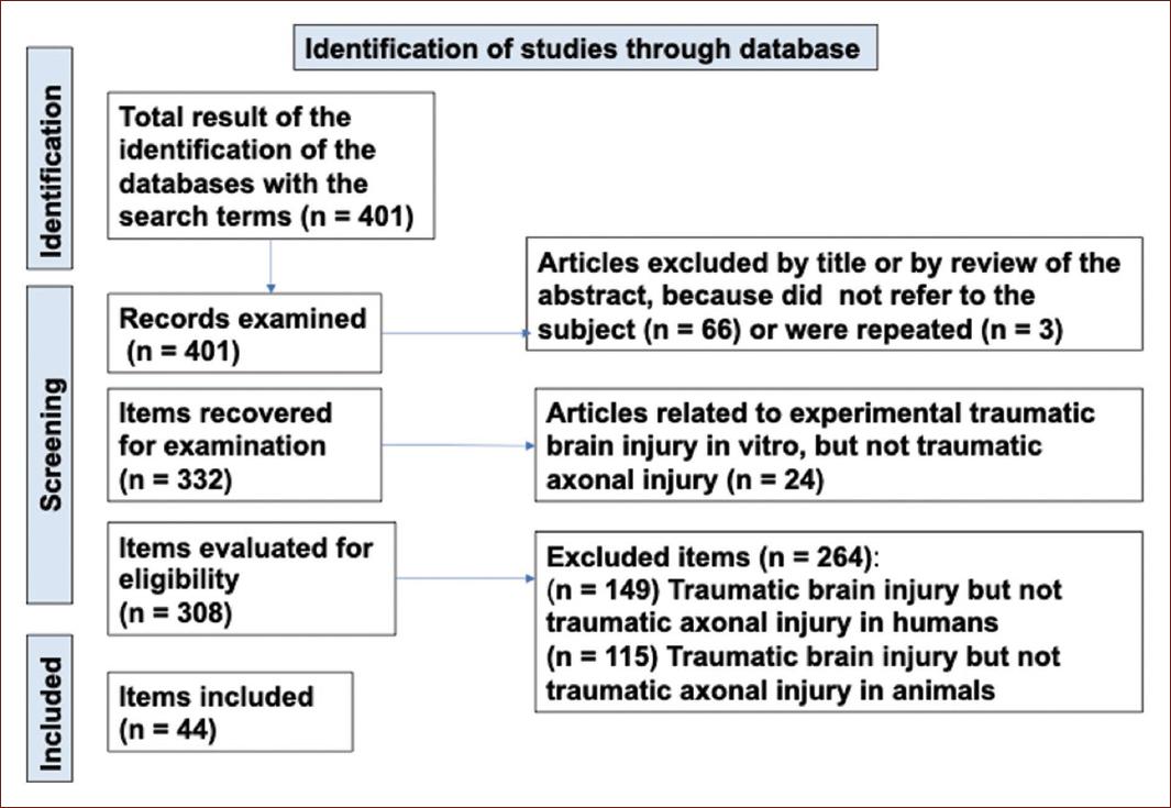
Figure 1 Flowchart diagram of the PRISMA (Preferred Reporting Items for Systematic reviews and Meta-Analyses) review on traumatic axonal injury.
Articles were excluded by two of us (ALCG, FGD) by title or by review of the abstract, because they did not refer to the subject (n = 66) or they were repeated (n = 3). Of the remaining 332 articles, 24 referred to experimental TBI, but not to TAI, and were eliminated. From the remaining 308 articles, 264 were excluded; 149 of them because they referred to TBI but not to TAI in humans. The remaining articles referred to TBI but not to TAI in animals. After discarding those articles that did not meet the inclusion criteria, 44 papers were included in this review (Fig. 1).3-47
This initial review was conducted independently by both authors, and those articles that were excluded by one author but not the other, were subsequently re-evaluated until an agreement was reached based on the inclusion and exclusion criteria.
Results
The articles published on TAI, in the period analyzed, were mostly reviews (n = 16), clinical observational studies (n = 18), postmortem studies (n = 4), case reports (n = 3), experimental studies in animals (n = 2), and a clinical experimental pilot study (n = 1).
TBI is a global public health problem. Although it affects between 800 and 1200 people/100,000, not all require hospitalization. The manifestations of TBI are multiple. The patient may have experienced a concussion or become comatose, with lesions that include fractured skull bones, dural laceration, epidural and subdural hematomas, subarachnoid hemorrhage, parenchymal hematomas, contusion, laceration and cerebral infarction. A common but not always recognized TBI is TAI.4
Pathophysiology
Damage is initiated by the tearing of axons in the brain white matter, usually due to a blunt trauma.3,4 It conditions a series of changes at the cellular level that magnify the damage, including alteration in ionic flow, especially of calcium; cytoskeletal disorganization; actin-spectrin complex degradation; alteration in neurofilament structure; mitochondrial damage; oxidative stress, and activation of apoptosis. Axonal transport disruption is evident after 24 h post-injury associated to deposit of amyloid protein pre-cursor (APP).4 Depending of the trauma intensity, petechial hemorrhages are also present in moderate-to-severe TAI, as a consequence of rapid acceleration and deceleration forces with damage of the capillaries, constituting the "micro-bleeds," an indirect marker of TAI.5
Clinical diagnosis and diagnostic imaging
How is TAI clinically suspected in a trauma patient? It is necessary to evaluate clinical and imaging characteristics. The severe form of TAI is considered in a patient with head trauma without demonstrable mass lesion but with a low GCS score (≤ 8) for more than 6 h, plus findings of multiple small white matter lesions in the grey-white matter junction.6 When a patient arrives at a shock-trauma room, the Glasgow scale is applied, which gives a quick idea of the severity of brain damage by evaluating three parameters: (A) Eye Opening (E): 4-spontaneous, 3-to voice, 2-to pain, 1-none; (B) Verbal response (V): 5-normal conversation, 4-oriented conversation, 3-words, but not coherent, 2-no words, only sounds, 1-none, and (C) Motor response (M): 6-normal, 5-localized to pain, 4-withdraws to pain, 3-decorticate posture, 2-decerebrate. A score of 13-15 is considered mild; 9-12 moderate, and ≤ 8, severe brain damage.6
Clinical severity of TAI correlated with three grades of progressive and cumulative damage: Grade 1, white matter injury of cerebral hemispheres; Grade 2, focal injury in the corpus callosum; Grade 3, lesion in the brainstem6. Even concussions may be associated with TAI. A concussion is a transient disturbance of brain function, usually < 15 minutes, secondary to mild TBI. Concussion studies in animal models and human subjects have shown TAI histopatologically.7 TAI causes disruption of brain functional networks.8 Although the computed tomography scan (CTS) is usually the first study available in patients with TBI, it has a low sensitivity for the detection of non-hemorrhagic lesions in the white matter, which constitute 80-90% of the lesions in TAI.9,10 However, it is possible to identify small, non-expansive hemorrhagic lesions in the white-grey matter, intraventricular blood and cerebral edema.10 In the initial CTS of a TBI patient, 24 h after the event, the presence of intraventricular hemorrhage and/or blood in the interpeduncular cistern is associated with a severe TAI, and this severity increases with the number of affected ventricles. This is explained because the corpus callosum and brain stem, located in the midline, are closely related to the ventricles. The forces involved that produce disruption and axonal rupture in these structures also produce vascular damage to small vessels, which conditions variable degrees of intraventricular hemorrhage and laminar subarachnoid hemorrhage in the midline, in the interhemispheric and peri-mesencephalic regions.10
Using magnetic resonance imaging (MRI), cases of TAI observed increase with the degree of severity of TBI, being 6% in mild cases, 70% in moderate, and 95% in severe cases.9 In fact, a Trondheim TAI-MRI grading (grades 1-5) has been proposed with bilateral TAI in mesencephalon or thalami, and bilateral TAI in pons as the worst grades.9
When the Mean Rotterdam computed tomography (CT) score, Injury Severity Scale, and admission GCS were used to evaluate patients with TAI, the results were similar; there was an association between TAI neuroanatomic injury pattern and neurologic recovery timing. This time was shorter in patients with grade 1-TAI, versus grades 2 and 3.11
Even in young subjects (≤ 24 years) with a diagnosis of mild BTI (13-15 GCS), the presence of involvement of the corpus callosum in the initial CTS is a poor prognostic factor for total recovery after 1-year post-TBI.12
CTS and MRI are used in the acute and also in the subacute and chronic phases to evaluate TAI.13,14 MRI lesion distribution maps have shown a higher frequency of TAI in the posterior corpus callosum; TAI in the splenium, mesencephalon, and pons is associated with poor outcome.14,15
Assessment of white matter integrity by diffusion MRI tractography is able to quantify white matter microstructure in vivo.16 This technique has given evidence that the pre-motor, temporal, and parietal sections of the corpus callosum are the most commonly damaged tracts.16 In TAI, the corpus callosum may show intraventricular hemorrhage on the initial CT study. Later on, post-traumatic corpus callosum damage can be evaluated by MRI sequences. Using diffusion tensor imaging (DTI) and tractography, a significant decrease in the number of corpus callosum fibers is reported, compared with those in healthy control patients.17
Imaging has taken on a very important role in the diagnosis and prognosis of TAI.18 The incorporation of artificial intelligence is already increasing diagnostic accuracy, and improvements in diagnosis are expected in the short term.48
Biomarkers
In cases of TBI and TAI, outcome predictive biological markers are required.19 In recent years, various biomarkers have been suggested in the diagnosis and prognosis of TAI, including tau protein, β-Amyloid precursor protein, S-100β, neurofilaments, myelin basic protein, neuron-specific enolase, hemopexin, peripherin, the neuronal protein ubiquitin carboxyl-terminal hydrolase-L1 (UCH-L1) and glial fibrillary acidic protein (GFAP) (these past two, included in the test Brain Trauma Indicator™).20 Two axonal proteins, neurofilament-light chain (NF-L) and phosphorylated neurofilament-heavy chain (pNF-H), have a serum level peak at day 20-30 post-moderate-severe TBI, and this finding is associated with patient outcomes and disability.21 NF-L is a not specific-disease biomarker of axonal damage.22 However, some authors have found that, in patients with initially moderate/severe TBI, persistently high NF-L levels are associated with worse outcome after TBI, reflecting ongoing brain injury.23 Furthermore, it seems that early levels of plasma NF-L may associate with the presence of TAI at a later phase of TBI, evaluated by DTI measurements of TAI.24 Neurofilaments are biomarkers of neuro-axonal damage in several neurological disorders, from multiple sclerosis to TBI.25 The usefulness of neurofilaments as a marker in TAI is still in process.25
Neuron-specific enolase, a glycolytic enzyme, is not normally present in extracellular fluids.26 If neuronal damage is suspected, its serum levels should be determined within 12-24 h post-injury. In adults, levels > 9 μg/L and, in children, levels > 15 μg/L within 24 h suggest poor prognosis when combined with CTS abnormalities.26
Peripherin is a class III-intermediate filament located in the axonal tract throughout the whole brain. In experimental animals and in humans, axonal injuries with peripherin inclusion are observed post-TBI. Furthermore, cerebral spinal fluid (CSF) and plasma levels are elevated at the early phase post-TBI. Hence, its usefulness as a biomarker in TAI is being investigated.27
Microtubule-associated protein tau is found to be hyperphosphorylated (p-tau) in injured axons and has been studied as a biomarker for brain injury and TAI. In rats with severe spinal cord injury at T8, the presence of p-tau in damaged axons showed a similar time course and distribution pattern to β-APP, a biomarker of axonal injury.28
Two glial biomarkers, the GFAP and the S100 protein, have been studied. GFAP is an intermediate filament protein (half-life of 24-48 h); its serum levels measured within 24 h post-injury are 10 times higher in CTS-positive patients than in CTS-negative patients.29 After TBI, serum levels of NF-L and GFAP seem to be associated with progression of brain atrophy,29 and early, persistently-elevated serum NF-L levels are associated with a worse outcome.29
On the other hand, S-100b has been associated with neuroinflammation and oxidative stress post-TBI because it induces microglia migration and activation through the S-100/ICaBP-type calcium-binding domain.30 Neurogranin has also been evaluated as a marker of TAI in rats.31 UCH-L1 is a protein found in the cytoplasm of neurons. There is an increased serum level after TBI.32
Biomarker research in TAI is ongoing (Table 1).20-22,26-33 There are no biomarkers yet approved for routine clinical use, because the determination in different matrices (CSF, blood, plasma) is not yet sufficiently standardized. Combining several biomarkers in CSF and blood, as well as image analysis, is expected to improve accuracy and help predict outcomes in TAI.30,32
Table 1 Biomarkers for diagnosis and prognosis in TAI
| References | Marker | Characteristics |
|---|---|---|
| Zhang et al.,20 2024 | UCH-L1 | Deubiquitinating enzyme. Regulates target protein degradation primarily in neurons and neuroendocrine cells in the brain. It is also a neuronal antioxidant |
| Zhang et al.,20 2024 | S100b | S100 proteins form a family characterized by having two calcium-binding sites. They are expressed in astrocytes, Schwann cells, melanocytes, adipocytes, macrophages, and chondrocytes. S100β is a calcium sensor protein that regulates cellular energy metabolism by controlling the phosphorylation of protein kinase C and calmodulin and ribonucleic acid synthesis |
| Wang et al.,21 2024 | NF | Neuronal intermediate filament, three subtypes based on molecular weight: light chain (NF-L), medium chain, and heavy chain. Structural role in the axonal cytoskeleton. Involved in the transport and docking of mitochondria, and intracellular signaling and transcription. It regulates large fast-conducting neurons' radial growth |
| Coppens et al.,22 2023 | NF | Neuronal intermediate filament, three subtypes based on molecular weight: light chain (NF-L), medium chain, and heavy chain. Structural role in the axonal cytoskeleton. Involved in the transport and docking of mitochondria, and intracellular signaling and transcription. It regulates large fast-conducting neurons' radial growth |
| Kim and Kim26 2024 | NSE | Cell-specific isoform (neurons and neuroendocrine cells) of enolase, a glycolytic enzyme. Role in glucose metabolism and adenosine triphosphate production. Half-life 24 h |
| Fang et al.,27 2023 | Peripherin | A class III-intermediate filament is predominantly expressed in the peripheral nervous system. Interacts with neurofilaments and kinases, influencing cytoskeletal dynamics and neuronal function |
| Caprelli et al.,28 2018 | Tau | Structural element of the axonal cytoskeleton. It maintains the stability of microtubules and coordinates the movement of molecules along the microtubules. After TBI, acetylated tau accumulates in the blood and plays a role in the acute phase of secondary brain injury, leading to axonal degeneration and neurological impairment |
| Shahim et al.,29 2024 | GFAP | Glial fibrillary acidic protein (GFAP) in a type III intermediate filament and structural protein of astrocytes and ependymal cells. It plays a role in cellular homeostasis, neurotransmitter regulation, and synaptic function. In TBI, GFAP is upregulated as part of astrogliosis |
| Zhang et al.,30 2024 | α-II spectrin | Cytoskeletal protein located in neurons and highly concentrated in axons and presynaptic terminals. A rise in α-II spectrin degradation is an early indication of calpain and caspase-3 action linked to cell necrosis |
| Zhang et al.,30 2024 | miRNAs (miR21, miR16, miR92) | miRNAs are non-coded, small-sized (20 nucleotides),
hairpin-like structures involved in the post-transcriptional
silencing of messenger RNA (mRNA) and downregulation protein
synthesis Levels 3 times higher, in the brain of patients that die from TAI |
| Baskurt et al.,31 2024 | Neurogranin | Neurogranin is a calmodulin-binding protein participating in the protein kinase C signaling pathway that controls long-term potentiation (LTP). Its expression has been corroborated in the brain, aortic endothelial cells, and cardiomyocytes |
| Castaño-Leon et al.,32 2022 | GFAP, t-Tau, UCH-L1, and NfL | Combination of biomarkers to assess prognosis |
| Sharma et al.,33 2023 | Amyloid precursor protein (APP) | Membrane protein expressed in neurons. APP is involved in forming synapses and neuronal transport |
TBI: traumatic brain injury; TAI: traumatic axonal injury.
Neuropathology
In TAI, there are three main findings: (1) different degrees of axonal damage (axonal swellings, axonal bulbs),33 (Figs. 1 and 2), (2) Vascular damage (hemorrhages covering the vessel and adjacent tissue and perivascular ring hemorrhages with dilated axons between the hemorrhage and the vessel),33 (Figs. 3-5) Hypoxic-ischemic changes (acidophilic degeneration of neurons), as well as perivascular infarcts with accumulations of macrophages.33 Based on these parameters, a histological grading system has been proposed in sections stained with Hematoxylin and Eosin (H&E).33
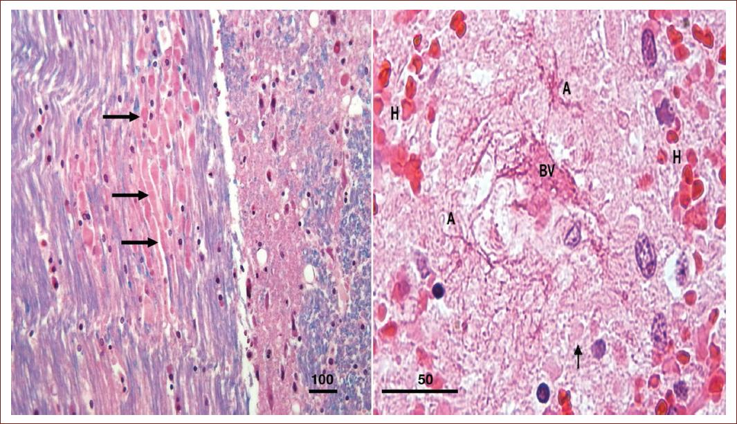
Figure 2 TAI in a 27-year-old motorcyclist. Left: pons. Eosinophilic axonal tracts with bulbous dilations (arrows) are observed in a longitudinal section. ×100, LFB-H&E. Right: high magnification of a central blood vessel (BV) surrounded by hypertrophic stellar astrocytes (A). Axonal spheroids (arrows) are visible inside the hemorrhagic ring (H). ×400, LFB-H&E (Autopsied case at our Institute). H&E: Hematoxylin and Eosin.
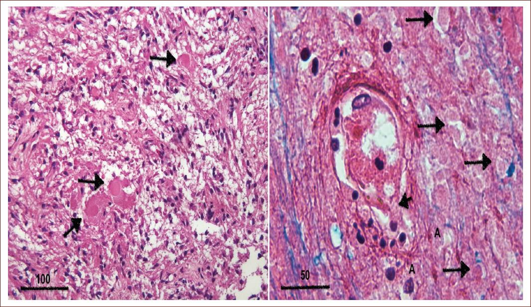
Figure 3 Neurohypophysis shows axonal spheroids (arrows). Left: H&E, ×100; Close-up of a vessel in the thalamus showing numerous perivascular axonal spheroids with loss of myelin (arrows). Loss of continuity is observed in the vessel wall (short arrow) and some extravasated inflammatory cells are observed. Right: LFB-H&E, ×400. H&E: Hematoxylin and Eosin.
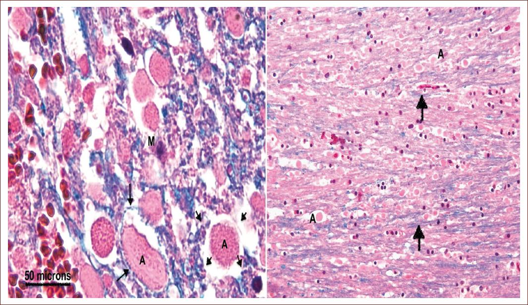
Figure 4 Left: axons (A) are observed in cross-section that show dilation and eosinophilia, as well as thinning and/or loss of the myelin sheath (arrows). Pons, luxol-fast-blue and hematoxylin, ×400. Right: few remaining myelin sheaths are observed, which stain blue with LFB&H (arrows), as well as numerous axonal bulbs devoid of their sheath (A). Putamen, ×100. H&E: Hematoxylin and Eosin.
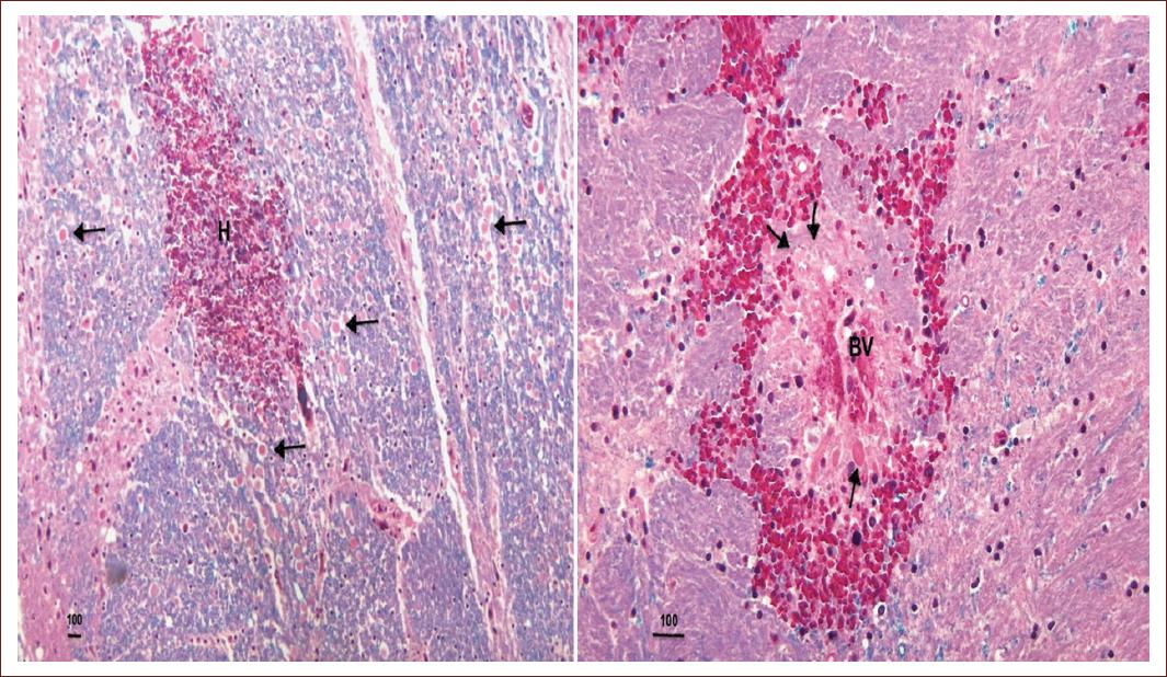
Figure 5 Parenchymal hemorrhage patterns Left: pons. The parenchymal hemorrhage (H) completely obscures the vessel. Numerous dilated eosinophilic axons are identified (arrows). LFB-H&E ×50. Right: pons. A blood vessel (BV) is centrally observed. A hemorrhagic ring is identified. Between this ring and the vessel, numerous dilated axons measuring between 3 and 50 microns are observed (arrows). LFB-H&E, ×100. H&E: Hematoxylin and Eosin.
A case of severe TAI, autopsied at our Institute (Figs. 2-5), with 5-day survival after trauma, is illustrated, showing diffuse axonal damage (Fig. 2), including involvement of the neurohypophysis (Fig. 3). The patient was unconscious on scene, with a Glasgow score of 8, and apart from the edema and parenchymal hemorrhages, there were no other injuries to the skull and brain. On the 5th day after the accident, the loss of the myelin sheath was evident in many of the dilated axons (Fig. 4). Axonal damage was evident on H&E stain and two types of hemorrhage were observed; one which completely obscured the vessel and the ring hemorrhages, where numerous spheroids were seen between the blood vessel and the hemorrhagic ring (Fig. 5).
A useful marker for postmortem evaluation of TAI is amyloid β protein, which is deposited in damaged axons within 30-40 min after trauma. Aβ peptides are cleaved from the APP, a membrane protein with a membrane-spanning domain, an extracellular glycosylated N-terminus and a cytoplasmic C-terminus, expressed in the synapses of neurons.34 It is not specific to trauma, but its presence implies axonal damage.
Hemorrhages in the corpus callosum are a marker of primary vascular injuries in TAI. Hemorrhage per rhexin refers to that caused by the rupture of a blood vessel, often due to mechanical force. In TAI, hemorrhagic lesions are small (4 × 0.8 mm), elongated, and in clusters.35
Generally, autopsies performed in forensic medical services are of individuals who either died instantly during the accident or survived a few hours or days. In these acute cases, excluding contusions and hypoxic-ischemic changes, the damage is predominantly axonal and vascular. However, there is a report of αb-crystallin-positive balloon neurons predominantly in the anterior cingulate, in three subjects with severe TAI who survived 5-10 weeks. These neurons were reminiscent of the phenomenon of chromatolysis.36
In this review on TAI, only four articles focused on neuropathology were found. The chronology of the lesions is important in acute and chronic states of this pathology. The sequence of events that occur in the different brain regions involving neurons, glia, and blood vessels is also important to understand the pathophysiology. Finally, neuropathological studies in humans are very valuable for comparing with the results of experimental studies in other species. It would be useful to apply this knowledge in the acute therapy and in the prevention of sequelae.
Initial diagnosis, treatment, rehabilitation, and prognosis
The initial evaluation of patients with TAI is very important. Although hemorrhage from the corpus callosum is generally associated with TBI and severe TAI, there are reports of such hematomas in children with concussion after minor blows.37 On the other hand, even TAI patients with favorable outcome may present different domains of cognitive impairment.38 Therefore, in TBI patients, a rapid and complete evaluation is of the outmost importance at admission to implement the best treatment.2
90% of the 4 million TBIs occurring in the world are mild or concussions,39 however, there are many health consequences of TBIs: disruptive behavior disorders in girls and boys,40 sleep disturbances,41 increased risk of developing Alzheimer's Disease,42 Parkinson's Disease, chronic traumatic encephalopathy with memory loss, aggression, confusion, depression, impaired judgment, difficulty controlling impulses, fatigue, anxiety disorders, headaches/migraines or vestibulo-ocular problems, and a nearly-doubled risk of suicide.43
In patients who survive and are admitted to a hospital, TAI findings and prognosis will depend on the severity of the initial insult based on the GCS on admission (mild, moderate, or severe injury) and the survival time.2
After a BTI, the situational diagnosis is important to determine therapeutic behaviors. In addition to the GCS, there are other evaluation models that can help predict an unfavorable evolution in the following 6 months, such as the Trauma and Injury Severity Score (TRISS) and International Mission for Prognosis and Analysis of Clinical Trials (IMPACT) models; or in the prediction of mortality, such as TRISS, Corticosteroid Randomization After Significant Head Injury, and IMPACT models.44
Supportive measures and ventilatory support, a surgical team to resolve concomitant problems, and having an intensive therapy unit for patients with shock-trauma are essential to ensure comprehensive, rapid, and effective medical care. Once the patient is stabilized, early physical therapy is highly recommended.45
In the acute phase, some medications have been tested, such as amantadine, which have demonstrated their usefulness in improving the cognitive disability associated with severe trauma.46 Even the mildest TAI injuries, such as concussion, require early rehabilitation and determining when it is appropriate to return to daily activities.47 The summary by topic of the articles analyzed can be found in table 2.
Table 2 Articles included in the review
| No. of paper | References | Main concepts | Article type |
|---|---|---|---|
| TAI general review | |||
| 1 | 3 | General concepts of TBI and TAI | Review |
| Physiopathology | |||
| 2 | 4 | Cytoskeletal and axolemmal changes in TAI.: Amyloid Precursor Protein (APP) accumulation, neurofilament alterations, calpain-mediated spectrin degradation, and sodium channel (NaCh) pathology | Review |
| 3 | 5 | Different techniques needed to identify axonal lesions and to understand lesion mechanisms | Review |
| Clinical diagnosis and diagnostic imaging | |||
| 4 | 6 | TBI and TAI | Review |
| 5 | 7 | Diagnosis of TAI in patients with concussion | Mini-review |
| 6 | 8 | Study of brain functional networks and cognitive impairment | Retrospective case-control study. MRI |
| 7 | 9 | Improvement of the TAI grading system to reflect the outcome. MRI-Glasgow Outcome Scale | Prospective clinical and imaging study |
| 8 | 10 | CTS-MRI in pediatric patients with TAI | Review |
| 9 | 11 | Systematic review and consensus process for inclusion of biological markers, including MRI, in patients with TAI | Systematic review |
| 10 | 12 | Global outcome in patients with TBI-TAI based in CTS and Glasgow Outcome Scale Extended (GOSE). Young adults and pediatrics | Prospective clinical and imaging study |
| 11 | 13 | Use of MRI in the acute phase of TAI/TBI | Review |
| 12 | 14 | The severity and location of TAI are associated with functional outcomes | Prospective cohort study Clinical and MRI |
| 13 | 15 | To determine the spatial distribution of TAI lesions on different MRI sequences and its associations to outcome with use of lesion mapping | Prospective cohort study Clinical and MRI |
| 14 | 16 | Use of an automated tractography pipeline to detect and quantify TAI | Case-control study Diffusion MRI tractography |
| 15 | 17 | Follow-up of 2 cases with traumatic intraventricular hemorrhage on admission CT and long-term corpus callosum impairment detected on MRI | Case reports |
| 16 | 18 | TAI classification to evaluate short-term prognosis in 19 patients | Retrospective study of case series |
| Biomarkers | |||
| 17 | 20 | Review of the 2022 research production in TBI, including biomarkers | Review |
| 18 | 21 | Review of the molecular biomarkers of TAI | Review |
| 19 | 22 | Serum NF-L levels were associated with global patient outcomes and disability after moderate-severe TBI | Multicenter study Two clinical cohorts |
| 20 | 23 | Review on neurofilament-light biomarker | Review |
| 21 | 24 | Persistently elevated NF-L levels were associated with worse outcome after TBI | Prospective clinical and biomarker study |
| 22 | 25 | To investigate the association between admission levels of plasma NF-L with white matter integrity, measured using DW-MR images 3 months from the injury | Prospective cohort study. Case and controls |
| 23 | 26 | Current value and potential clinical applications of neurofilaments as a biomarker of neuro-axonal damage in a range of neurological disorders, including TAI | Review |
| 24 | 27 | Review in biomarkers in TAI, including NSE | Review |
| 25 | 28 | To determine whether peripherin can be useful as a biomarker in TAI | Experimental study Mice |
| 26 | 30 | NfL and GFAP as biomarkers of progressive neurodegeneration in TBI | Case and control study in humans (cross-sectionally longitudinal |
| 27 | 31 | Review of biomarkers in TAI, including S-100b | Review |
| 28 | 32 | The article provides an impression of serum and CSF NRGN levels in a rat DAI model in consecutive time periods | Experimental study Rats |
| 29 | 33 | The prognostic utility of the biomarkers | Prospective, observational study |
| Neuropathology | |||
| 30 | 34 | To study the post-mortem axonal changes and develop a grading system to identify DAI on the basis of histopathological and immunoreactive β-amyloid precursor protein | Prospective study. Postmortem study |
| 31 | 35 | The study compared the characteristics of head injury cases with (n = 17) and without dTA (n = 40). | Retrospective study Postmortem study |
| 32 | 36 | To describe the vascular changes in the corpus callosum in acute cases of TAI | Retrospective study Postmortem study |
| 33 | 37 | To describe ballooned neurons in TAI | Case reports Postmortem study |
| Initial diagnosis, treatment, rehabilitation, and prognosis | |||
| 34 | 38 | To describe a trivial trauma causing a well-defined corpus callosal hematoma | Case report |
| 35 | 39 | To analyze the functional status of nine cognitive domains in 79 patients with TAI at 6 months after injury | Prospective cohort study |
| 36 | 40 | Summarize the symptomatology and current diagnostic parameters of mTBI, and pre-clinical concussion models of axonal damage, and examine new therapeutic targets under investigation | Review |
| 37 | 41 | Children with DBDs were more likely to have sustained TBIs that were associated with additional, sex-specific alterations of white matter microstructure | Comparative study, case and controls |
| 38 | 42 | Axonal injury, specifically to the left anterior internal capsule, is robustly associated with sleep disturbances in multiple TBI populations | Comparative study Two cohorts |
| 39 | 43 | Patients after TBI had abnormal brain atrophy involving the corpus callosum and other WM tracts, including corticospinal tracts, in a pattern that was specific and distinct from AD and aging | Comparative longitudinal study Two groups |
| 40 | 44 | Individuals with TBI had higher rates of suicide attempt | Nationwide cohort study |
| 41 | 45 | Testing different scales used in the evaluation of TAI to predict mortality at 14 days and 6 months, and unfavorable outcomes at 6 months | Prospective cohort study |
| 42 | 46 | Early physiotherapy exercises help patients achieve their highest level of functional independence and also enhance their quality of life | Case report |
| 43 | 47 | In patients with severe TBI, amantadine use in the acute post-injury period may be associated with an improvement in cognitive disability | Prospective, clinical- experimental study |
| 44 | 48 | Consensus-based protocols to conduct clinical exams in concussion are provided. Appropriate therapeutic and rehabilitation measures are also reviewed | Review |
MRI: magnetic resonance image; TBI: traumatic brain injury; TAI: traumatic axonal injury; CSF: cerebral spinal fluid; GFAP: glial fibrillary acidic protein; CT: computed tomography.
Limitations
Using only one database (PubMed) may limit the type and number of articles found. Not all peer-reviewed scientific articles are found in PubMed and its MEDLINE database. PubMed does not include grey literature, such as doctoral theses, which are generally very current and detailed. Since PubMed focuses primarily on biomedical literature, it is less useful for interdisciplinary topics. Not all articles are available in full for free.
Conclusion
TAI is a relatively common pathology in subjects with brain trauma, but unless it is a patient without specific or localized brain injuries, who was found unconscious at the scene, this diagnosis is generally not considered in trauma patients. When it is considered, it is generally in severe cases of TAI. With the development of imaging, this entity is rarely documented histologically in forensic autopsies, as we were able to verify in this review. This fact represents a loss of valuable information that would be very useful to compare to and to strengthen the information obtained in animal models. It is necessary to study this entity in forensic protocols of polytraumatized patients. The postmortem histological examination using H&E and immunohistochemistry in a TBI patient with TAI will provide us with realistic information on the extent of damage at the axonal level, and will help to identify and understand the damage mechanisms and morphological alterations at different stages after a traumatic event.

