


Servicios Personalizados
Revista
Articulo
 Inglés (pdf)
 Artículo en XML
 Referencias del artículo
 Enviar artículo por email
Indicadores
-
 Citado por SciELO
 -
 Accesos
Links relacionados
-
 Similares en
SciELO
Similares en
SciELO
Compartir

 Permalink
Hidrobiológica
versión impresa ISSN 0188-8897
Hidrobiológica vol.13 no.4 Ciudad de México dic. 2003
Article
Further observations on a hypothecal pore in the genus Protoperidinium Bergh (Dinoflagellata)
Observaciones adicionales del poro hipotecal en el género Protoperidinium Bergh (Dinoflagellata)
Yuri B. Okolodkov
Laboratorio de Fitoplancton Marino y Salobre, Departamento de Hidrobiología, División CBS, Universidad Autónoma Metropolitana-Iztapalapa, Av. San Rafael Atlixco No. 186, Col. Vicentina, A.P. 55-535, 09340 México, D.F.
Recibido: 25 de noviembre de 2002.
Aceptado: 28 de septiembre de 2003.
Abstract
The data on 21 Protoperidinium species (including 8 species assigned to the genus Peridinium, which supposedly also belong to the genus Protoperidinium) with a hypothecal pore on the first postcingular plate are summarized. The hypothecal pore is shown to be present not only in the species with the first apical plate of the para-type, but also in some species with the meta-type. It is confirmed that the pore is almost exclusively associated with the hexa second intercalary plate. The pore is thought to be present in closely related species and to be a reliable diagnostic feature to differentiate Protoperidinium species. At least five groups of morphologically similar and supposedly related taxa can be distinguished: (1) Protoperidinium affine, P. pallidum and P. pellucidum; (2) P. curvipes and P. variegatum; (3) P. diabolum and P. dodgei; (4) P. cruciferum, P. cf. cruciferum, Protoperidinium sp. 1 (meta-hexa) and Peridinium acutum; (5) P. ovum and Protoperidinium sp. 2 (para-penta).
Key words: Dinoflagellates, hypothecal pore, Protoperidinium, thecal morphology.
Resumen
Se presentan los datos resumidos de 21 especies de Protoperidinium (incluyendo 8 especies asignadas al género Peridinium, las cuales supuestamente también pertenecen al género Protoperidinium) con un poro hipotecal en la primera placa postcingular. El poro hipotecal se encuentra presente no sólo en las especies con la primera placa apical del tipo para, sino también en algunas especies con el tipo meta. Esto confirma que el poro está casi exclusivamente asociado siempre con la segunda placa intercalar hexa. El poro, sin embargo, puede presentarse en especies cercanamente relacionadas y es una característica diagnóstica confiable para diferenciar especies de Protoperidinium. Al menos cinco grupos, morfológicamente similares y taxa supuestamente relacionados, pueden ser distinguidos: (1) Protoperidinium affine, P. pallidum y P. pellucidum; (2) P. curvipes y P. variegatum; (3) P. diabolum y P. dodgei; (4) P. cruciferum, P. cf. cruciferum, Protoperidinium sp. 1 (meta-hexa) y Peridinium acutum; (5) P. ovum y Protoperidinium sp. 2 (para-penta).
Palabras clave: Dinoflagelados, poro hipotecal, Protoperidinium, morfología tecal.
Introduction
A hypothecal pore is a peculiar structure in the first postcingular plate (1"') in some species of the genus Protoperidinium Bergh. It was first described in P. cruciferum Balech by Balech (1971a). In this species, the hypothecal pore is formed by four big dots forming a cross or by three dots (Balech, 1971a, 1973, 1974, 1988). In other species in which the hypothecal pore has been found, the pore looks like an opening on the first postcingular plate. The first species with the hypothecal pore as an opening, P. diabolus (Cleve) Balech, was described by Balech (1976a). The presence of the hypothecal pore allowed Balech (1988) to describe a new subspecies, P. pellucidum Bergh subsp. stellatum Balech. Abé (1981) reported the presence of the hypothecal pore under the name "ventral pore" in nine species ascribed to the group Paraperidinium of the section Pellucida. Dodge (1987) revised the results obtained by Balech and Abé, finding the hypothecal pore in eight species, of which he identified seven. To avoid the confusion with the ventral pore characteristic of some species of the genera Alexandrium Halim and Gonyaulax Diesing, which is located on the first apical plate (1'), Dodge offered the term "hypothecal pore". With the use of the scanning electron microscopy, he showed that the hypothecal pore consists of 9-16 small perforations of about 0.1 µm diameter. Also, he stressed that all species possessing the hypothecal pore have a six-sided, or the para type, first apical plate. Besides, the hypothecal pore was described in a new species from the NE Atlantic, P. dodgei, which is morphologically similar to P. diabolus and P. longipes (Karsten) Balech, and was reported for P. affine and P. variegatum (Peters) Balech (Okolodkov, 1997, 2002).
In this paper, new data on other Protoperidinium species, which have the hypothecal pore are presented, and the data on all species of the genera Protoperidinium and Peridinium (also supposedly belonging to the genus Protoperidinium) possessing the pore are summarized.
Material and methods
Samples in which the thecal morphology of Protoperidinium species was studied came from the Eurasian Arctic (1980-1997), the Ross Sea (1998), the NE Atlantic (1988-1990), the Mexican Pacific (2000) and the Gulf of Mexico (1983). The material was collected by a plankton net, mesh 20 µm or 70 µm, and fixed with 2-4% formalin. To examine the thecal morphology in detail, Trypan Blue was added to water mounts (Lebour, 1925). The following number of cells has been studied in detail: Protoperidinium sp. 1 (meta-hexa) - 10, Protoperidinium sp. 2 (para-hexa) - 7, P. dodgei Okolodkov - 3, P. pallidum (Ostenfeld) Balech - 5, P. pellucidum - 7, P. affine (Balech) Balech - 30, P. variegatum (Peters) Balech - 15. A Zeiss Photomicroscope supplied with the objectives 10/0.22, 16/0.35, 25/0.45, Ph2 40/0.65 and the phase contrast was used in combination with a Sony color digital video camera ExwaveHAD and the program KS 300, version 3.0, Carl Zeiss Vision GmbH. The maximum magnification under which the cells were studied under the microscope reached 800. Measurements of the hypothecal pore size were performed in digital images based on the cell length and width measured during microscope observations.
Results and discussion
Among 23 studied endemic Antarctic Protoperidinium species, only 2 of them were found to have the hypothecal pore, P. affine and P. variegatum (Okolodkov, 2002). Although the Antarctic Protoperidinium species were studied in much detail by Balech (1957, 1958, 1962, 1968, 1976b), it seems that he overlooked the hypothecal pore in these two species. Another four species with the hypothecal pore were found in the Mexican Pacific and the Gulf of Mexico (Table 1). The taxa assigned to the genus Peridinium Ehrenberg described by different authors, most likely, belong to the genus Protoperidinium which is predominant in the marine environment (Table 2). Although Balech (1994) discusses the synonymy of Peridinium brachypus Abé, P. parvum Abé and P. rhomboidale Abé mentioned in table 2, he does not propose any nomenclatural changes. According to him, P. brachypus "es especie muy difícilmente separable de Protoperidinium capurroi" (Balech, 1994: 63), "homónimo posterior y, probablemente = P. capurroi" (Balech, 1994: 78); Peridinium parvum "parece ser mi P. cruciferum"(Balech, 1994: 64) and it is "casi seguro" synonymous to Protoperidinium cruciferum (Balech, 1994: 79); Peridinium rhomboidale "corresponde a mi Protoperidinium pallidum daedalum y P. cerasiformis" (Balech, 1994: 64). Besides, the problem is that to transfer the discussed Peridinium into the genus Protoperidinium, one should be sure that these Peridinium species have a certain number of the cingular plates - the main diagnostic feature, which distinguishes the two genera. The original drawings and descriptions of the Peridinium species included into table 2 do not permit us to make comparison. Thus, based on the general external morphology of the theca, we can only suggest that all Peridinium species mentioned in table 2 belong to the genus Protoperidinium.
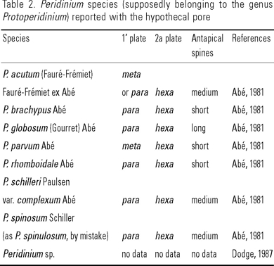
Most species with the hypothecal pore have the 1' plate of the para type, although six species with the meta first apical plate were observed. Thus, it seems unlikely that the hypothecal pore is exclusively or nearly exclusively associated with the para type, as was emphasized by Dodge (1987). Rather, the hypothecal pore is almost exclusively associated with the hexa second intercalary plate (Tables 1 and 2). Within the Protoperidinium and Peridinium species with the hypothecal pore, at least five groups of morphologically similar and supposedly related taxa can be distinguished: (1) Species with the para 1' plate, the hexa 2a plate and short or medium antapical spines, cells are usually slightly longer than wide - Protoperidinium affine, P. pallidum and P. pellucidum; (2) Species with the para or meta 1' plate, variable 2a plate, and short antapical spines, cells are slightly compressed apically-antapically - P. curvipes (Ostenfeld) Balech and P. variegatum; (3) Species with the para 1' plate, the hexa 2a plate and long antapical spines, cells are usually notice-ably longer than wide - P. diabolum and P. dodgei; (4) Species with the meta 1' plate and hexa 2a plate and short antapical spines, cells are slightly wider than long - P. cruciferum, P. cf. cruciferum, Protoperidinium sp. 1 (meta-hexa) and Peridinium acutum (Fauré-Frémiet) Fauré-Frémiet ex Abé; (5) Species with the para 1' plate, the hexa 2a plate and one or two short or medium antapical spines and a prominent sulcal list formed by the right sulcal (Sp) plate, cells are slightly longer than wide - P. ovum (Schiller) Balech and Protoperidinium sp. 2 (para-hexa). The latter is morphologically similar to P. ovum, however, it has only one (right) antapical spine, more displaced cingulum (usually about one-width of the cingulum), more rounded cell shape, smaller size and the hypothecal pore located closer to the cingulum. The 1' and 2a plates are similar in these two species. In general, the features described above allow to ascribe our Protoperidinium sp. 2 (para-hexa) to the species designated by Balech (1988: 122, pl. 52, fig. 13-15) as Protoperidinium sp. K, although it is also very similar to P. capurroi (Balech) Balech subsp. subpellucidum Balech (Balech, 1971a: 155, pl. 33, fig. 240-242) and P. aequatoriale (Balech) Balech (Balech, 1971b: 26, pl. 6, fig. 112-114). However, in none of these species the hypothecal pore was described or illustrated, which makes correct identification of our specimens difficult.
The pore is thought to be present in closely related species and to be a reliable diagnostic feature to differentiate Protoperidinium species. Similarities in morphology of the sulcal plates, especially of the anterior (Sa), left (Ss) and right sulcal plates (Sd) between species of group 1 (Balech, 1988) and to a lesser extent of group 2 (Balech, 1975) and group 5 (Balech, 1988) substantiate our suggestion. It is important to note that in all studied cells of the same species the hypothecal pore had the same location on the 1"' plate. Groups 1 and 2, which include the species inhabiting the regions north of the Antarctic Convergence or the Antarctic Ocean, to which they are endemic (Protoperidinium affine and P. variegatum), seem to represent the geographical vicariants (Okolodkov, 2000). Among all species given in Tables 1 and 2, only Protoperidinium pellucidum subsp. stellatum sometimes demonstrates 2 or more hypothecal pores described as star-like structures (Balech, 1978, 1988).
In our material, under the objective 40/0.65, the hypothecal pore was clearly seen in all species but Protoperidinium ovum, in which the pore was hardly discernible. However, it was easily distinguishable in the empty thecae of P. ovum, especially when the negative phase contrast was used. The pore size including a rim around the pore varied between species (1.0 to 3.5 µm) and within the same species. For example, the pore size in Protoperidinium variegatum ranged from 1.8 to 3.0 µm. The largest hypothecal pores were observed in P. pallidum and P. variegatum.
It is interesting to note that groups of dots on the first antapical plate (3 or 4 dots in Protoperidinium cruciferum and Peridinium acutum), which can be interpreted as the hypothecal pore, were observed by Balech (1971a) in Protoperidinium mite (Pavillard) Balech. For this species, two groups of 4 or 5 dots, similar to those in P. cruciferum, have been known (the pores are pictured but no comments are given).
In addition, there is another Protoperidinium species, P. cf. retiferum (Matzenauer) Balech, which has one or two rather large pores on the hypotheca (Balech, 1978, 1988). However, these are located at the base of the antapical horns, on the ventral side of the postcingular plates 1"' and 2"': on the left horn if only one pore is present and on each horn if two pores.
Abé (1981) suggested a secretory function of the hypothecal pore such as that of connecting the pusule system to the exterior. However, Dodge (1987), who analyzed the possible connection, concluded that at present it was not possible to suggest any function for the hypothecal pore.
Apart from the hypothecal pore located on the 1"' plate, another structure consisting of a group of pores, designated as "a distinctive zone of honey-combed pores" and located on the antapical plate (1"") of the hypotheca of the Peridiniales has been observed. Carbonell-Moore (1994) described it in the three new genera of the family Podolampaceae, Mysticella Carbonell-Moore, Gaarderia Carbonell-Moore and Heterobractum Carbonell-Moore (in the genus Heterobractum, a group of pores is pictured but no comments are given).
Acknowledgements
I am grateful to Alain Couté, who hosted my visit to the Natural History Museum in Paris to study the thecal morphology of Antarctic dinoflagellates. The present research was also supported by CONACyT in 2001-2002 during my stay at the Department of Hydrobiology, UAM-Iztapalapa. My thanks are extended to John D. Dodge, the Royal Holloway, the University of London, Ma. Esther Meave del Castillo of UAM-Iztapalapa, Sergio Licea Durán of the Institute of Marine Sciences and Limnology, UNAM, and to David L. Garrison, the U.S. National Science Foundation, for making the collections available for studies and for laboratory facilities. Critical comments of the three referees and one of the editors are very much appreciated. My participation in the RVIB Nathaniel B. Palmer cruise 98-03 and sampling phytoplankton in the Ross Sea was supported by an award from International Programs at the NSF given to me and by a grant from the NSF, Office of Polar Programs, to D.L.Garrison (OPP 9614201). Martin McLennan from the Technological Institute of Monterrey, Mexico City, kindly improved the writing style. Boris Y. Okolodkov is thanked for his technical assistance with the illustrations.
References
ABÉ, T. 1981. Studies on the family Peridiniidae. An unfinished monograph of the armoured Dinoflagellata. Special Publications of the Seto Marine Biological Laboratory, Special Publication Series 6. viii+412 p. [ Links ]
BALECH, E. 1957. Dinoflagellés et Tintinnides de la Terre Adélie, Secteur Français Antarctique. Vie et Milieu 8 (4): 382-408. [ Links ]
BALECH, E. 1958. Plancton de la Campaña Antártica Argentina 1954-1955. Physis 21 (60): 75-108. [ Links ]
BALECH, E. 1962. Plancton de las campañas oceanográficas Drake I y II. Servicio de Hidrografía Naval, Buenos Aires, H 627: 1-57. [ Links ]
BALECH, E. 1968. Dinoflagellates. In: Antarctic Map Folio Series, Folio 10, American Geographic Society: 8-9, pl. 11 and 12. [ Links ]
BALECH, E. 1971a. Microplancton de la Campaña Productividad III. Revista del Museo Argentino de Ciencias Naturales "Bernardino Rivadavia", Hidrobiología 3(1): 1-202, láms. 1-39. [ Links ]
BALECH, E. 1971b. Microplancton del Atlántico Equatorial Oeste (Equalant I). Servicio de Hidrografía Naval, Buenos Aires, H 654: 1-103, lams. 1-12. [ Links ]
BALECH, E. 1973. Segunda contribución al conocimiento del microplancton del Mar de Bellingshausen. Contribución del Instituto Antártico Argentino 107: 1-63. [ Links ]
BALECH, E. 1974. El género Protoperidinium Bergh, 1881 (Peridinium Ehr., 1831, partim). Revista del Museo Argentino de Ciencias Naturales "Bernardino Rivadavia", Hidrobiología 4(1): 1-78. [ Links ]
BALECH, E. 1975. Clave ilustrada de dinoflagelados antárticos. Publicación del Instituto Antàrtico Argentino No. 11, Buenos Aires. 99 p. [ Links ]
BALECH, E. 1976a. Sur quelques Protoperidinium (Dinoflagellata) du golfe de Lion. Vie et Milieu B26(1): 27-46. [ Links ]
BALECH, E. 1976b. Clave ilustrada de dinoflagelados antárticos. Publicación del Instituto Antártico Argentino 11, Buenos Aires. 99 p. [ Links ]
BALECH, E. 1978. Microplancton de la Campaña Productividad IV. Revista del Museo Argentino de Ciencias Naturales "Bernardino Rivadavia", Hidrobiología 5(7): 137-201. [ Links ]
BALECH, E. 1988. Los dinoflagelados del Atlántico Sudoccidental. Publicación Especial del Instituto Español de Oceanografía 1, Madrid. 310 p. [ Links ]
BALECH, E. 1994. Contribución a la taxonomía y nomenclatura del género Protoperidinium (Dinoflagellata). Revista del Museo Argentino de Ciencias Naturales "Bernardino Rivadavia", Hidrobiología 7(4): 61-80. [ Links ]
CARBONELL-MOORE, M. C. 1994. On the taxonomy of the family Podolampaceae Lindemann (Dinophyceae) with descriptions of three new genera. Review of Palaeobotany and Palynology 84: 73-99. [ Links ]
DODGE, J. D. 1987. A hypothecal pore in some species of Protoperidinium (Dinophyceae). British Phycological Journal 22: 335-338. [ Links ]
LEBOUR, M. 1925. The dinoflagellates of northern seas. Marine Biological Association, Plymouth. vii+250 p. inc. pl. 1-35. [ Links ]
OKOLODKOV, Y. B. 1997. A new combination and new species of Gymnodinium, Gyrodinium and Protoperidinium (Dinophyceae) from the Arctic and N. E. Atlantic. Botanical Journal, Russian Academy of Sciences 82(5): 97-106. [ Links ]
OKOLODKOV, Y. B. 2002. The genus Protoperidinium Bergh in the Mexican Pacific. Resúmenes del XII Reunión Nacional de la Sociedad Mexicana de Planctología y V International Meeting of the Mexican Society of Planktology, [ Links ] p. 68. [ Links ]

