




Serviços Personalizados
Journal
Artigo
 texto em
 Inglês (pdf)
 Artigo em XML
 Referências do artigo

 Tradução automática
 Enviar este artigo por email
Indicadores
-
 Citado por SciELO
-
 Acessos
Links relacionados
-
 Similares em
SciELO
Compartilhar

 Permalink

Revista mexicana de fitopatología
versão On-line ISSN 2007-8080versão impressa ISSN 0185-3309
Rev. mex. fitopatol vol.42 no.2 Texcoco Mai. 2024 Epub 24-Fev-2025
https://doi.org/10.18781/r.mex.fit.2309-5
Scientific Article
Morphological characterization, phylogeny and pathogenesis of Setophoma terrestris causing corky and pink roots of tomato (Solanum lycopersicum) in Sinaloa, Mexico
1 Centro de Investigación en Alimentación y Desarrollo, Coordinación Regional Culiacán, 80110, Sinaloa, México;
2 Universidad Veracruzana, Facultad de Ciencias Biológicas y Agropecuarias, Tuxpan, 92895, Veracruz, México
3 Universidad Nacional de Catamarca, Departamento de Ingeniería Agronómica, 4700, Catamarca, Argentina
Objective / Background.
Tomato (Solanum lycopersicum) is one of Mexico’s main crops. In the years 2017 and 2018, symptoms of corky and pink roots were observed with an incidence of 10 to 20% in Culiacan, Sinaloa, Mexico. In the foliage, plants presented a generalized chlorosis, with stunted growth and senescence in the leaves. In the roots, brown and pink lesions were formed, as well as a corky texture. The objective of this study was to morphologically and molecularly characterize fungal isolates associated to corky and pink root in tomato orchards in Culiacan, Sinaloa, as well as to evaluate their pathogenicity.
Materials and methods.
Monoconidial isolates were obtained and they were identified as Setophoma terrestris, based on their morphological characteristics. To confirm the identity, the area of the internal transcribed spacers (ITS) of the rDNA was amplified and sequenced, along with a fragment of the gene 28S of the rRNA (LSU).
Results.
Using the sequences obtained, a phylogenetic tree was created using the Bayesian Inference and it was found that the sequences were grouped with the ex-type sequences of Setophoma terrestris. The pathogenicity of the isolates was verified by inoculating mycelial discs into the root of 10 one-month-old tomato seedlings. The roots of the seedlings inoculated with PDA discs without mycelium served as a control. Thirty days after inoculation, corky and pink root symptoms appeared, whereas the roots of control plants remained healthy.
Conclusion.
According to the morphological characterization, the molecular identification and the pathogenicity tests, Setophoma terrestris was confirmed to be the causal agent of corky and pink root in agricultural tomato orchards in Culiacan, Sinaloa.
Keywords: Setophoma; fungus; tomato; bayesian inference; root disease
Objetivos/antecedentes
El jitomate (Solanum lycopersicum) es uno de los principales cultivos en México. En los años 2017 y 2018, se observaron síntomas de raíz corchosa y rosada con una incidencia de 10 a 20 % en Culiacán, Sinaloa, México. En el follaje, las plantas presentaron clorosis generalizada, con crecimiento raquítico y senescencia de las hojas. En las raíces, se desarrollaron lesiones de color café oscuro y rosa, además de textura corchosa. El objetivo de este estudio fue caracterizar morfológica y molecularmente a los aislados fúngicos asociados a los síntomas de raíz corchosa y rosada en campos de jitomate de Culiacán, Sinaloa, así como evaluar su patogenicidad.
Materiales y Métodos.
Se obtuvieron aislados monoconidiales y se identificaron como Setophoma terrestris con base en los caracteres morfológicos. Para la confirmación de la identidad, se amplificó y se secuenció la región de los espaciadores transcritos internos (ITS) del ADNr, así como un fragmento del gen 28S del ARNr (LSU).
Resultados.
Con las secuencias obtenidas se construyó un árbol filogenético con el método de Inferencia Bayesiana y se encontró que las secuencias se agruparon con las secuencias ex-tipo de Setophoma terrestris. La patogenicidad de los aislados se verificó mediante la inoculación de discos miceliales en raíces de 10 plántulas de jitomate de un mes de edad. Las raíces de plántulas que se inocularon con discos de PDA sin micelio, sirvieron como control. Treinta días después de la inoculación se observaron síntomas de raíz corchosa y rosada, mientras que las raíces de las plantas de control permanecieron sanas.
Conclusión.
De acuerdo con la caracterización morfológica, la identificación molecular y las pruebas de patogenicidad, se confirmó que Setophoma terrestris es el agente causal de la raíz corchosa y rosada en campos agrícolas de jitomate distribuidos en Culiacán, Sinaloa.
Palabras clave: Setophoma; hongo; jitomate; inferencia bayesiana; enfermedad radical
Introduction
Mexico is the ninth most important producer of tomato (Solanum lycopersicum) in the world. In the 2018-2019 agricultural cycle, the country produced a total of 938,009 t, with Sinaloa being the state at the top of the list of nationwide producers with 605,261 t (SIAP, 2020). However, pests and diseases are the main problems that impact this crop (Jones et al. 2016).
Out of the diseases in tomato, corky root is scarcely studied, despite being very troublesome; it creates brown lesions and corky roots. Initially, the lesions are limited and later extends progressively and surrounds the roots, developing lesions that range from a few milimeters to several centimeters (Blancard, 2011). In this context, the disease called corky root is one of the most sever problems in recent years in agricultural plots dedicated to the tomato crop in Mexico (Pardo, 1999; Martínez-Ruiz et al., 2016).
Although the corky root disease is often found in greenhouse crops and its distribution was initially reported in Europe (Schneider, 1966), United States (Campbell et al., 1982), Lebanon (Davet, 1973) and Chile (Olavarria, 1991), there are not enough studies related with the causal agent of the disease (USDA, 2017). Generally, corky root in tomato is associated with Pseudopyrenochaeta lycopersici (sin. Pyrenochaeta lycopersici) as its causal agent (Golzar, 2009). However, a study carried out in Mexico proved, with morphological bases, phylogenetic analyses and pathogenicity tests, that corky root in Sinaloa fields is caused by Setophoma terrestris and additionally, it could cause pink root in the crop (López-López et al., 2020).
According to De Gruyter et al. (2010), S. terrestris and P. lycopersici share very similar morphological characteristics, which were taxonomically differentiated according to phyogenetic analyses with linked sequeneces from the ITS area and fragments of the LSU gene (Valenzuela-López et al., 2018).
Setophoma is a fungal genus that forms pycnidia, which are asexual reproductive structures. The pycnidium can have a globular to semi-globular structure, almost setae-like, with an ostiolar neck around it. Inside the pycnidia are the spores called conidia, which are hyaline, cylindrical, septated and mainly guttulate (De Gruyter et al., 2010).
Setophoma terrestris has been mostly studied in onion (Allium cepa) crops, since they are its main host, causing the disease known as pink root, which is important and has a large global economic impact. This fungus attacks the root system first, and progressively affects the entire plant until its death (Castillo and Plata, 2010). It has also been reported to cause damage in other hosts such as canola (Brassica napus) in Canada (Yang et al., 2017), zucchini (Curcubita maxima) in the USA (Rivedal et al., 2018), as well as garlic (Allium sativum) and tea (Camellia sinensis) plants in China (Liu et al., 2019; Zhang et al., 2019).
An earlier study (López-López et al., 2020) proved, for the first time, the infectious nature of Setophoma terrestris in tomato plants (S. lycopersicum) in Mexico, based on one single isolate. The aim of this study was to morphologically and molecularly characterize the fungal isolations associated with the symptoms of corky root and pink root in tomato orchards in Culiacán, Sinaloa, as well as to evaluate their pathogenicity.
Materials and Methods
Sample collection. Samples were taken from five sites (Figure 1), which were distributed in five locations in the valley of Culiacán, as well as on the limits of Navolato, Sinaloa, with an intensive tomato production with undetermined growth in the production stage (age three months). These sites had a history of corky and pink root in tomato. In some plants at the harvest stage, different types of symptoms were observed in the foliar area, such as chlorosis and reduction in growth. Therefore, targeted sampling was conducted according to the symptoms mentioned. In addition, the roots of the plants were examined by extraction and observation of the damage along them. Once the symptoms related to the disease of the crop were confirmed, 15 samples of diseased tomato roots were gathered from each site. The collection sites were shade house-tye greenhouses, each one approximately 5 ha. Finally, they were placed in plastic bags and transported to the Phytopathology Laboratory of the Coordinación Culiacán of CIAD for analysis.
Isolation, purification and conservation of fungi. The diseased roots were submerged in drinking water to remove excess soil without hariming the plant tissue. Subsequently, from necrotic lesions developed in tomato roots, fragments of 5 mm in length were cut, then surface-disintested by immersion in a 2% sodium hypochlorite solution for 2 minutes. They were rinsed in sterile distilled water and placed in Petri dishes containing acidified potato dextrose agar (APDA) and acidified V8 agar medium (V8A) and incubated at 25 ºC in continuous darkness. Afterwards, the fungal isolates were transfered to new Petri dishes with V8A,
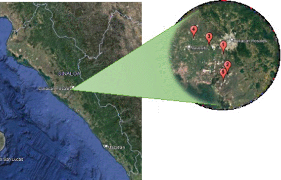
Figure 1 Location of the sites of recollection of plants with corky root symptoms in agricultural systems with tomato production in the valley of Culiacan, Sinaloa. Site 1 (24°42’56.58”N, 107°26’42.86”W), Site 2 (24°35’21.67”N, 107°24’56.50”W), Site 3 (24°46’2.77”N, 107°32’51.30”W), Site 4 (24°48’47.44”N, 107°39’26.19”W) and Site 5 24°32’34.07”N, 107°26’44.93”W).
and after 10 days in incubation at 25 ºC, the isolations were purified using the monosporic culture technique (Crous et al., 2019), which consisted of placing the pycnidia on microscope slides and the conidia were placed under a compound microscope. From these conidia, monospore cultures were obtained in Petri dishes with V8A medium, and 7 days after planting, a pink mycelial growth was observed. The fungal isolates were preserved by placing discs, 4 mm in diameter, from colonies with 5 days of growth into sterile 2 mL tubes containing sterile distilled water and stored at room temperature in the strain collection of the Phytopathology Laboratory in the Culiacán Unit of CIAD.
Morphological characterization. In order to stimulate the production of asexual structures, approximately three tomato roots were placed in moist chambers at a temperature of 20 ºC. After 30 days, they were removed and observed under a sterescopic microscope, locating the pycnidia and placing them on miscroscope slides with sterile distilled water for their observation and characterization.
The morphological characterization of the isolates was performed using keys and descriptions reported by De Gruyter et al. (2010). In order to do this, the fungus was cultivated in Petri dishes with an V8A medium for 10 days. The isolations that did not form pycnids in the V8A medium were stimulated using the method described by Clerjeau (1973), which consists in placing melon (Cucumis melo) seedling roots on the mycelium of the fungus of interest with 5-7 days of growth and later incubate once again, at room temperature (25-30 ºC) for 15 to 20 days for the artificial production of pycnidia. Once produced, the characteristics of the pycnidia, conidia and setae were examined under a compound microscope (Zeiss®, Germany) with a built-in digital camera (AxioCam) and 50 measurements were taken of each one of the different asexual fungal structures of each isolate.
DNA extraction, PCR amplification and sequencing. Total DNA extraction as carried out from seven-day-old fungal colonies, cultivated in an V8A medium. The mycelium of each isolation was scraped with a sterile microscope slide, macerated with a sterile mortar using liquid nitrogen and transferred to a 1.5 mL microcentrifuge tube with 500 µL of hexadecyltrimethylammonium bromide solution, CTAB (Tris 100 mM, pH 8; 20 mM of EDTA, pH 8; NaCl, 1.4 M and 3% of CTAB), mixed in a vortex for 10 s and incubated for 60 min at 65 °C. Later, 700 µL of isolamilic clorophorm-alcohol (24:1v/v) was added, run through a vortex for 10 s and placed in a 5810 R centrifuge (Eppendorf®, EE. UU.) at 13000 g for10 min. Next, the supernatant was transfered to a 1.5 mL microcentrifuge tube and 700 µL of isopropanol were added. The tubes were mixed by inversion 4 to 5 times and left at -20 °C for 10 min, and were then centrifuges at 13000 g for 10 min to precipitate the DNA. Subsequently, the precipitate was washed twice with 500 µL of ethanol at 70%, centrifuging at 13000 g for 5 min and the supernatant was discarded every time. Afterwards, the DNA pellet was left to dry by placing the inverted tubes on absorbent paper for no more than 15 min to allow the evaporation of the ethanol and finally, the DNA was resuspended to allow the ethanol to evaporate and finally, the DNA was resuspended in 100 µL of sterile water, DNAse- and RNAse-free. The quality and concentration of the DNA was evaluated in a Q 3000 UV spectrophotometer (Quawell, EE. UU.) and electrophoresis in 1% agarose gel, with DNA being considered adequate when it had purity levels (ratio of A260/280) ranging between 1.8 and 2.0 and a concentration (A260) of approximately 2.0 µg µL-1, as well as intact high-molecular weight bands, respectively. The DNA was stored at -20 °C for later use.
The amplification of the ITS region of the rDNA and the long subunit (LSU) was performed using endpoint polymerase chain reaction (PCR). The ITS region (ITS1- 5.8S-IT2) was amplified using primers ITS1 (TCCGTAGGTGAACCTGCGG) and ITS4 (TCCTCCGCTTATTGA TATGC) with 480 pb, approximately (White et al., 1990), whereas the amplified band of the LSU gene (including domains D1 and D2) was performed with a nested PCR using primers PM3 (Takamatsu and Kano, 2001) /TW14 (Mori et al., 2000) and NL1/TW14 (Mori et al., 2000) for the first and second reaction, respectively. The reaction mixture was prepared at a final volume of 25 µL, containing 1X PCR buffer, 2.5 mM of MgCl2, 0.5 mM of dNTPs, 1 µM of each primer, 1U of polymerase DNA (Promega®, USA) and 100 ng of DNA. The PCR was carried out under the following thermocycling conditions (C-1000 thermocycler, Bio-Rad®, USA): initial denaturalization at 95 ºC for 5 min, 35 cycles at 95 ºC for 30 s, 54 ºC for 30 s, 72 ºC for 30 s, with a final extension of 72 ºC for 10 min. For the emplification of the 28S gene, the same thermocycling conditions were used, except for the temperature of alignment, which was 52 ºC. The amplified products were anayzed by electrophoresis in 1% agarose gel dyed with ethidium bromide. Electrophoresis was carried out at 90 V for 40 min and the results were viewed in an M-26X transluminator in a GelDoc-ItTM 300 image system (UVP®, USA). The PCR products were purified using the DNA Clean & Concentrator kit (Zymo Research®, USA), following the recommendations by the manufacturer and they were sequenced in the Macrogen company (www.macrogen. com) in Seoul, South Korea.
Phylogenetic analysis. The quality of the nucleotide sequences and the assembly method were carried out using the Staden package (Staden et al., 2000). The sequences were cleaned and thos generated from the iTS region and the large sububit (LSU) of the rRNA were aligned with sequences from different species of the Setophoma genus obtained from the GenBank and from published reference data (De Gruyter et al., 2010; Liu et al., 2019) (Table 1). Multiple alignments were generated from sequences using ClustalW as implemented in MEGA 11 (Tamura et al., 2021) and they were improved manually to allow the maximim similarity between the dataset.The Bayesian phylogenetic estimations were infered with MrBayes 3.2.6 (Ronquist et al., 2012) implementes in the cluster CIPRES (https:// www.phylo.org/portal2/home.action) and selecting the nucleotide substitution model that best fit the data of this investigation, according to the Akaike Information Criterion (AIC) obtained with MrModeltest 2.3 (Nylander, 2004). The number of generatiosn was 10,000,000 and the subsequent probabilities were calculated after discarding the first 25% of the iteracions. The phylogenetic trees were viewed using TreeView (Page, 1996).
Prathogenicity tests. The pathogenicity of seven fungal isolates were determined by inoculating them in the main roots of 30-day-old cv. 8444 tomato seedlings. Ten plants were used for each one of the isolations, along with 10 non-inoculated plants there served as a control. V8A plugs, 4 mm in diameter, with an active growth
Table 1 Information of fungal isolates and GenBank accession numbers of Setophoma species used in the phylogeneti analysis.
| Species | Collection code | Host | Country | Access number LSU | Access number ITS |
|---|---|---|---|---|---|
| Setophoma antiqua | LC6594 | Camellia sinensis | China | MK511947 | MK511909 |
| LC6595 | C. sinensis | China | MK511948 | MK511910 | |
| S. chromolaenae | CBS 135105x | Chromolaena odorata | Brazil | KF251747 | KF251244 |
| S. cyperi | CBS 141450x | Cyperus sphaerocephala | South Africa | KX228337 | KX228286 |
| S. endophytica | CGMCC 3.19528 | C. sinensis | China | MK511956 | MK511931 |
| LC3164 | C. sinensis | China | MK511957 | MK511932 | |
| S. longinqua | LC6593x | C. sinensis | China | MK511946 | MK511908 |
| S. poaceicola | MFLUCC 16-0880x | - | Thailand | KY550386 | KY568988 |
| Setophoma sp. | CGMCC 3.19526 | Carbonatite in a cave | China | MK511965 | MK511944 |
| S. sacchari | MFLUCC 12-0241 | Saccharum officinarum | Thailand | KJ476147 | KJ476145 |
| CBS 333.39x | Brazil | MH867535 | MH856038 |
xisolates ex-type. The accession numbers of the sequences generated in this study are in bold.
CBS: Collection of isolates in the Centraalbureau Voor Schimmelcultures, Fungal Biodiversity Centre, Utrecht, Netherlands; IMI: Collection of isolates in CABI Europe UK Centre, Egham, United Kingdom; CPC: Pedro W. Crous work collection, stored in CBS. CGMCC: General Center for the Collection of Microbiological Cultures of China, Institute of Microbiology, Chinese Academy of Sciences, Beijing, China; LC: Lei Cai wok collection, stored in the Institute of Microbiology, Chinese Academy of Sciences, Beijing, China; MUCL: Mycothèque de l’Université catholique de Louvain, Louvain-la-Neuve, Belgium.
of the fungus per isolate were used, which were placed on a lesion made earlier with a toothpick. The mycelial disk was attached to the lesion of the root using Parafilm® paper (USA). In the control plants, the lesion was made and a disc with V8A culture medium was placed, without fungal growth. The plants were kept in a greeenhouse at 20-35 ºC for 30 días. The inoculated roots were then removed and the fungi were reisolates in an V8A medium. Once the mycelium was developed, the morphological and cultural characteristics were considered, such as pigmentation of the V8A medium, as well as the qualitative and quantitative characteristics of mushrooms, pycnidia and conidia, according to earlier descriptions. The test was repeated twice.
Results and Discussion
Symptoms of chlorosis and leaf senescence were observed, as well as the reduction in tomato plant growth (Figures 2A-C) in an incidence that varied from 10 to 20% in the five different commercial agricultural fields sampled in the valley of Culiacan, Sinaloa, Mexico. The symptomatic roots developed dark brown lesions, swollen and with a corky texture (Figure 2D-E) and pink (Figure 2F). These symptoms in the foliar area and root system coincide with those reported by López-López et al. (2020), whereas the same causal agent affected tomato plants reported in Brazil, Canada, USA and Venezuela (Farr and Rossman, 2022). Likewise, these symptoms were reported in other hosts such as scallions (Allium fistulosum) in China (Luong et al., 2008). Similarly, the symptoms of the corky and pink root diseases coincide with those described for S. terrestris in onion (Schwartz and Mohan, 2008).
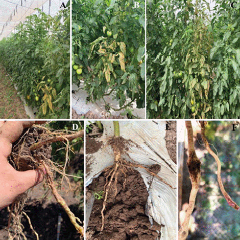
Figure 2 Symptoms of corky and pink roots in tomato. A-C) Chlorosis, deficient growth and senescence in plants. D-F) Dark brown lesions on the root, with swelling, a corky texture and a pink color.
Pink root symptomatology may be similar to that caused by Pseudopyrenochaeta lycopersici, which was found in a wide number of hosts such as sorghum (Sorghum), maize (Zea mays), sugarcane (Saccharum officinarum) (Sprague, 1950), grass (Cynodon dactylon) (Conners, 1967), leek (Allium porrum) (Hall et al., 2007), cantaloupe (Cucumis melo), zucchini (Curcubita pepo), wheat (Triticum) and spinach (Spinacia oleracea) (USDA, 1960), as well as willow trees (Salix) (Pfleger and Vaughn, 1972). On the other hand, in tomato, it has been reported in Australia (Golzar, 2009), Africa (Testen et al., 2019), Germany (Valenzuela-López et al., 2018) and in the United States as a pathogen associated to the brown rot in tomato root (Vrisman et al., 2017).
Isolation and morphological identification. Out of the 15 samples (plants) of tomato roots with symptoms of corky and pink roots by sites, seven fungal isolates were used, which came from the five sites previously indicated on the map with their respective coordenates in the valley of Culiacan. The technique that worked for the isolation of fungi was to place sections of infected tissue in a humid chamber to stimulate the pycnidia in the root tissues at 4 ºC (Gilchrist-Saavedra et al., 2005). The isolates obtained correspond to Setophoma terrestris, and the colonies were pink on both sides, with a root growth (80 mm) after 7 days of growing (Figures 3A-B). These characteristics of growth of the fungus in V8A medium were similar to those reported by De Gruyter et al. (2010).
The morphological examination of the asexual structures displayed black to dark brown globular to obpyriform pycnidia, (Figure 3C) and measuring 142- 220 × 104-140 µm. The presence of dark brown septated setae was observed, measuring 52-114 × 2-8 µm and that surrounded the neck ostilar (Figure 3D). The conidia were hyaline, cyllindrical, measuring 2-8 × 1-3 µm, and with two very characteristic biguttulate on its tips (Figure 3E-F). All the morphological characteristcis coincided with those reported by other authors (Koenning et al., 2007; Wiriyajitsomboon, 2015; Yang et al., 2017; Rivedal et al., 2018; Zhang et al., 2019) (Table 2). Despite some measurements being variable between each other, these data are not forceful to differentiate between species, therefore molecular tools and phylogenetic analyses are required to confirm at the level of species.
Regarding the ranges of the morphometric values, the seven S. terrestris of this study were distributed into three groups, according to upstream measures: group
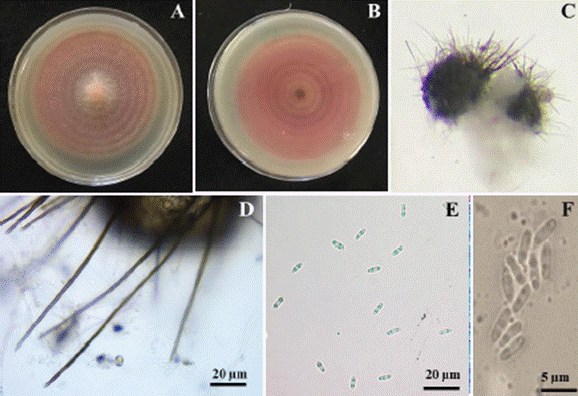
Figure 3 Colonies and asexual reproductive structures of Setophoma terrestris. A) Colony of S. terrestris in a V8A medium with 7 days of growth. B) Colony growth on the reverse of the dish. D) Pycnidium. D) Setae. E-F) Biguttulate conidia.
Table 2 Measurements of asexual structures in Setophoma terrestres isolates obtained from tomato plants.
| Setae (µm) | Pycnidium (µm) | Conidia (µm) | Reference |
|---|---|---|---|
| yNA | yNA | 4.5-5.5 × 1.8-2.3 | Koenning et al., 2007 |
| ≈ 100-120 | ≈ 200 | ≈ 4-5 | Wiriyajitsomboon, 2015 |
| ≈ 100 | ≈ 300 | ≈ 5 | Yang et al., 2017 |
| yNA | yNA | 5.0 × 2.0 | Rivedal et al., 2018 |
| 55-150 | 160-230 | 4.0-5.0 × 1.5-2.0 | Zhang et al., 2019 |
| 68-84 × 2-3 | 237-345 × 105-250 | 5-7 × 2-3 | López-López et al., 2020 (CCLF01) |
| 52-114 × 3-6 | 150-183 × 133-156 | 6-7 × 2-3 | zCCLF02 |
| 60-108 × 2-5 | 142-160 × 124-132 | 6-8 × 2-3 | zCCLF03 |
| 46-102 × 3-5 | 200-233 × 110-130 | 4-6 × 2-3 | zCCLF04 |
| 55-106 × 3-4 | 220-231 × 105-133 | 2-3 × 1-1.5 | zCCLF05 |
| 67-90 × 2.5-4 | 216-255 × 104-140 | 3-4 × 1-2 | zCCLF06 |
| 90-107 × 3-8 | 175-180 × 140-155 | 5-8 × 2-3 | zCCLF07 |
| 60-101 × 3-5 | 160-175 × 137-153 | 5-6 × 2-3 | zCCLF08 |
yNA: Not applicable / Not reported.
zThe measurements generated in this study are highlighted in bold in references.
1, composed ot isolate CCLF05; group 2, by CCLF06 and CCLF04, and finally, group 3, with isolates CCLF07, CCLF08, CCFL02 and CCLF03. It is important to mention that group 3 also includes the isolate (CCLF01) reported previously in another study in Sinaloa, Mexico (López-López et al., 2020).
In turn, Luong et al. (2008) identified S. terrestris as the causal agent of pink root in onion, using characteristics of the colony and they observed the pink pigment in the culture medium, as well as the setae in the pycnidia and conidia, typical of this root in some pink roots. Additionally, large guttulates were observed in the conidia of their islates, which coincide with those reported in this study. These same microscopic characteristics in the conidia were described by De Gruyter and Boerema (2002).
Similarly, Ikeda et al. (2012) identified S. terrestris as the causal agent of pink root in zucchini and they obtained two isolates that formed brown to dark black pycnidia, subglobular to globular in shape and with a neck ostilar with setae around it. These were found to be cyllindrical in shape and brown to dark brown, and measured 104-262 × 3.5-6.9 µm. These characteristics coincide with those obtained in this study.
On the other hand, measurements may vary slightly between the isolates and the above may be due to multiple environmental factors that affect the development of the pathogen, as mentioned by Burton et al. (1997), making a relationship with the symptoms of the disease in cantaloupe and watermelon and identifying the causal agent based on morphological structures and analysis of ITS sequences. Unllike our isolates, the pycnidia reported by Duarte and Barreto (2015) do not have the typical setae around the ostiole, indicating that this attribute is not a generic characteristic. Likewise, the characteristics shared by the fungi P. lycopersici and S. terrestris are very similar to the difference that in P. lycopersici the setae of its pycnidia are over 200 µm long, wereas S. terrestris regularly range between 50-150 µm, approximately (De Gruyter, 2010).
Phylogenetic analysis. The aligned sequences corresponding to the ITS region and the long subunit (LSU) resulted in a total length of 1,345 nucleotides. The phylogenetic analysis generated with MrBayes based on the data set (Figure 4) indicated that all the isolations obtained in this study were grouped with the species Setophoma terrestris, with a high posterior probability support (1.0).
On the other hand, the phylogenetic analysis shows two groupings, which mostly addresses all isolates with 0.88 subsequent probability, except for isolate CCLF05, which was grouped separately and displayed the largest phylogenetic difference. Likewise, with the previously mentioned and according the the morphometry, was grouped in only one group. Based on this, it was possible to verify, once again, that Pseudopyrenochaeta lycopersici is not the species that causes the disease known as corky and pink root in Sinaloa, Mexico.
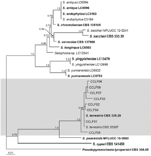
Figure 4 Bayesian Tree obtained with combined data from sequences ITS and LSU. The tree shows the phylogenetic relations of the Setophoma species. The Bayesian posterior probability values of over 0.70 are shown in the nodes. The species Pseudopyrenochaeta lycopersici was used as an external group and the scale bar indicates the number of expected changes per site.
The phylogenetic of this study coincides with the molecular identity reported by Rivedal et al. (2018), who used the ITS region, finding that the DNA of all its isolates displayed a 99% similarity with Setophoma terrestris. Likewise, those sequences were also compared with those reported by De Gruyter et al. (2010) and similar results were obtained. On the other hand, Duarte and Barreto (2015) used the same Bayesian inference analysis criterion, with the difference that they relied only on LSU sequences, whereas in this study, concatenated ITS and LSU
sequences were used. This is of great importance, since in recent years, it has been proven and supported that these genera are very closely related and can only be distinguished through phylogenetic analysis with concatenated sequences (De Gruyter et al., 2010). This information is extremely important, since the Phoma genus has grown to encompass over 3000 species that form pycnidia with hyaline and septated conidia (Aveskamp et al., 2008), thereby reevaluating several studies related to this genus upon correct identification (Boerema et al., 2004).
Pathogenicity tests. Thirty days after inoculation, the leaves displayed yellowing (Figures 5A-B). The inoculated tomato roots displayed a corky texture and pink coloring (Figure 5C), which were similar to the symptoms observed on the field.
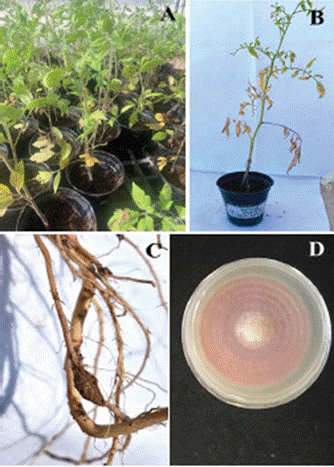
Figure 5 Pathogenicity tests in tomato plants. A-B) Tomato plants of the 8444 variety inoculated with S. terrestris and showing symptoms of yellowing. C) Corky root symptom 30 days after inoculation with S. terrestris. D) Colony obtained from the in vitro reisolation of S. terrestris.
From the reisolations, colonies (Figure 5D) and fungal structures identical to those of the inoculated fungus were obtained, which confirmed the pathogenicity of the seven inoculated isolates, while the roots of the control plants remained asymptomatic.
The results of the pathogencity tests of this study coincide with those reportd by Zhang et al. (2019), who observed typical pink root symptoms in garlic. This pathogen has also been found damaging other hosts such ascanola (Brassica napus) in Canada (Yang et al., 2017), zucchini (Curcubita maxima) in the USA (Rivedal et al., 2018), and tea plants (Camellia sinensis) in China (Liu et al., 2019).
The symptoms observed in the pathogenicity tests in the study conductes by Zhang et al. (2019) showed that S. terrestris caused chlorosis, wilting and death in garlic plants within a span of 40 days, similar to descriptions by López-López et al. (2020), which also coincides with the results in this study. This proves that Pseudopyrenochaeta lycopersici is not the causal agent of corky root in tomato fields in Sinaloa, Mexico, as it was considered for a long time.
Furthermore, S. terrestris was proven to be a slow-growing pathogen, since the pathogenicity tests by Rivedal et al. (2018) presented pink root symptoms 12 weeks after inoculation in winter squa, whereas in our tests it was 4 weeks, probably due to the pathogen-host relation being different. Similarly, Yang et al. (2017) used mycelial S. terrestris plugs on canola, and found symptoms of corky and pink root after 21 days, which coincides with the approximate time of this study. The time taken for the pathogen to colonize the plant tissue may be affected by the conditions it is in, i.e., after multiple reactivations in culture medium, its growth rate varies (de Gruyer and Borema, 2002), possibly thereby losing the original degree of virulence of each isolation, consequently delaying the visibility of disease symptoms under study.
Conclusions
According to the morphological characterization, molecular identification and pathogenicity tests, Setophoma terrestris was confirmed to be the causal agent of the disease known as corky and pink root in agricultural tomato fields distributed in Culiacan, Sinaloa. The morphological analysis showed that in Setophoma terrestres presented pycnidia measuring 142-220 × 104-140 µm, dark brown setae measuring 52-114 × 2-8 µm and conidia measuring 2-8 × 1-3 µm. Likewise, three groups of isolates were obtained, in hich variations of morphometric and phylogenetic values were observed. The symptoms presented by infected plants were corkiness, pink root and a reduction in root quantity.
Literatura Citada
Allescher, A. (1896). Diagnosen einiger neuer, im Jahre 1895 gesammelter Arten bayerischer Pilze, aus der Abteilung der Fungi imperfecti. Berichte der Bayerischen Botanischen Gesellschaft, 4, 31-40. https://biotanz.landcareresearch.co.nz/references/ea66d275-f7ff-47c8-8c62-0b41c359618f [ Links ]
Aveskamp, M. M., Gruyter, J. y Crous, P. W. (2008). Biology and recent developments in the systematics of Phoma, a complex genus of major quarantine significance. Fungal Divers, 31, 1-18. https://citeseerx.ist.psu.edu/viewdoc/download?doi=10.1.1.516.3365&rep=rep1&type=pdf [ Links ]
Blancard, D., Laterrot, H., Marchoux, G. y Condresse, T. (2011). Enfermedades del tomate: identificar, conocer y controlar. Ediciones Mundi-Prensa. Madrid - México. 978-84-8476-427-4 [ Links ]
Bruton, B. D., Biles, C. L. y Duthie, J. A. (1997). Pink root of muskmelon and watermelon caused by Phoma terrestris. Subtropical Plant Science Journal, 49, 34-41. http://www.subplantsci.org/wp-content/uploads/2016/02/SPSJ-49-34-41-Bruton-et-al.pdf [ Links ]
Boerema, G. H., Gruyter, J. de, Noordeloos, M. E. y Hamers, M. E. C. (2004). Phoma identification manual. Wallingford, UK: CABI Digital Library. 0851997430, 9780851997438 [ Links ]
Castillo, J. A. y Plata, G. (2010). Diagnóstico de raíz rosada utilizando técnicas convencionales y moleculares. Revista de Agricultura, 62, 56-61. https://Diagnosis_of_pink_root_by_using_conventional_and_molecular_techniques [ Links ]
Campbell, R., Schweers, V. y Hall, D. (1982). Corky root in California caused by Pyrenochaeta lycopersici and control by soil fumigation. Plant Disease, 66, 657-661. https://doi.org/10.1094/PD-66-657 [ Links ]
Clerjeau, M. (1973). Methode rapide de contamination des jeunes plantules de tomate par Pyrenochaeta lycopersici Schneider & Gerlach. Annales de Amelioration des Plantes, 5(2), 143-150. [ Links ]
Conners, I. L. (1967). An Annotated Index of Plant Diseases in Canada and Fungi Recorded on Plants in Alaska, Canada and Greenland. Canada Department of Agriculture Publication, 1251, 1-381. urn:oclc:record:1039510843 [ Links ]
Davet, P. (1973). Distribution et évolution du complexe parasitaire des racines de tomate dans une région du Liban ou prédomine le Pyrenochaeta lycopersici Schneider et Gerlach. Annales de Phytopathologie, 5, 53-63. https://www.documentation.ird.fr/hor/fdi:06471 [ Links ]
Gruyter, J. y Boerema, G. H. (2002). Contributions towards a monograph of Phoma (Coelomycetes) VIII. Section Paraphoma: Taxa with setose pycnidia. Persoonia Molecular Phylogeny and Evolution of Fungi, 17(4), 541-561. https://repository.naturalis.nl/pub/532020 [ Links ]
Gruyter, J., Woudenberg, J. H. C., Aveskamp, M. M., Verkley, G. J. M., Groenewald, J. Z. y Crous, P. W. (2010). Systematic reappraisal of species in Phoma section Paraphoma, Pyrenochaeta and Pleurophoma. Mycotaxon, 102, 1066-1081. https://doi.org/10.3852/09-240 [ Links ]
Duarte, L. L. y Barreto, R. W. (2015). Phoma canadensis Allesch.: a synonym of Pyrenochaeta cava. Mycotaxon, 130(3), 731-738. https://doi.org/10.5248/130.731 [ Links ]
Farr, D. F. y Rossman, A. Y. (2022). Funga Databases, Syst. Mycol. Microbiol. Lab., ARS, USDA. Google Scholar. https://nt.ars-grin.gov/fungaldatabases/ [ Links ]
French, A. M. (1989). California plant disease host index. California Department of Food and Agriculture, Sacramento, 394. http://www.cdfa.ca.gov/plant/ [ Links ]
Gilchrist-Saavedra, L. G., Fuentes-Dávila, C., Martínez-Cano, R. M., López-Atilano, E., Duveiller, R. P., Singh, M. H. y García, A. (2005). Guía práctica para la identificación de algunas enfermedades de trigo y cebada. Segunda edición. México, D.F.: CIMMYT. http://hdl.handle.net/10883/3217 [ Links ]
Golzar, H. (2009). First report of Pyrenochaeta lycopersici, causal agent of tomato corky root rot in Australia. Australasian Plant Disease Notes, 4, 126-128. https://doi.org/10.1071/DN09051 [ Links ]
Hall, B. H., Hitch, C. J., Oxspring, E. A. y Wicks, T. J. (2007). Leek diseases in Australia. Australasian Plant Pathology, 36, 383-388. https://doi.org/10.1071/AP07038 [ Links ]
Ikeda, K., Kuwabara, K., Urushibara, T., Soyai, P., Miki, S. y Shibata, S. (2012). Pink root rot of squash caused by Setophoma terrestris in Japan. Journal of General Plant Pathology, 78(5), 372-375. https://doi.org/10.1007/s10327-012-0398-2 [ Links ]
Jones, J. B., Zitter, T. A., Momol, T. M. y Miller, S. A. (2016). Compendium of tomato diseases and pests. Second edition. American Press Society. 978-0-89054-434-1, 15-119. https://doi.org/10.1094/9780890544341 [ Links ]
Liu, F., Wang, J., Li, H., Wang, W. y Cai, L. (2019). Setophoma spp. on Camellia sinensis. Fungal Systematics and Evolution, 4, 43-57. https://doi.org/10.3114/fuse.2019.04.05 [ Links ]
López-López, A. M., León-Félix, J., Allende-Molar, R., Lima, N. B., Tovar-Pedraza, J. M. y García-Estrada, R. S. (2020). First report of Setophoma terrestris causing corky and pink root of tomato in Sinaloa, Mexico. Plant Disease, 104(5), 1553. https://doi.org/10.1094/PDIS-10-19-2226-PDN [ Links ]
Luong, T. M., Huynh, L. M. T., Tran, U., Dau, V. T., Burgess, L. W., Phan, H. T., Aveskamp, M. M. y Vo, A. D. (2008). First report of Phoma terrestris causing pink root rot of Chinese onion in Vietnam. Australasian Plant Disease Notes, 3, 147-149. https://doi.org/10.1007/BF03211273 [ Links ]
Martínez-Ruiz, F. E., Cervantes-Díaz, L., Aíl-Catzím, C. E., Hernández-Montiel, L. G., Del Toro Sánchez, C. L. y Rueda-Puente, E. O. (2016). Hongos fitopatógenos asociados al tomate (Solanum lycopersicum L.) en la zona árida del noroeste de México: la importancia de su diagnóstico. European Scientific Journal, 12(18), 1857-7881. https://doi.org/10.19044/esj.2016.v12n18p232 [ Links ]
Mori, Y., Sato, Y. y Takamatsu, S. (2000). Evolutionary analysis of the powdery mildew fungi using nucleotide sequences of the nuclear ribosomal DNA. Mycologia, 92(1), 74-93. https://doi.org/10.1080/00275514.2000.12061132 [ Links ]
Nylander, J. (2004). MrModeltest v2. Program distributed by the author. Bioinformatics, 24, 581-583. https://www.researchgate.net/publication/285805344 [ Links ]
Olavarria, L. (1991). Caracterización e identificación del agente causal de la enfermedad raíz corchosa en tomate (Lycopersicon esculentum Mill). Taller de licenciatura Ing. Agr., Universidad Católica de Valparaíso, Facultad de Agronomía. Quillota. 59. http://repositorio.ucv.cl/handle/10.4151/78293 [ Links ]
Page, R. D. (1996). TREEVIEW: An application to display phylogenetic trees on personal computers. Computer Applications in the Biosciences, 12(4), 357-358. https://doi.org/10.1093/bioinformatics/12.4.357 [ Links ]
Pfleger, F. L. y Vaughn, E. K. (1972). Pyrenochaeta terrestris as a naturally occurring component of the soil in Oregon. Plant Disease Reporter, 56(1), 180-182. sim_plant-disease_1972-01_56_1 [ Links ]
Quaedvlieg, W., Verkley, G. J. M., Shin, H.-D., Barreto, R. W., Alfenas, A. C., Swart, W. J., Groenewald, J. Z. y Crous, P. W. (2013). Sizing up Septoria. Studies in Mycology, 75, 307-390. http://dx.doi.org/10.3114/sim0017 [ Links ]
Rivedal, H. M., Stone, A. G. y Johnson, K. B. (2018). First report of Setophoma terrestris causing pink root rot of winter squash (Cucurbita maxima) in Oregon. Plant Disease, 102(12), 2661. https://doi.org/10.1094/PDIS-06-18-0921-PDN [ Links ]
Rivedal, H. M., Tabima, J. F., Stone, A. G. y Johnson, K. B. (2022). Identity and pathogenicity of fungi associated with root, crown, and vascular symptoms related to winter squash yield decline. Plant Disease, 106(6), 1660-1668. https://doi.org/10.1094/PDIS-0920-2090-RE [ Links ]
Ronquist, F., Teslenko, M., Van der Mark, P., Ayres, D. L., Darling, A., Höhna, S., Larget, B., Liu, L., Suchard, M. A. y Huelsenbeck, J. P. (2012). MrBayes v. 3.2: efficient Bayesian phylogenetic inference and model choice across a large model space. Systematic Biology, 61(3), 539-542. https://doi.org/10.1093/sysbio/sys029 [ Links ]
Schneider, R. y Gerlach, W. (1966). Pyrenochaeta lycopersici nov. spec., der Erreger der Korkwurzelkrankheit der Tomate. Journal of Phytopathology, 56(2), 117-122. https://doi.org/10.1111/j.1439-0434.1966.tb02251.x [ Links ]
Schwartz, H. F. y Mohan, S. K. (2008). Compendium of onion and garlic diseases. 2nd ed. American Phytopathological Society Press: St Paul, MN. https://doi.org/10.1094/9780890545003 [ Links ]
SIAP (2020). https://www.gob.mx/siap/acciones-y-programas/produccion-agricola-33119. Fecha de consulta: junio del 2020. [ Links ]
Sprague, R. (1950). Diseases of Cereals and Grasses in North America. Agronomy Journal, 43(3), 152. https://doi.org/10.2134/agronj1951.00021962004300030013x [ Links ]
Staden, R., Beal, K. F. y Bonfiel, J. K. (2000). The Staden package, 1998. Bioinformatics Methods and Protocols. Methods in Molecular Biology, 132, 115-130. https://doi.org/10.1385/1-59259-192-2:115 [ Links ]
Takamatsu, S. y Kano, Y. (2001). PCR primers useful for nucleotide sequencing of rDNA of the powdery mildew fungi. Mycoscience, 42(1), 135-139. http://doi.org/10.1007/BF0246387 [ Links ]
Tamura, K., Stecher, G. y Kumar, S. (2021). MEGA11: Molecular Evolutionary Genetics Analysis Version 11. Molecular Biology and Evolution, 38(7), 3022-3027. https://doi.org/10.1093/molbev/msab120 [ Links ]
Testen, A. L., Chala, A., Azerefegne, F. y Miller, S. A. (2019). First report of corky root rot of tomato caused by Pyrenochaeta lycopersici in Africa. Plant Disease, 103(5), 1032. https://doi.org/10.1094/PDIS-10-18-1887-PDN [ Links ]
USDA (1960). Index of Plant Diseases in the United States. Agric. Handb. 165, 1-531. Fecha de consulta: mayo del 2017. https://nt.ars-grin.gov/fungaldatabases/ [ Links ]
Valenzuela-Lopez, N., Cano-Lira, J. F., Guarro, J., Sutton, D. A., Wiederhold, N., Crous, P. W. y Stchigel, A. M. (2018). Coelomycetous Dothideomycetes with emphasis on the families Cucurbitariaceae and Didymellaceae. Studies in Mycology, 90(1), 1-69. https://doi.org/10.1016/j.simyco.2017.11.003 [ Links ]
Vrisman, C. M., Testen, A. L., Elahi, F. y Miller, S. A. (2017). First report of tomato brown root rot complex caused by Colletotrichum coccodes and Pyrenochaeta lycopersici in Ohio. Plant Disease, 101(1), 247. https://doi.org/10.1094/PDIS-05-16-0623-PDN [ Links ]
Yang, Y., Zuzak, K., Harding, M., Neilson, E., Feindel, D. y Feng, J. (2017). First report of pink root rot caused by Setophoma (Pyrenochaeta) terrestris on canola. Canadian Journal of Plant Pathology, 39(3), 354-360. https://doi.org/10.1080/07060661.2017.1355849 [ Links ]
Zhang, F. B., Zheng, H. L., Zhang, M. Q., Yin, Y. S., Cui, M. y Gao, M. (2019). First report of Setophoma terrestris causing pink root of garlic in China. Plant Disease, 103(3), 584. https://doi.org/10.1094/PDIS-03-18-0530-PDN [ Links ]
Received: September 27, 2023; Accepted: January 28, 2024
 Este es un artículo publicado en acceso abierto bajo una licencia Creative Commons
