




Serviços Personalizados
Journal
Artigo
 texto em
 Inglês (pdf)
 Artigo em XML
 Referências do artigo

 Tradução automática
 Enviar este artigo por email
Indicadores
-
 Citado por SciELO
-
 Acessos
Links relacionados
-
 Similares em
SciELO
Compartilhar

 Permalink

Revista mexicana de fitopatología
versão On-line ISSN 2007-8080versão impressa ISSN 0185-3309
Rev. mex. fitopatol vol.42 no.2 Texcoco Mai. 2024 Epub 24-Fev-2025
https://doi.org/10.18781/r.mex.fit.2310-7
Scientific Article
Epidemiological etiology of Erysiphe sp. and putative viral and phytoplasma-like symptoms in Ayocote bean (Phaseolus coccineus)
1 Programa de Fitosanidad-Fitopatología. Departamento de Fitotecnia, Universidad Autónoma Chapingo, Carretera Federal México-Texcoco Km 38.5, Texcoco, CP 56230. Estado de México, México.
2 CP-LANREF. Colegio de Postgraduados, Km 36.5 Carretera México-Texcoco, Montecillo, CP 56230; Estado de México, México.
3 Programa de Genética. Colegio de Postgraduados, Km 36.5 Carretera México-Texcoco, Montecillo, CP 56230; Estado de México, México.
Introduction/Objective.
Ayocote bean (Phaseolus coccineus) has potential as a source of resistance in breeding programs because it exhibits greater tolerance to plant pathogens than P. vulgaris. However, its sanitary characterization is insipient; therefore, the purpose of this work was to carry out an etiological-epidemiological diagnosis, with emphasis on presumptive symptoms of viral and phytoplasmic organisms, and a typical fungal signs of powdery mildew.
Materials and Methods
A plot (50 x 62 m) of flowering Ayocote bean was selected. It was divided into 80 (8 x 10) quadrats (6 x 6 m) and 720 subquadrats (2 x 2 m). From 25 plants with powdery-mildew-type leaf symptoms, mycelium was collected with adhesive tape for light microscopy observation and taxonomic identification. Length-width measurements were made on 60 conidia. Pure mycelium collected in situ and ex situ from 1-5 leaflets/plant was used for genomic analysis by PCR with universal primers ITS1 and ITS4. Samples were sequenced in Macrogen Inc. Korea. A total of 63 plants and 121 trifoliate leaves with viral and phytoplasmic symptoms were collected by direct sampling. In 88/121 samples, genomic analysis was performed by PCR with universal primers for Potyvirus (1), Begomovirus (2), and Phytoplasmas (1). Sequence editing and analysis were performed in SeqAssem and BLASTn/GenBank. Phylogenetic constructions were developed in Mega 11 with MUSCLE, Maximum Likelihood (ML), and HKY substitution model (1000-Bootstrap). Putative powdery mildew severity (%), flower damage (%), Macrodactylus sp. adult density, and plant vigor (%) were evaluated in 80 quadrats (3subquadrats/quadrat) with App-Monitor®v1.1 configured with a 5-class scale. In GoldenSurfer® v10, Kriging geostatistical analysis was performed to determine the spatial interrelationship between these variables.
Results
Erysiphe vignae was identified as associated with powdery mildew of P. coccineus. The fungus, with hyaline, ovoid to ellipsoid conidia measuring 31.74 ± 0.3419 µm x 15.11 ± 0.1579 µm, without the presence of fibrosin bodies, had 100% genomic homology. This is the first report in Mexico. With average July-August temperature and relative humidity of 16.3 °C (±5.8) and 92.8 % (±10.7), respectively, powdery mildew leaf incidence and severity were 65.3 and 22.7 % (±16.9, range: 0 - 66.5 %), respectively. The most inductive focus (60- 80 % severity) had an aggregate e 4-quadrat pattern (96 m2, lag = 4 and σ2-s = 450). Inoculum dispersal was significantly associated with dominant North-South winds and plant vigor (lag = 4 and σ2-s = 470). Flower damage was inconclusive in its spatial association with powdery mildew and Macrodactylus sp. suggesting uncorrelated events. No Potyvirus, Begomovirus, or Phytoplasmas were detected associated with yellowing, leaf distortion, mosaic, internode shortening, and other symptoms observed in situ. This confirms the relative tolerance/resistance reported for P. coccineus.
Conclusion
E. vignae (Erysiphales: Erysiphaceae) associated with P. coccineus is reported for the first time in Mexico with moderate to intense epidemic level, which indicates its susceptible condition to this fungus. However, negative results for Potyvirus, Begomovirus, and Phytoplasmas, validate the apparent tolerance/ resistance of P. coccineus to these organisms.
Key words: Erysiphe vignae; Macrodactylus sp. powdery mildew; Potyvirus; Begomovirus
Introducción/Objetivo
El frijol Ayocote (Phaseolus coccineus) tiene potencial como fuente de resistencia en programas de fitomejoramiento al exhibir mayor tolerancia a fitopatógenos que P. vulgaris. Sin embargo, su caracterización sanitaria es insipiente por lo que este trabajo tuvo como propósito realizar un diagnóstico etiológico-epidemiológico, con énfasis en síntomas presuntivos a organismos virales y fitoplásmicos, y a signos fungosos típicos de cenicilla polvosa.
Materiales y Métodos.
Se seleccionó una parcela (50 x 62 m) de frijol Ayocote en floración. Se dividió en 80 (8 x 10) cuadrantes (6 x 6 m) y 720 subcuadrantes (2 x 2 m). A partir de 25 plantas con síntomas foliares tipo cenicilla se colectó micelio con cinta adhesiva para observación en microscopia de luz e identificación taxonómica. Se realizaron mediciones longitud-ancho en 60 conidios. Micelio puro colectado in situ y ex situ de 1-5 foliolos / planta se empleó para análisis genómico mediante PCR con iniciadores universales ITS1 e ITS4. Las muestras se secuenciaron con Macrogen Inc. Corea. Un total de 63 plantas y 121 hojas-trifoliadas con síntomas tipo viral y fitoplásmico se colectaron mediante muestreo dirigido. En 88/121 muestras se realizó análisis genómico mediante PCR con iniciadores universales para Potyvirus (1), Begomovirus (2) y Fitoplasmas (1). La edición y análisis de secuencias se realizó en SeqAssem y BLASTn/GenBank. Construcciones filogenéticas se realizaron en Mega 11 con MUSCLE, Máxima verosimilitud (ML) y modelo de sustitución HKY (1000-Bootstrap). Severidad putativa a cenicilla (%), daño en flor (%), adultos de Macrodactylus sp. y vigor de planta (%) se evaluaron en 80 cuadrantes (3subcuadrantes/cuadrante) con App-Monitor®v1.1 configurada con escala de 5-clases. En GoldenSurfer® v10, se realizó análisis geoestadístico Kriging para determinar la interrelación espacial entre estas variables.
Resultados.
Se identificó a Erysiphe vignae asociado a cenicilla de P. coccineus. El hongo, con conidios hialinos, ovoides a elipsoides de 31.74 ± 0.3419 µm x 15.11 ± 0.1579 µm, sin presencia de cuerpos de fibrosina, tuvo homología genómica del 100 %. Este constituye el primer reporte en México. Con temperatura y humedad relativa promedio julio-agosto de 16.3 °C (±5.8) y 92.8 % (±10.7), respectivamente, la cenicilla exhibió incidencia y severidad foliar de 65.3 y 22.7 % (±16.9, rango: 0 - 66.5 %), respectivamente. El foco más inductivo (60-80 % severidad) tuvo un patrón agregado de 4-cuadrantes (96 m2, lag = 4 y σ2-s = 450). La dispersión de inóculo se asoció significativamente con vientos dominantes Norte-Sur y con vigor de planta (lag = 4 y σ2-s = 470). Daño en flor no fue conclusivo en su asociación espacial con cenicilla y Macrodactylus sp., sugiriendo eventos no correlacionados. No se detectaron Potyvirus, Begomovirus o Fitoplasmas asociados a presuntivos síntomas tipo amarillamiento, distorsión foliar, mosaico, acortamiento de entrenudos, y otros observados in situ. Esto confirma la relativa tolerancia/resistencia reportada para P. coccineus.
Conclusión.
Se reporta por primera vez en México a E. vignae (Erysiphales: Erysiphaceae) asociado a P. coccineus con niveles epidémicos de moderado a intenso, lo cual indica su condición susceptible a este hongo. Sin embargo, resultados negativos a Potyvirus, Begomovirus y Fitoplasmas, validan la aparente tolerancia/ resistencia de P. coccineus a estos organismos.
Palabras clave: Erysiphe vignae; Macrodactylus sp.; cenicilla; Potyvirus; Begomovirus
Introduction
Phaseolus genus (Fabaceae: Papilionoideae) is widely distributed in the world, although the origin centers are Mesoamerica and South America (Graham y Ranalli, 1997). Among the 56 landrace species to Mesoamerica in this genus (Mora-Aguilera et al., 2023), ‘Ayocote’ bean (Phaseolus coccineus) is the third most economically important after the ‘ancho’ bean (P. lunatus), and common bean (P. vulgaris) (De Ron and Santalla, 2013; Santalla et al., 2004). Other landrace materials or cultivars of P. coccineus are cultivated in Central America, South America, Africa, Asia, and Europe, in addition to Mexico (Giurca, 2009; Watanabe and Tojo, 2006).
The Ayocote bean is a vigorous and semiperennial plant that provides 18.4 % of protein, an amount lower than average of common beans (Pérez et al., 2002), but high tolerance to cold and plant pathogens such as Colletotrichum sp., Uromyces sp., Sclerotinia sclerotiorum y Xanthomonas campestris (Vargas et al., 2014; Giurca, 2009; Schwartz et al., 2006). Notoriously, since Japanese genotypes were introduced, this bean has been susceptibility to Pythium myriotylum showing stem and root necrosis in commercial plantations, reaching 93-100 % incidence under controlled inoculations (Watanabe and Tojo, 2006). However, this fungus has not been reported in Mexico for this crop. Regarding viral diseases, Cowpea mild mottle virus (Betaflexiviridae: Carlavirus) has been reported in symptomatic conditions (chlorotic lesions, moderate and severe mottling, moderate mosaic, and tissue deformation), and asymptomatic from Jalisco y Nayarit (Chiquito-Almanza et al., 2018); however, the viral agent prevalence in Ayocote bean is lower 20 %, whereas in common bean it is over 97 % in western Mexico (Chiquito-Almanza et al., 2021).
These studies support that P. coccineus exhibits greater tolerance to pathogens than P. vulgaris. This tolerance condition can be due to restricted genetic manipulation of the specie, allowing natural adaptation to parasitic stress factors. On the other hand, P. coccineus exhibits greater allogamy compared to the common bean, leading to greater genetic diversity expressed as phenotypic plasticity, productive cycle duration, color, and weight of bean-grain (Vargas et al., 2014). The parasitic tolerance of Ayocote bean has justified the incorporation into breeding programs in Mexico, although emphasizing to improve productive features. The first cultivar developed by Instituto Nacional de Investigaciones Forestales, Agrícolas y Pecuarias (INIFAP) was ‘Blanco Tlaxcala’ with this approach, keeping natural tolerance to rust and anthracnose (Vargas et al., 2014; Pérez et al., 2002). Nonetheless, P. coccineus has been of concern as a resistance source for Sclerotinia sclerotiorum y Bean golden yellow mosaic virus management (Osorno et al., 2007; Schwartz et al., 2006).
In the experimental field from Colegio de Postgraduados, Campus Montecillo, Estado de México, in a plot of P. coccineus with 3100 m2 planted for seed production and genetic improvement purposes, high incidence of symptoms presumptive to powdery mildew was detected, in addition to presence of distortion and leaf yellowing putatively associated to virus or phytoplasmic infection. Due to the recognized tolerance of Ayocote bean to plant pathogens, in order to characterize the parasitic behavior for future breeding programs, this work aimed to determine the etiology of described symptomps; to analize the spatial contagious behavior of presumptive powdery mildew, the most prevalent disease, for determining the epidemiological fitness considering that study region falls within the origin and diversification center of P. coccineus.
MATERIALS AND METHODS
Experimental plot. On July 2022, summer-autumn crop cycle, a plot (50 x 62m) of Ayocote bean on flowering stage from Colegio de Postgraduados, Campus Montecillo Texcoco, Estado de México (19°46´80´´N, 98°89´76´´W) was selected. The plants, second-year resprouts, exhibited severe symptoms of presumptive powdery mildew and viral/phytoplasmic type (Figure 1). The characterization plot showed irregular density crop, discontinue canopy, moderate agronomic management, weeds control, and flood irrigation. A 13mpx image was captured through a 50 m vertical flight using a DJI® Phantom-3 drone for spatial sampling design. The plot was divided into 80-quadrants and 720-subcuadrants with 6x6 m and 2x2 m, respectively, to assess the powdery mildew severity type and other prevalent symptoms (Figure 1A). Field quadrant marking was carried out using peripheral wooden-stakes and slat-net made (Figure 1B). A HOBO u23 Pro v2 weatherproof data logger to measure temperature (°C) and relative humidity (HR) setting at 30 min intervals between 16-July and 11-August 2022 was placed at plot centroid.
Etiological and genomic identification of the fungus
Morphological identification of the fungus. A targeted sampling was performed in 25 plants with different severity levels of the powdery mildew symptom, the only fungal symptom detected. On symptomatic leaves, using the adhesive tape- aqueous solution technique, microscope slides in situ and laboratory were prepared for structure review under a compound microscope (40x) and fungal identification at genus level using taxonomic keys (Barnet and Hunter, 1998). Sixty conidia were measured with ImageJ® software and the average ± standard deviation of conidia length and width were calculated using a sample determined by Muestra-N Cuanti v.1.0 (CP-LANREF, 2020. Unpublished). Microscopic observations were compared with specialized taxonomic descriptions (Takamatsu, 2014; Cook and Braun, 2009; Glawe, 2008).
DNA extraction and genomic identification of the fungus. A targeted sampling was performed in 10 plants spatially distributed on the plot. Per plant, 3-5 leaves with signs of the fungus were collected and placed independently in ‘polipapel’ bags for transporting to laboratory. Leaves immediately were photographed. Only one leaflet was selected by quantity and quality of symptom-sign criteria. In order to avoid fungal contaminants, one leaf per plant was scraped from surface mycelium in early infections showing active signs and severe growth. Collected mycelium was placed in an Eppendorf tube with 600 µL of AP buffer and two 1/8”
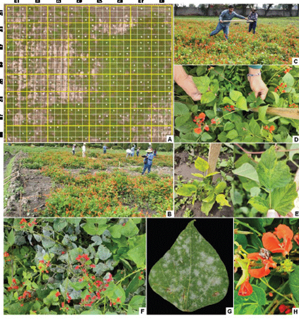
Figure 1 Sampling methodology to identify and assess severity of fungal, presumtive viral/phytoplasmal symptoms, and entomological signs on Phaseolus coccineus. A. 13mpx image at 50m using DJI® Phantom-3 drone, showing quadrantization of experimental plot. Yellow-lines correspond to 6x6 m quadrants, and white-lines to 2x2 m subquadrants. Asterisks indicate randomly selected subquadrants/quadrants; B. Field quadrant marking with wooden-stakes and a slat-net; C. Selection of subquadrant by placing wooden frame 1x1 m for assessment guidance; D. Dotted mosaic symptoms (left) and vein clearing (right), putative to virosis. Plants marked with stakes for traceability samples; E. Generalized yellowing with growth reduction (left), mosaic with leaf deformation (right) presumptive viral; F. Leaf symptom with white fungal mycelial growth putative to powdery mildew; G. Front leaflet showing white mycelial growth. H. Macrodactylus sp. adults, and flowering color- morphology of P. coccineus. Note some petals showing small white-spots (see arrows).
pellets. Each tube was considered as a sample/plant. The mycelium in tube was macerated in a disruptor at 30 frequencies for 15 min. The pellets were removed. One µL of β-mercaptoethanol was added, vortexed for 10 s and incubated at 56°C for 45 min. After that, 400 µL of phenol:chloroform:isoamyl alcohol (25:24:1) was added, homogenized by vortex, and centrifuged at 13,500 rpm for 10 min at laboratory temperature. Five hundred µL of aqueous phase was recovered, 500 µL of isopropanol and 50 µL of ammonium acetate were added, mixed by immersion, and incubated at -20 °C overnight. Subsequently, it was centrifuged at 13,500 rpm for 10 min. The supernatant was discarded and washed with 500 µL of ethanol at 80%. It was centrifuged at 13,500 rpm for 3 min. The supernatant was discarded, and the pellet was dried for 1 h and finally resuspended in 50 µL of nuclease-free water and stored at -20 °C. Per sample, the concentration and purity of total nucleic acids were quantified with NanoDrop 2000 (Thermo Fisher Scientific 2000, EUA). For PCR, ITS1 and ITS4 primers amplifying the ITS region of the DNA were used (Table 1). The reaction mixture consisted of 10 µL of each primer, 0.2 µL of Taq DNA Polymerase (Invitrogen), 2 µL of DNA (40 ng µL-1) and 16.5 µL of nuclease-free water. PCR conditions consisted initial denaturation at 95 °C for 5 min, 31 cycles with denaturation at 94 °C for 1 min, alignment at 55 °C for 1 min, extension at 72 °C for 90 s, and a final extension at 72 °C for 5 min. PCR products were analyzed by 1.5% agarose gel electrophoresis stained with ethidium bromide and visualized in a photodocumenter (UVP, Biolmaging Systems, Epi Chemi II Darkroom). Five of 10 samples were amplified and sent for sequencing to
Table 1 Primers, sequences, and amplicon size for genomic identification of Potyvirus, Begomovirus, Phytoplasmas and eukaryotic microorganisms in P. coccineus plants exhibiting signs of powdery mildew and putative virus and phytoplasma symptoms.
| Organism | Primer | Sequence | Size (bp) | Reference |
|---|---|---|---|---|
| Fungus | ITS1 ITS4 | TCCGTAGGTGAACCTGCGG TCCTCCGCTTATTGATATGC | ~500 | White et al., 1990 |
| Potyvirus | NIb2F NIb3R | GTITGYGTIGAYGAYTTYAAYAA TCIACIACIGTIGAIGGYTGNCC | 350 | Zheng et al., 2008 |
| Begomovirus | PBL1v2040 PCR1c | CARTGRTCKATCTTCATACA CATATTTACRARWATGCCA | 500~650 | Rojas et al., 1993 |
| AV494 AC1048 | GCCYATRTAYAGRAAGCCMAG GGRTTDGARGCATGHGTACATG | ⁓550 | Wyatt y Brown, 1996 | |
| Phytoplasmas | P1 P7 | AAGAGTTTGATCCTGCAGGAT CGTCCTTCATCGGCTCTT | 1,800 | Deng y Hiruki, 1991; Samart et al., 1996 |
| FU5 RU3N | CGGCAATGGAGGAAACT TTCAGCTACTCTTTGTAACA | 890 | Lorenz et al., 1995 |
x Universal primers were used.
Macrogen®, South Korea. Four sequences of five amplicons had quality required for identification studies (FAC6, FAC7, FAC8 and FAC9). All sequences were edited with SeqAssem software (https://acortar.link/r0FdRx) and compared in BLASTn (https://blast.ncbi.nlm.nih.gov/Blast.cgi) with GenBank sequences to determine pathogen identity based on percent identity and e-value.
Phylogenetic analysis. Once Erysiphe sp. was identified, in order to determine the phylogenetic relationship among other species of this genus, as well as to confirm identity, the sequences obtained were aligned against sequences downloaded from GenBank using the MUSCLE algorithm in Mega 11 (Table 2). The phylogenetic analysis was performed with the same software, using the Maximum Likelihood (ML) statistical method, and Hasegawa-Kishino-Yano substitution model with 1000 Bootstrap replications. Oidium sp. (accession number: EU377475) was included as an outgroup. The analysis included 12 sequences of approximately 561 bp.
Table 2 Sequences obtained from the NCBI Genebank used to construct the phylogenetic tree for comparison with amplicon sequences obtained from four samples of powdery mildew fungus present in P. coccineus plants.
| Powdery mildew causing species | Genbank accession | Plant Host | Location and Date of report |
|---|---|---|---|
| Erysiphe diffusa | FJ378880 | Glycine max | Australia, 2006 |
| E. fallax | LC228618 | Carica papaya | USA, 2017 |
| E. manihoticola | MT106660 | Manihot esculenta | Argentina |
| E. pisi | KR912079 | Pisum sativum | China, 2016 |
| E. vignae | MW293895 | Vigna radiata | Australia, 2019 |
| E. vignae | MT628286 | V. radiata | Australia, 2019 |
| Erysiphe sp. | JQ730709 | Senna septemtrionalis | Mexico, 2012 |
| Oidium sp. | EU377475 | Knautia arvensi | USA, 2009 |
Genomic identification of viruses and phytoplasma
Plant sample collection. Plants exhibiting presumptive symptoms of Potyvirus, Begomovirus and Phytoplasma were selected in situ. Symptoms included faint mosaics, moderate leaf deformation, generalized yellowing, leaf chlorosis, vein clearing, leaf reduction, and violaceous coloration on leaves, peduncles and stems. Plants selected were marked with wood-stakes and labeled for later sampling. The sampling was conducted at two different periods; in the first, two trifoliate leaves were collected in 32 plants, each leaf as a replicate (64 samples), and in the second, 31 plants were collected.
Nucleic acids extraction (RNA and DNA). Leaf tissue of the 64 samples from first sampling was macerated in liquid nitrogen. A total of 0.1 g was weighed in 32 samples selected by symptoms and total RNA was extracted using the modified Green and Sambrook method (2012). For the second sampling, total RNA and DNA extraction was performed using the 2 % CTAB method (Yu, 2012; modified by CP-LANREF, 2021). Per sample, concentration and purity of nucleic acids were quantified with NanoDrop 2000 (Thermo Fisher Scientific 2000, USA).
Potyvirus genomic identification. Nucleic acid of 28 and 18 samples from the first and second sampling, respectively, were analyzed. NIb2F and NIb3R universal primers (Table 1) which amplify part of the coding region of the Potyvirus genome (350 bp) for the nuclear inclusion protein B (NIb) were used (Zheng et al., 2008). Firstly, cDNA synthesis of total RNA was performed and subsequently RT-PCR according to Gonzalez-Cruces et al. (2022) protocol. A pre-RT-PCR reaction mixture was performed with 9.75 µL of nuclease-free H2O, 500 nM per primer and 2.5 µL of total RNA, incubated at 85 °C for 3 min. For RT-PCR, a mixture with 2 mM mix dNTP’s, 1X buffer-RT and 100 U of reverse transcriptase enzyme (M-MLV-RT), all from Promega Corp. USA was added to the pre-RT-PCR reaction obtaining a total reaction volume of 25 µL. RT-PCR conditions were 44 °C for 60 min for incubation, followed of 92 °C for 10 min. The final reaction volume was 25 µL consisted of: 12.5 µL of Gotaq® G2 Hot star mix, 500 nM of each primer and 2.5 µL of cDNA. A program with initial denaturation at 95 °C for 2 min, 35 cycles of denaturation at 95 °C for 45 s, alignment at 45 °C for 45 s, extension at 72 °C for 45 s, and final extension at 72 °C for 5 min was performed.
Begomovirus genomic identification. Leaf tissue from 17 samples (second sampling) was analyzed. Two universal primer pairs for the Begomovirus genus were used: PBL1v2040 / PCR1c (Rojas et al., 1993) and Av494 / Ac1048 (Table
1) (Wyatt and Brown, 1996). PCR mix with PBL1v2040 / PCR1c primers were performed with a final volume of 25 µL: 1X GoTaq® buffer, 0.2 mM dNTP mix, 300 nM of PBL1v2040 - PCR1c primers, 0.5 U of GoTaq® G2 and 2 µL of DNA. The final volume was measured with nuclease-free water to 25 µL. The program included initial denaturation at 95 °C for 3 min, 30 cycles with denaturation at 94 °C for 30 s, alignment at 50 °C for 30 s, extension at 72 °C for 45 s, and a final extension at 72 °C for 5 min. The PCR was performed on the Arktik Thermal Cycler (Thermo Fisher Scientific). For AV494 / AC1048 primers, the PCR mix consisted of a final volume of 25 µL: 15 μL of H2O, 5 μL of 5X Buffer Green, 2 μL of MgCl2 (7. 5 mM), 0.5 μL of dNTPs (10 mM), 0.5 μL of each primer (10 mM), 0.25 μL of GoTaq® DNA Polymerase (U μL-1) (Promega) and 2 μL of DNA.
PCR conditions included initial denaturation at 95 °C for 4 min, 30 cycles with denaturation at 95 °C for 1 min, alignment at 55 °C for 2 min, extension at 72 °C for 60 s, and a final extension at 72 °C for 10 min.
Phytoplasma genomic identification. Leaf tissue from 42 samples (second sampling) was analyzed. The primers P1 / P7 were used for the first nested PCR step (Samart et al., 1996; Deng and Hiruki, 1991). The PCR mix was prepared for a final volume of 25 µL consisting of: 12 μL of nuclease-free H2O, 2.5 μL of 1X Buffer, 0.6 μL of MgCl2 (50 mM), 0.5 μL of dNTPs (10 mM), 2 μL of each primer (10 mM), 0.4 μL of Taq DNA Polymerase (U μL-1) (Invitrogen), and 5 μL of DNA. An initial denaturation program was performed at 95 ºC for 5 min and 35 cycles at 95 ºC for 30 s, alignment at 52 ºC for 75 s, extension at 72 ºC for 90 s, and final extension at 72 ºC for 10 min. The PCR was performed in the T-100 thermal cycler (BioRad).
Primers FU5 and RU3N targeting the amplified 16S region were used in the second nested PCR step (Lorenz et al., 1995). PCR-direct products obtained were diluted (1:30) in sterile distilled water and used as template DNA with the same concentrations described above. An initial denaturation program was performed at 94 ºC for 2 min and 40 cycles at 94 ºC for 30 s, alignment at 56 ºC for 40 s, extension at 72 ºC for 90 s, and final extension at 72 ºC for 5 min. PCR was performed on the T-100 thermal cycler (BioRad). PCR product was analyzed by 1.5% agarose gel electrophoresis stained with ethidium bromide and visualized with UV light in photodocumenter (UVP, Biolmaging Systems, Epi Chemi II Darkroom).
Epidemiological Analysis
Sampling and assessment. Per quadrat, 3/9 subquadrats were selected as sampling units to assess variables of disease intensity and Macrodactylus sp. adults incidence (Coleoptera: Scarabaeidae) as a potential agent spread of presumptive powdery mildew on flower structures. Subquadrant selection was randomized using MS Excel Aleatoriza v1.0 (CP-LANREF, 2022. Unpublised) (Figure 1A). A 1x1 m portable wooden-frame was placed at centroid by subquadrant to delimit the area of assessment variables (Figure 1C-D). For sampling units having less than 25% leaf canopy, sampling was addressed to a different section inside the respective subquadrant.
Assessment variables. App-Monitor® v1.1 Android®, available in PlayStore® (CP- LANREF, 2022) was used to characterize the experimental plot and assesement variables. Geographic coordinates, state, municipality, locality, crop name, crop area, phenology, technological level, irrigation, and planting density were entered. A severity sampling of powdery mildew, flower damage and plant canopy was set up through a 5-classes arithmetic scale: 1=0%, 2=0.01-25%, 3=25.01-50%, 4=50.01- 75% and 5=75.01-100% (Figure 1F-G). Furthermore, Macrodactylus sp. adults were counted (Figure 1H). Regardless of plant number available, assessments were performed considering of canopy of visible plant tissue within sampling portable wooden-frame.
Geospatial analysis. The records in App-Monitor® v1.1 were exported in MS Excel®. Data matrix was structured in spatial array: X = rows (quadrant-i), Y = columns (subquadrant-j), and Z = variables-k for geostatistical analysis in Golden Surfer® v10. The maximum value obtained with the 5-class arithmetic scale for powdery mildew severity, flower damage and plant canopy were chosen to calculate a value for each quadrant based on three subquadrants assessed. For Macrodactylus sp. the cumulative insects per quadrant were determined. Geostatistical analysis by variable was performed using the Kriging method and displayed as contour maps. Spatial dependence and autocorrelation were calculated using omnidirectional variograms (360°) fitted to Spherical model. Variographic indicators lag-distance and sill or plateau (σ2-s) to determine spatial dependence at plot level were obtained (Gonzalez-Cruces et al., 2021).
Results and Discussion
Fungal morphological and genetic identification. Symptoms observed in situ were irregular white powdery spots on the leaf surface, predominantly on middle and lower leaves of the plant. Yellowish spots were identified in advanced symptoms. Slight distortion and reduction of leaf area was observed on young leaves due to a greater fungus abundance. Flowers showed slight leaf distortion and whitish spots but no powdery appearance. An abundant presence of Macrodactylus sp. was also observed, mainly in flowers. This pest affects several crops such as maize and bean (Aragón et al., 2021), and therefore could be a potential spreader of fungi due to feeding and mobility habits.
The powdery appearance was confirmed by compound microscopy corresponding to signs of fungus characterized by conidia, conidiophores, and hyphae (Figure 2A-C). Fungal structures were not obtained in flowers. The conidiophores were cylindrical and erect, with straight or slightly curved basal cell followed by two shorter apical cells. Conidiogenesis Pseudoidium-type was exhibited (asynchronous conidial maturation, one at a time) (Figure 2B-C). Hyaline, ovoid to ellipsoid conidia of 31.74 ± 0.3419 µm x 15.11 ± 0.1579 µm, and absence fibrosine bodies (Figure 2A-E). Germ tubes with 1 to 2-fold the length of conidia, terminal or subterminal development, and lobed apices (Figure 2D-E).
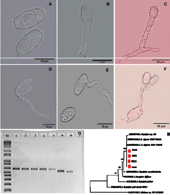
Figure 2 Morphological and genomic identification of powdery mildew in Ayocote bean (Phaseolus coccineus). A. hyaline, ovoid to ellipsoid conidia; B-C. cylindrical and erect conidiophores; D-F. germinating conidia; G. Amplification of the internal transcribed spacer region (ITS) of nuclear ribosomal DNA (~500 bp) of five samples of cenicilla DNA (1-5), two positive PCR controls (+) belonging to the ITS region of Alternaria and Fusarium genera, 1 kb molecular weight marker (M) plus Invitrogen and negative PCR control (-); H. phylogenetic tree performed by Maximum Likelihood (ML) and the Hasegawa-Kishino-Yano substitution model with 1000 Bootstrap replications, based on ITS region of fungal sequences belonging to Erysiphe genus (Table 2). The study sequences are: FAC6, FAC7, FAC8 and FAC9 (red dot). Oidium sp. (accession number: EU377475) was included as outgroup.
Morphological characteristics determined that the fungus belongs to the Erysiphe genus (Bradshaw et al., 2020, Tovar-Pedraza et al., 2018; Takamatsu, 2014; Cook y Braun, 2009; Glawe, 2008). Sexual structures were not observed. Agarose gel electrophoresis confirmed amplification of internal transcribed spacer region (ITS) of nuclear ribosomal DNA (~500 bp) in five fungal DNA samples, including DNA from positive PCR controls belonging to Alternaria and Fusarium genera (Figure 2G). Four sequences were suitable for BLASTn comparative analysis with GenBank collections. The genus was confirmed by the BLASTn, hence Erysiphe vignae was determined as causal agent of powdery mildew in Ayocote bean with 100 % homology (e-value = 0). Sequences were registered in GenBank with accession numbers OQ448664 (FAC6), OQ448665 (FAC7), Q448666 (FAC8), and OQ448667 (FAC9). Furthermore, at 94 % reliability, the four sequences clustered closely to two sequences belonging to Erysiphe vignae (MW293895, MT628286) associated to Vigna radiata (Fabaceae) from Australia (Figure 2H). Morphological and genomic results in this study were consistent with reported features by Kelly et al. (2021) y Deng et al. (2022) for this specie.
In addition to reports of E. vignae infecting Vigna radiata (Kelly et al., 2021), in other fabaceae such as black bean (V. mungo) in Australia, espelon (V. unguiculata) in Brazil, and common bean (Phaseolus vulgaris) in South America and China have also been reported (Deng et al., 2022; Kelly et al., 2021). In Sinaloa, Mexico, E. diffusa has been reported as causal agent of common bean powdery mildew disease reaching incidences up to 10% in the planted area (Félix-Gastélum et al., 2017; Félix-Gastélum et al., 2011). However, E. vignae is not reported affecting fabaceae in Mexico (Schoch et al., 2020). Therefore, this study provides the first report of E. vignae associated to Ayocote bean (Phaseolus coccineus) in conditions of the central Mexican Altiplano. This genus, as well as Erysiphales in overall, constitute an obligate parasite having relative host-specificity (Deng et al., 2022; Félix-Gastélum et al., 2017), thus, fungal-plant association can be assumed as causality indicator. However, in the future, etiological studies conducting to determine the pathogenesis and to evaluate the behavior of P. coccineous stocks to fungal infection, analogously to common bean studies (Deng et al., 2022).
Overall, Ayocote bean has been recognized for the resistance and tolerance to pests, diseases, and cold (Chiquito-Almanza et al., 2021; 2018; Schwember et al., 2017; Vargas et al., 2014; Giurca, 2009; Osorno et al., 2007; Schwartz et al., 2006). For instance, Ruíz-Salazar et al. (2019) highlight resistance to common blight (Xanthomonas axonopodis pv. phaseoli) and anthracnose (Colletotrichum lindemutianum) in Ayocote bean. However, E. vignae is not reported by some research. Powdery mildew, caused by other Erysiphe species, represents a yield restricting factor for some common bean growing regions worldwide, reaching losses up to 69 % (Deng et al., 2022; Félix-Gastélum et al., 2017).
Genomic identification of viruses and phytoplasmas. Potyvirus was not detected with the universal primers used (Zheng et al., 2008) in a total of 46 leaves showing symptoms of mosaic, yellowing, leaf deformation and vein clearing (Figure 1D, F). Analogous, the genus Begomovirus was not detected using two pairs of universal primers (Wyatt and Brown, 1996; Rojas et al., 1993) in 17 samples showing symptoms of yellowing and slight deformation leaf (Figure 1E). In common bean, both genera were reported in Mexico, and therefore were included for this study, as well as by the coincidence with some origin centers and cultivation between common bean and Ayocote bean (Mora-Aguilera et al., 2023). Particularly, the sampling for Potyvirus was increased due to the higher prevalence of Bean common mosaic virus, probably associated to seed transmission efficiency and several aphid species present (Hemitera: Aphididade) (Gonzalez-Cruces et al., 2022; Chiquito- Almanza et al., 2021). Begomovirus occurrence, i.e. Bean golden yellow mosaic virus, has decreased since the 90’s when it had an epidemic impact on bean crop (Chiquito-Almanza et al., 2021; Rojas et al., 1993). Worldwide, 23 viral species belonging to nine genera have been reported in common bean (Potyvirus, Comovirus Nepovirus, Sobemovirus, Alfamovirus, Cucumovirus, Tobamovirus, Begomovirus y Curtovirus), which 11 predominant species belonging to Potyvirus genus are the main causing viral diseases (Meziadi et al., 2017).
The Ayocote bean viral etiological researchs in Mexico are scarce (Chiquito-Almanza et al., 2021; 2018). Osorno et al. (2007) suggests that P. coccineus exhibits resistance to Bean golden yellow mosaic virus, due two genes. A recessive gene conferring resistance to leaf chlorosis and a dominant gene avoiding deformation of virus-infected seed pods. However, Cowpea mild mottle virus (Betaflexiviridae: Carlavirus), transmitted by Bemisia tabaci (Hemitera: Aleyrodidae), has been reported in Jalisco and Nayarit under symptomatic (chlorotic lesions, severe and moderate mottling, moderate mosaic, and leaf deformation) and asymptomatic conditions (Chiquito-Almanza et al., 2018). This virus was not analyzed in this study and therefore must be considered in future studies. However, findings in this and other studies suggest tolerance and/or resistance of P. coccineus to viral pathogens. For instance, a regional study in western Mexico revealed less than 20 % incidence of plants exhibiting viral symptoms in this species, reporting only Cowpea mild mottle virus, whereas more than 97 % in common bean were associated to at least 1/9 species of five identified virus genera (Chiquito-Almanza et al., 2021).
Diagnosis was also negative for phytoplasma in 42 samples analyzed using universal primers and nested PCR (Samart et al., 1996; Lorenz et al., 1995; Deng y Hiruki, 1991). The analysis was repeated several times, with PCR protocol setting and resampling in situ, due to the symptoms observed, overall associated with these organisms. These symptoms included leaf deformation, brown or violet coloration, evident purplish coloration on the veins of back younger leaves, stunting and internodes shortening, slight leaves yellowing in middle stratum, and abortion and/or delay of flower emissions (Figure 3A-B). However, agarose gel electrophoresis of the PCR products obtained only showed bands of the positive control (Figure 3C). Currently, ‘Candidatus Phytoplasma asteris’ has only been reported on fabaceae Macroptilium lathyroides (Alves et al., 2018) and Crotalaria juncea (Bianco et al., 2014), exhibiting leaf reduction, yellowing, and stunting symptoms. The leafhoppers Empoasca kraemeri, E. fabae, and E. papayae (Hemiptera: Cicadellidae) have also been reported associated with a viral and phytoplasmic symptom complex in common bean (Lozano-Gutiérrez et al., 2017; Sánchez-Castro et al., 2016). The presence of leafhopper adults was reported in this study, although identification was not performed. Therefore, future researches are needed to discard phytoplasma(s) presence in Ayocote bean using a greater number of production units and plant samples, in order to determine the association of these mollicutes as potential insect-vectors. Rather than productive, the main relevance of P. coccineus might be the potential use as resistance source in breeding studies of common bean, for which a comprehensive phytosanitary characterization of this specie is required.
Geospatial analysis. Geostatistical analysis by Kriging method and omnidirectional variograms of powdery mildew epidemic inductivity, flower damage, Macrodactylus sp. density and plant canopy, clearly evidenced the high infective capacity of powdery mildew (E. vignae) in Ayocote beans, even though the presumptive viral and phytoplasmic symptoms were not etiologically confirmed (Figure 4A-C). Powdery mildew incidence was 65.3% (subquadrant unit) and average severity 22.7% (±16.9, range: 0 - 66.5%). These severity levels are moderate to high, therefore E. vignae must be considered a high-risk pathogen for P. coccineus. Spatial contagion of powdery mildew exhibited an aggregate pattern conform by four quadrant as maximum spatial dependence in the main outbreak (96 m2, lag = 4, and σ2-s = 450), and 1-2 quadrants in secondary outbreaks (48 m2, lag = 2 and σ2- s= 350, Figure 4A). The main outbreak, exhibited by the red color continous in the scale, had a severity range of 60 - 80% and a noticeable border effect associated with highest plant canopy (range 50-75 %) (Figure 4D). This statement is evidenced by overall coincidence of the highest value colorimetric contours on the scale between the quadrants (c) 6-10 and blocks (b) 2-6 (Figure 4A and 4D). The infectious foliar of these fungi is consistent with their biological nature, which ensuring re-infection at higher biomass with microclimatic conditions suitable for inductive pathogenic, such as reduced exposure to direct sunlight, which inhibits spore germination (Craig and Weyne, 2012; Hückelhoven and Panstruga, 2011). In this work, powdery mildew occurred at average temperatures and relative humidity (July-August) of 16.3 °C (±5.8) and 92.8 % (±10.7), respectively. However, the directional spatial
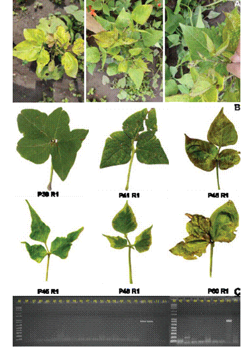
Figure 3 Presumptive field symptoms of phytoplasma infection in Ayocote bean (Phaseolus coccineus). A. Putative symptoms to phytoplasmas. Including leaf deformation, brown or violet coloration, obvious purplish coloration on veins of back young leaves, stunting, slight yellowing of leaves; B. Examples of six trifoliate leaves showing presumptive symptoms of phytoplasmas, included in PCR diagnosis; C. Agarose gel electrophoresis of 24 samples processed by nested PCR. PCR-amplified positive controls only.
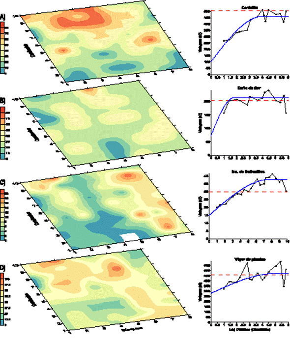
Figura 4 Geostatistical Kriging contour maps and variograms of phytosanitary variables and vigor in Ayocote bean. A. Powdery mildew severity, B. Flower damage, C. Density of Macrodactylus sp. adults and D. Plant canopy. For adult analysis, cumulative of three subquadrants per quadrant was calculated. For the analysis of powdery mildew severity, flower damage, and plant canopy, the maximum damage obtained per quadrant was calculated. Omnidirectional variograms were obtained by the Spherical method. X-axis = distance-lag in quadrants and Y-axis= variance (σ2).
dependence, and aggregate structure; also validated that dominant North-South winds effect in the gradient conformation. Moreover, the high epidemiological fitness of Erysiphales is well known by the abundant and intermittent conidial production with easily released and spread by air (Deng et al., 2022; Glawe, 2008). Plant canopy heterogeneity, probably due to the second growing year or phenotypic plasticity (Vargas et al., 2014), explained the conformation of differentiated plant canopy aggregates and identification of spatial dependence (lag = 4 and σ2-s =470) (Figure 4D). In epidemiological terms, flower damage was not associated conclusively with powdery mildew incidence. This agreed with signs absent at the microscopic level. The spatial distribution of this damage had randomly distributed, moderate outbreaks with dependence upon 1-2 quadrants and 30 - 40 % severity (lag = 1.5 y σ2-s = 250) (Figure 4B). No colorimetric correspondence between flowers damage and powdery mildew incidence maps (Figure 4A) suggested events without infectious correlation.
Similarly, the spatial density of Macrodactylus sp. adults, although heterogeneous, two main large aggregates were located between 1-10 c and 6-8 b, and between 5-10 c and 1-3 b, containing one and four foci (> 18 insects/centroid), respectively (Figure 4C). A contour map of flower damage dispersion or powdery mildew on leaf, as originally hypothesized, was not associated with Macrodactylus sp. spatial distribution. No feeding damage was detected in flowers, where adults were more prevalent (Figura 1H). The Macrodactylus sp. role as potential pollinator, important in P. coccineus, was not evaluated. The adults have been reported feeding on flowers, fruits, pollen grains, and sweet sap secretions of several crops such as maize and common bean (Aragón et al., 2021). Macrodactylus sp. involvement in E. vignae dissemination is not conclusive. For this purpose, comprehensive spatio- temporal studies need to be conducted.
Conclusions
The pathogen associated with powdery mildew on Phaseolus coccineus was Erysiphe vignae (Erysiphales: Erysiphacea). This is the first report on Ayocote bean in Mexico. Powdery mildew exhibited an incidence of 65.3 % and average severity of 22.7 % (±16.9, range: 0 - 66.5 %). These severity levels were from moderate to high, and therefore E. vignae must be considered a pathogen of high-risk for P. coccineus. Aggregate pattern of E. vignae infection was associated with plant canopy and directionality of dominant winds. The pattern was not associated with other damage such as whitish spots on flowers and occurrence of Macrodactylus sp. (Coleoptera: Scarabaeidae). E. vignae can also be a risk for common bean (P. vulgaris) in Mexico because it has not been reported. Potyvirus and Begomovirus species and phytoplasma-type mollicutes associated with yellowing symptoms, leaf distortion, moderate mosaic, internode shortening, and purple coloration of leaves and stems were not detected, validating the tolerance/resistance reported for P. coccineus. Potential nutritional importance of the Ayocote bean, as well as the implementation in breeding resistance programs, requires the encouragement of etiological-epidemiological research in P. coccineus.
Acknowledgments
The authors are grateful for the support of the CP-LANREF team and participants of COLPOS course on Agricultural Epidemiology FIT612; to MC Jorge Valdez for his guidance in microscopic measurements and photographic editing; to CONAHCYT for grants awarded; and to COLPOS for the opportunity to conduct postgraduate studies.
REFERENCES
Alves, M.S., de Souza, A.N., Ribeiro, G.M. y Carvalho, C.M. (2018). First report of a ‘Candidatus Phytoplasma asteris’ isolate associated with Macroptilium lathyroides yellow leaf disease in Brazil. New Disease Reports, 37, 11. https://doi.org/10.5197/j.2044-0588.2018.037.011 [ Links ]
Aragón-Sánchez, M., Aragón-García, A., Arce-Pérez, R., Pérez Torres, B.C., Cuate-Mozo, V.A. y López-Olguín, J.F. (2021). Actualización de las especies del género Macrodactylus Dejean (Coleoptera: Melolonthinae: Macrodactylini) para el estado de Puebla, México. Acta zoológica mexicana, 37, e3712403. https://doi.org/10.21829/azm.2021.3712403 [ Links ]
Bianco, L.F., Martins, E.C., Toloy, R.S., Coletti, D.A.B., Teixeira, D.C. y Wulff, N.A. (2014). First report of phytoplasmas groups 16SrI and 16SrXV in Crotalaria juncea in Brazil. Diseases Notes. https://doi.org/10.1094/PDIS-11-13-1190-PDN [ Links ]
Bradshaw, M., Braun, U., Götz, M., Takamatsu, S., Brand, T., Cabrera, M.G., Dirchwolf, P., Kummer, V., Medina, R., Moparthi, S. y Salcedo-Sarmiento, S. (2020). Contributions to the knowledge of the phylogeny and taxonomy of the Erysiphaceae (powdery mildews)-part 1. Sydowia, 73, 89-112. https://doi.org/10.12905/0380.sydowia73-2020-0089 [ Links ]
Barnet, H.L. y Hunter, B.B. (1998). Illustrated genera of imperfect fungi. American Phytopathological Society. USA. 240p. [ Links ]
Chiquito-Almanza, E., Caballero-Pérez, J., Acosta-Gallegos, J.A., Montero-Tavera, V., Mariscal-Amaro, L.A. y Anaya-López, J.L. (2021). Diversity and distribution of viruses infecting wild and domesticated Phaseolus spp. in the mesoamerican center of domestication. Viruses, 13(6), 1153. https://doi.org/10.3390/v13061153 [ Links ]
Chiquito-Almanza, E., Caballero-Pérez, J., Guevara-Olvera, L., Acosta-García, G., Pérez-Pérez, M.C.I., Acosta-Gallegos, J.A. y Anaya-López, J.L. (2018). First report of Cowpea mild mottle virus infecting cultivated and wild Phaseolus in the Central-Western region of Mexico. Plant Disease, 102, 1047. https://doi.org/10.1094/PDIS-07-17-0990-PDN [ Links ]
Cook, R.T.A. y Braun, U. (2009). Conidial germination patterns in powdery mildews. Mycological Research, 113(5), 616-636. https://doi.org/10.101 6/j.mycres.2009.01.010 [ Links ]
Craig, A. y Weyne, W.F. (2012). Effects of sunlight exposure on grapevine powdery mildew development. Phytopathology, 102(9), 857-866. https://doi.org/10.1094/PHYTO-07-11-0205 [ Links ]
CP-LANREF (2022). App-EMonitor. Ver 2.1. Autores: Acevedo-Sánchez G, Coria-Contreras JJ, Flores-Colorado OE, Guzmán-Hernández E, Mora-Aguilera G. No. Registro INDAUTOR: 03-2022-061611452600-01. https://shorturl.at/hiRS5 [ Links ]
De Ron, A.M. y Santalla, M. (2013). Phaseolus vulgaris (Beans). En: Maloy, S. y Hughes, K. (Eds.), Brenner’s Encyclopedia of Genetics. Second Edition. Academic Press, Elsevier. [ Links ]
Deng, D., Sun, S., Wu, W., Duan, C., Wang, Z., Zhang, S. y Zhu, Z. (2022). Identification of causal agent inciting powdery mildew on common bean and screening of resistance cultivars. Plants, 11(7), 7. https://doi.org/10.3390/plants11070874 [ Links ]
Deng, S. y Hiruki, C. (1991). Amplification of 16S rRNA genes from culturable and nonculturable mollicutes. Journal of microbiological methods, 14(1), 53-61. [ Links ]
Félix-Gastélum, R., Maldonado-Mendoza, I.E., Beltran-Peña, H., Apodaca-Sánchez, M.A., Espinoza-Matías, S., Martínez-Valenzuela, M.C., Longoria-Espinoza, R.M. y Olivas-Peraza, N.G. (2017). Powdery mildews in agricultural crops of Sinaloa: Current status on their identification and future research lines. Mexican Journal of Phytopathology, 35, 106-129. https://doi.org/10.18781/R.MEX.FIT.1607-4 [ Links ]
Félix-Gastélum, R., Maldonado-Mendoza, I.E., Herrera-Rodríguez, G., Martinez-Valenzuela, C., Espinosa-Matías, S., Cordero-Ramírez, J.D. y Martínez-Álvarez, J.C. (2011). Powdery mildew on common bean (Phaseolus vulgaris L.) in Northern Sinaloa, Mexico. Sydowia, 63(2), 169-182. https://www.scopus.com/record/display.uri?eid=2-s2.0-84856681084&origin=inward&txGid=ff970fdfb3ec9dc061b2e27296e844a4 [ Links ]
Glawe, D.A. (2008). The powdery mildews: A review of the World’s most familiar (yet poorly known) plant pathogens. Annual Review of Phytopathology, 46(1), 27-51. https://doi.org/10.1146/annurev.phyto.46.081407.104740 [ Links ]
González-Cruces, A., Arista-Carmona, E., Díaz-Arias, K.V., Ramírez-Razo, K., Hernández-Livera, A., Acevedo-Sánchez, G., Mendoza-Ramos, C. y Mora-Aguilera, G. (2022). Epidemiology of Bean common mosaic virus and Alternaria alternata in 12 Phaseolus vulgaris genotypes. Mexican Journal of Phytopathology, 40(2), 188-220. https://doi.org/10.18781/R.MEX.FIT.2202-8 [ Links ]
Giurca, D.M. (2009). Morphological and phenological differences between the two species of the Phaseolus genus (Phaseolus vulgaris and Phaseolus coccineus). Cercetari Agronomice Moldova, 42, 39-45. http://www.uaiasi.ro/CERCET_AGROMOLD/ro/fisiere/2009/V2/CA2-09-04.pdf [ Links ]
Graham, P.H. y Ranalli, P. (1997). Common bean (Phaseolus vulgaris L.). En: Ranalli, P. y Graham, P.H. (Eds.), Field Crops Research, 53(1-3), 131-146. https://doi.org/10.1016/S0378-4290(97)00112-3 [ Links ]
Hückelhoven, R. y Panstruga, R. (2011). Cell biology of the plant-powdery mildew interaction. Current Opinion in Plant Biology, 14(6), 738-746. https://doi.org/10.1016/j.pbi.2011.08.002 [ Links ]
Kelly, L.A., Vaghefi, N., Bransgrove, K., Fechner, N.A., Stuart, K., Pandey, A.K., Sharma, M., Németh, M.Z., Liu, S.Y., Tang, S.R., Nair, R.M., Douglas, C.A. y Kiss, L. (2021). One crop disease, how many pathogens? Podosphaera xanthii and Erysiphe vignae sp. nov. identified as the two species that cause powdery mildew of mungbean (Vigna radiata) and black gram (V. mungo) in Australia. Phytopathology, 111(7), 1193-1206. https://doi.org/10.1094/PHYTO-12-20-0554-R [ Links ]
Lorenz, K.H., Schneider, B., Ahrens, U. y Seemüller, E. (1995). Detection of the apple proliferation and pear decline phytoplasmas by PCR amplification of ribosomal and nonribosomal DNA. Phytopathology, 85, 771-776. [ Links ]
Lozano-Gutiérrez, J., España-Luna, M.P., Lara-Herrera, A., Álvarez-Robles, F. y Martínez-Contreras, C.A. (2017). La chicharrita Empoasca kraemeri (Ross y Moore) (Hemiptera: Cicadellidae) y su parasitoide nativo Anagrus sp. Haliday, 1833 (Hymenoptera: Mymaridae) en el cultivo de frijol en Zacatecas. Agroecología, 46-49. https://www.repositorionacionalcti.mx/recurso/oai:localhost:20.500.11845/1438 [ Links ]
Meziadi, C., Blanchet, S., Geffroy, V. y Pflieger, S. (2017). Genetic resistance against viruses in Phaseolus vulgaris L.: State of the art and future prospects. Plant Science, 265, 39-50. https://doi.org/10.1016/j.plantsci.2017.08.009 [ Links ]
Mora-Aguilera, G., Acevedo-Sánchez, G., González-Cruces, A. (2023). Riesgos a la diversidad de razas y producción de frijol común por incidencia de fitopatógenos. En: Ávila-Alistac, N., Rivas-Valencia, P. y Espinosa-Calderón, A. (Eds.), El Maíz y Frijol en México: Etiología, Epidemiología y Mejoramiento Genético. Sociedad Mexicana de Fitopatología (SMF) - Consejo Nacional de Ciencias Humanidades y Tecnología. Primera Edición. https://doi.org/10.18781/R.MEX.FIT.2024-1 [ Links ]
Osorno, J.M., Muñoz, C.G., Beaver, J.S., Ferwerda, F.H., Bassett, M.J., Miklas, P.N., Olczyk, T. y Bussey, B. (2007). Two genes from Phaseolus coccineus confer resistance to Bean golden yellow mosaic virus in common bean. Journal American Society Horticulture Science, 132(4), 530-533. https://doi.org/10.21273/JASHS.132.4.530 [ Links ]
Pérez, H.P., Esquivel, E.G., Rosales, S.R. y Acosta-Gallegos, J.A. (2002). Caracterización física, culinaria y nutricional de frijol del altiplano subhúmedo de México. Archivos Latinoamericanos de Nutrición, 52, 172-180. https://ve.scielo.org/scielo.php?script=sci_arttext&pid=S0004-06222002000200009 [ Links ]
Rojas, M.R., Gilbertson, R.L., Russel, D.R. y Maxwell, D.P. (1993). Use of degenerate primers in the polymerase chain reaction to detect whitefly-transmitted geminiviruses. Plant Disease, 77, 340-347. https://doi.org/10.1094/PD-770340 [ Links ]
Ruíz-Salazar, R., Vargas-Vázquez, M.L.P., Hernández-Delgado, S., Muruaga-Martínez, J.S. y Mayek-Pérez, N. (2019). Detección de marcadores genéticos asociados a la resistencia a patógenos en frijol Ayocote de Puebla, México. Revista Mexicana de Ciencias Agrícolas, 10(7), 1591-1602. https://doi.org/10.29312/remexca.v10i7.1956 [ Links ]
Sánchez-Castro, A., Miranda, I., Quiñones, M.L., Piñol, B.E. y Fernández, B.M. (2016). Saltahojas (Typhlocybinae) y su relación con los síntomas de enfermedades en un campo de frijol (Phaseolus vulgaris L.). Revista Protección Vegetal, 31, 153-158. https://n9.cl/uoxd1 [ Links ]
Santalla, M., Monteagudo, A.B., González, A.M. y De Ron, A.M. (2004). Agronomical and quality traits of runner bean germplasm and implications for breeding. Euphytica, 135, 205-215. https://link.springer.com/article/10.1023/B:EUPH.0000014912.07993.e7 [ Links ]
Schoch, C.L., Ciufo, S., Domrachev, M., Hotton, C.L., Kannan, S., Khovanskaya, R., Leipe, D., Mcveigh, R., O’Neill, K., Robbertse, B., Sharma, S., Soussov, V., Sullivan, J.P., Sun, L., Turner, S. y Karsch-Mizrachi, I. (2020). NCBI Taxonomy: A comprehensive update on curation, resources and tools. Database, 2020, baaa062. https://doi.org/10.1093/database/baaa062 [ Links ]
Schwartz, H.F., Otto, K., Terán, H., Lema, M. y Singh, S.P. (2006). Inheritance of white mold resistance in Phaseolus vulgaris × P. coccineus crosses. Plant Disease, 90(9), 1167-1170. https://doi.org/10.1094/PD-90-1167 [ Links ]
Schwember, A.R., Carrasco, B. y Gepts, P. (2017). Unraveling agronomic and genetic aspects of runner bean (Phaseolus coccineus L.). Field Crops Research, 206, 86-94. https://doi.org/10.1016/j.fcr.2017.02.020 [ Links ]
Takamatsu, S. (2014). Molecular phylogeny reveals phenotypic evolution of powdery mildews (Erysiphales, Ascomycota). Journal of General Plant Pathology, 79. https://doi.org/10.1007/s10327-013-0447-5 [ Links ]
Tovar-Pedraza, J.M., Rosas-Saito, G.H., García-Ruiz, M.T., Camacho-Tapia, M. y Leyva-Mir, S.G. (2018). Observaciones con MEB de cinco especies de Erysiphales en plantas ornamentales y frutales en México. Scientia fungorum, 48, 33-43. https://doi.org/10.33885/sf.2018.48.1197 [ Links ]
Vargas, V.M.L.P., Muruaga, M.J.S., Mayek, P.N., Pérez, G.A. y Ramírez-Sánchez, S.E. (2014). Caracterización de frijol Ayocote (Phaseolus coccineus L.) del Eje Neovolcánico y la Sierra Madre Oriental. Revista Mexicana de Ciencias Agrícolas, 5(2), 191-200. https://www.scielo.org.mx/scielo.php?script=sci_arttext&pid=S2007-09342014000200002 [ Links ]
Watanabe, K. y Tojo, M. (2006). Stem and root rot of scarlet runner bean (Phaseolus coccineus) caused by Pythium myriotylum. Journal Genetic Plant Pathology, 72, 126-128. https://doi.org/10.1007/s10327-005-0258-4 [ Links ]
White, T.J., Bruns, T.D., Lee, S.B. y Taylor, J.W. (1990). Amplification and direct sequencing of fungal ribosomal RNA genes for phylogenetics. En: PCR Protocols: A guide to methods and applications . Academic Press, California, USA. 460 p.. 315-322 [ Links ]
Wyatt, S.D. y Brown, J.K. (1996). Detection of subgroup III geminivirus isolates in leaf extracts by degenerate primers and polimerasa chain reaction. Phytopathology, 86, 1288-1293 [ Links ]
Zheng, L., Wayper, P.J., Gibbs, A.J., Fourment, M., Rodoni, B.C. y Gibbs, M.J. (2008). Accumulating variation at conserved sites in potyvirus genomes is driven by species discovery and affects degenerate primer design. PLoS ONE, 3(2), 1586. https://doi.org/10.1371/journal.pone.0001586 [ Links ]
Received: October 30, 2023; Accepted: January 25, 2024
 Este es un artículo publicado en acceso abierto bajo una licencia Creative Commons
