


 nueva página del texto (beta)
 Inglés (pdf)
 Artículo en XML
 Referencias del artículo
 Enviar artículo por email
 Citado por SciELO
  Similares en
SciELO
Similares en
SciELO 
 Permalink
Nowadays, neural tube defects (NTDs - encephalomyelodysraphies) are a growing entity in developing countries. The spectrum for this group of malformations ranges from “benevolent” entities (occult spinal dysraphisms), to alterations incompatible with life for the carrier (anencephaly with exencephaly). 1-5 These defects usually have a multifactorial origin (environmental, genetic, among others).1-3
The embryological process of formation of the neural tube begins on the 18th day of gestation; during this period the neural plate invaginates on its longitudinal axis, which originates the neural groove with its corresponding folds on both sides. Around week three of gestation, the folds begin their fusion to give rise to the neural tube, during days 21-23 approximately.6,7 This fusion occurs irregularly, starting from the fourth somite and continues in both directions. As a result, the neural tube is totally or partially open at the ends during different stages and maintains communication with the amniotic cavity. The rostral end or anterior neuropore (cranial orifice) undergoes closure around day 24-25 of intrauterine life, with subsequent closure of the lumbar opening (posterior neuropore) around the end of week four (days 26-27, approximately); these processes are known as primary and secondary neurulation, respectively.6-8
NTDs are divided into cephalic and lumbar defects, and can be open or closed; however, there is a neural tube malformation whose magnitude is total, that is, that presents with encephalic absence in conjunction with spina bifida, which generally presents in an open form, which is called craniorachischisis.2,6 The incidence for this defect is currently unknown and is the rarest form of NTD, which is incompatible with life.2 According to Rodríguez-Partida2 and Jiménez-Guerra et al,9 data provided by the Morbidity Yearbook of the General Directorate of Epidemiology of the Mexican Ministry of Health, an incidence rate of 0.27 cases per 100,000 inhabitants was reported in 2016. 2,9 Likewise, Johnson et al,10 noted a prevalence of 0.51 per 10,000 live births along the Texas-Mexico border.
The following are images of a 37-week gestation newborn from Córdoba, Veracruz, whose mother reported adequate prenatal care and folic acid supplementation, who was diagnosed by ultrasound with craniorachischisis, and whose pregnancy was carried to term (Figure 1).
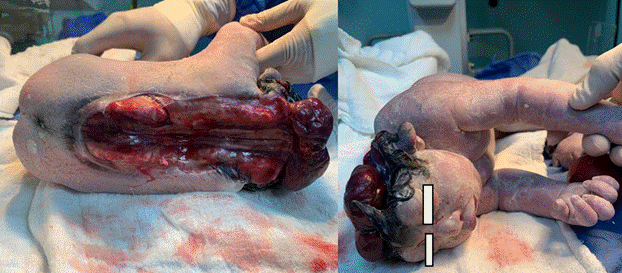
Figure 1 Term newborn, presenting complete neural tube closure defect. Absence of cranial vault with presence of proptosis is observed, in addition to complete opening from the cervical spine to the lumbar region (total absence of neural tube closure). On the other hand, there are stigmata of lumbar dysraphism such as local hypertrichosis in the area and dermal dimpling (dermal sinus), associated with total cutis aplasia.
Ethical responsibilities
The author states that no experiments on humans or animals have been performed for this research.
The author declares that he has followed his facility’s protocols on the publication of patient data.
The author declares that no patient data appears in this article.
Conflicts of interest
The author declares no conflict of interest.
Financing
This research has not received specific support from public sector agencies, commercial sector or non-profit entities.

