




 nueva página del texto (beta)
 Inglés (pdf)
 Artículo en XML
 Referencias del artículo
 Traducción automática
 Enviar artículo por email
 Citado por SciELO
  Similares en
SciELO
Similares en
SciELO 
 Permalink
Introduction
Swallowing is a complex neuromuscular process and evento1 that requires precise coordination of over 25 pairs of muscles, intact pharyngeal sensation, and central control in the brainstem and cerebral cortex,2-4 along with intact cognition, adequate sensory processing, reward and motivation mechanisms, sensorimotor control, protection of the airway, and intact involuntary functions.5 Neurophysiological studies have shifted the perception of swallowing from a purely reflexive and automatic mechanism to one that involves multiple levels of the nervous system, both cortical and subcortical, 6 and various brain regions with anticipatory, preparatory, and execution functions.1 Unlike other voluntary movements, a successful swallow concludes with a reflexive stage.1
Dysphagia is the disorder and disruption of swallowing that arises from any difficulty in one or more of the four stages of the swallowing process.1 It represents an impairment of the swallowing process, leading to problems in safely moving food bolus from the oral cavity to the stomach without the entry of food, liquids, saliva, or secretions into the respiratory system.8 The wide variety of structures involved in the swallowing process suggests that different pathophysiological mechanisms may result in dysphagia depending on the underlying disease and associated structural and functional impairment.3
The prevalence of dysphagia in the general population ranges from 8.4% to 16%.9 It is present in 5% to 8% of individuals aged 50 years or older10 and has a prevalence of 26% in individuals aged 76 years or older.11,12 However, the magnitude and frequency of dysphagia can be discriminated based on its clinical classification as oropharyngeal and esophageal or based on its etiology as structural, motor, or functional causes.13,14 Oropharyngeal dysphagia involves difficulty in moving the food bolus due to impairment in the oral, oral preparatory, or pharyngeal phases of swallowing.15 It can affect between 27% and 91% of individuals over the age of 70, with a frequency of approximately 47% in hospitalized patients and 91% in patients with community-acquired pneumonia.16 In patients with neurological and neurodegenerative diseases, the frequency of oropharyngeal dysphagia is between 30% and 82%,17,18 with neurological disorders accounting for 70% to 80% of its etiology.19
Oropharyngeal dysphagia is often caused by structural factors, which include conditions that narrow the lumen in the oral, pharyngeal, and/or esophageal cavities, and functional factors, where there is impaired physiology of swallowing.15 These functional causes can be neurologic and neuromuscular in nature, hence the terms neurogenic dysphagia and neurogenic oropharyngeal dysphagia (NOD).20 The global incidence of neurogenic dysphagia is reported to be between 400,000 and 800,000 people per year.10
In adults, NOD is primarily caused by stroke, followed by neurodegenerative disorders such as Parkinson's disease (PD), multiple sclerosis (MS), and amyotrophic lateral sclerosis (ALS),21,22 as well as traumatic brain injuries (TBI). It is a potentially dangerous condition that leads to various secondary complications,15 including respiratory, cardiovascular, nutritional, metabolic, and neurological disorders, and motor and cognitive developmental delays in cases of developmental dysphagia.23,24 Post-stroke dysphagia, and possibly other neurological and neuromuscular conditions, are reported to be underdiagnosed,25 emphasizing the need for greater awareness and training among all healthcare professionals.26
This narrative review aims to provide an updated overview of NOD, offering clinically relevant information on the neurophysiology of swallowing and its conceptualization, as well as the pathophysiological, clinical, and therapeutic characteristics of its main causes.
Methods
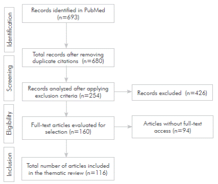
A systematic literature search was conducted following the PRISMA guidelines in PubMed, using the terms "swallowing neurology" and "neurogenic oropharyngeal dysphagia" in combination with the terms "etiology", "adults", "pathophysiology", "clinical", and "therapeutic." Studies conducted in humans and published in the last 29 years, from 1992 to 2021, in English and Spanish were considered. Included articles consisting of review articles, meta-analyses, systematic reviews, and original research. Additionally, four specialized reference texts on swallowing and dysphagia in gastrointestinal physiology, internal medicine, and clinical neurology were consulted. Case reports and protocols that did not align with the objective of the review, duplicate articles, and articles focused on neuropathic, neuromuscular, or myopathic causes of dysphagia, as well as articles on iatrogenic, mechanical, or structural etiologies of dysphagia and systemic causes of oropharyngeal dysphagia were excluded. The article selection process, from the initial search to the final selection, is depicted in Figure 1.
Neurophysiological aspects of swallowing
The initial studies utilizing functional magnetic resonance imaging (fMRI) to investigate swallowing showed bilateral activations in a wide cortical network, including the precentral gyrus, postcentral gyrus, supplementary motor cortex, prefrontal cortex, cingulate gyrus, insula cortex, Broca's area, precuneus, and frontal operculum up to the superior temporal gyrus with Heschl's gyrus.27,28 This extensive network encompassed areas and circuits involved in the motor and sensory innervation of the tongue, larynx, pharynx, face, and neck.6,28 However, subsequent studies with greater specificity regarding areas solely involved in swallowing demonstrated that the pharyngeal components of swallowing and laryngeal closure rely on subcortical circuits, while the oral components and lingual elevation are more represented in the primary motor and sensory cortices.29,30
Several brain regions have been implicated in the phases of swallowing, including the lower parts of both primary motor (M1) and somatosensory (S1) cortices, bilateral insula, cingulate gyrus,31,32 and supplementary motor area. The latter appears to be relevant in the pathophysiology of neurogenic dysphagia.33 Supratentorial structures such as M1 and S1 cortices, somatosensory association area, insula, frontal operculum, and superior temporal gyrus are more involved in the preparation and execution of voluntary phases of swallowing.34
Beyond cortical areas, supramedullary centers, such as the dorsal zone of the medulla oblongata and the nucleus of the solitary tract, ventral area of the nucleus ambiguus, and surrounding reticular formation, are other central components of swallowing, which are also involved in various functions such as arousal, phonation, respiration, and taste pathway. The nucleus of the solitary tract and the nucleus ambiguus are adjacent to the bulbar respiratory centers, allowing for the coordination between swallowing and breathing, a proximity and necessary connections for coordination between swallowing and breathing.35 The reticular formation is responsible for inhibiting respiration until the bolus is propelled from the pharynx to the esophagus.14 Brainstem structures generate the central pattern associated with the reflexive or involuntary phases of swallowing.36
In summary, the synchronization of the initiation and phases of swallowing, as well as the protection of the airway, are regulated by the swallowing pattern generator in the brainstem, while the regulation and execution of the motor response occur in cortical centers associated with the swallowing process.36,37
Concept of neurogenic oropharyngeal dysphagia Dysphagia can anatomically occur due to oropharyngeal or esophageal dysfunctions, while pathophysiologically it can result from structural (mechanical), motor (propulsive), or functional (swallowing physiology) causes,15,38 considering that more than one mechanism can be operational in a patient with oropharyngeal or esophageal dysphagia. From a clinical and epidemiological perspective, swallowing disorders are more likely to have a neurological basis.22,39 Following brain damage, somatic and visceral sensorimotor function, including swallowing, may be affected,40 resulting in functional causes of oropharyngeal dysphagia. Neurological diseases can affect swallowing with varying severity, ranging from mild to profound,5 and are primarily responsible for oropharyngeal dysphagia.41
Oropharyngeal dysphagia is a clinical form and comorbidity of various diseases, particularly neurological and neuromuscular disorders, in both children and adults. It is frequently associated with secondary pulmonary and nutritional complications.42,43 It commonly occurs due to structural and functional causes, with functional causes related to central neurological and neuromuscular impairments that disrupt the central neurological control of the oropharyngeal phase of swallowing, modulation of peristalsis, neuromuscular coordination of sphincters, and the action of orofacial, masticatory, lingual, soft palate, and pharyngeal muscles.14
The terms neurogenic dysphagia and neurogenic oropharyngeal dysphagia (NOD) are used to encompass functional oropharyngeal dysphagia of neurological and neuromuscular causes, also known as myogenic causes.20 Neurological causes refer to disruptions in swallowing mechanisms due to central nervous system (CNS) impairment, while neuromuscular causes involve the effector mechanisms of swallowing due to alterations in the nerves, neuromuscular junction, or muscles of the pharynx and esophagus.10,14
NOD can result from lesions anywhere in the central circuits and structures between the cerebral cortex and the peripheral components that reach the swallowing effectors,44 especially when the lesions involve the cerebral cortex, basal ganglia, central neuromuscular pathways of voluntary swallowing control, and lesions affecting the nucleus tractus solitarius and nucleus ambiguus,45 which impact the swallowing pattern generator in the brainstem.
Neurological lesions affecting cortico-subcortical connectivity are more likely to result in aspiration.37 Lesions in the right hemisphere lead to longer pharyngeal transit time, increased pharyngeal residue retention of the bolus, higher penetration and aspiration rates, and a higher need for non-oral nutrition due to a high frequency of pharyngeal dysmotility across all consistencies.37 Lesions in the left hemisphere result in greater dysfunction of oral motility, reduced coordination of lingual musculature, poor bolus organization, and delayed oral transit.47
The presence of residue in the valleculae and pyriform sinuses after swallowing suggests impairment in the oropharyngeal phase of swallowing, and pharyngeal residue is a characteristic finding of neurogenic dysphagia. Dysphagia for liquids may indicate functional dysphagia. Nasal regurgitation, the need for multiple swallows for a small bolus, and a history of recurrent respiratory infections suggest neurogenic dysphagia.48 NOD is one of the main reasons why patients in the ICU remain tracheostomized after being weaned off mechanical ventilation.49
A wide range of CNS disorders and lesions, such as stroke, neurodegenerative and inflammatory diseases, and traumatic brain injury, are common causes of NOD.50 In conditions such as ALS, PD, dementia, MS, and other neuromuscular disorders, dysphagia often has an insidious onset and slow progression, making early diagnosis critical for the prevention and management of associated complications.51,52
A systematic review from 2021 proposes a classification of seven phenotypes of neurogenic dysphagia based on functional endoscopic evaluation of swallowing (FEES):3 premature spillage, delayed swallowing reflex, predominant residue in the valleculae, predominant residue in the pyriform sinuses, pharyngolaryngeal movement disorder, fatigable weakness in swallowing, and complex (heterogeneous) disorder. These phenotypes help elucidate the pathophysiological mechanism and etiology in patients with oropharyngeal dysphagia.3 For example, the premature spillage phenotype is commonly found in supratentorial stroke and acute stroke patients, while the delayed swallowing reflex and predominant residue in the pyriform sinuses phenotypes are observed more frequently in infratentorial stroke patients. The phenotypes of predominant residue in the valleculae and pharyngolaryngeal movement disorder are more frequently seen in patients with PD, and the fatigable weakness in swallowing phenotype is common in patients with ALS.3
Pathophysiological, clinical, and therapeutic aspects of the main causes of neurogenic oropharyngeal dysphagia
Stroke
Stroke is the most frequent and studied cause of NOD.53 During the acute phase, the frequency of dysphagia can be higher than 50%, with 50% to 90% of patients showing improvement in the first two weeks post-stroke.54 However, about half of the patients may develop chronic dysphagia.20 Other studies indicate that at least one-third of patients experience persistent dysphagia.55 NOD is a prognostic marker in stroke patients,20,56 and signs of aspiration within the first 72 hours post-stroke can predict the presence of dysphagia in the next three months.20
Neurogenic dysphagia following a stroke can coexist with communication and language impairments.57 A study conducted in 59 stroke patients with lesions in the pons showed that stroke severity, as assessed by the National Institutes of Health Stroke Scale (NIHSS), was more severe in those with dysphagia, along with higher rates of dysphonia, dysarthria, and facial paralysis.58 Another study involving 687 patients with confirmed ischemic or hemorrhagic stroke, as diagnosed by neuroimaging, demonstrated a positive correlation between NIHSS score and dysphagia severity as measured by FEES.
A proposed ideal cut-off point for NIHSS score is >9 for supratentorial strokes and >5 for infratentorial strokes.25 Additionally, these patients often experience a higher frequency of hemiparesis, facial paralysis, and neglect.25 Long-term (>6 weeks) indicators of oropharyngeal dysphagia in post-stroke patients include altered voice, dysarthria, impaired gag reflex, voluntary cough, and changes in voice after swallowing.59
Patients with stroke and dysphagia require longer hospital stays, sometimes to the point of requiring nursing home care,60-62 compared to those without dysphagia. Neurogenic dysphagia is one of the causes of subacute death after stroke63 and is associated with disability at discharge.55
FMRI studies conducted in patients with acute stroke versus healthy controls show variability in findings, ranging from increased contralesional activation in areas related to swallowing to higher ipsilateral activations in areas suggesting possible compensatory recruitment through post-lesional cortical neuroplastic mechanisms.64
Regarding the regional location of the stroke, dysphagia is most commonly observed in infarcts of the medulla oblongata65 and pons,66 while it is infrequent in midbrain infarcts.67
The clinical manifestations and associated complications of dysphagia may vary depending on the location of the stroke. For example, strokes in the left hemisphere are associated with higher rates of aspiration68 compared to strokes in the right hemisphere.69 Posterior or vertebrobasilar territory strokes are more likely to result in aspiration and penetration,68 while lesions in the insular cortex, frontal operculum, and primary motor cortex are associated with impaired swallowing execution.70-72 There is also an association between motor deficits, dysphagia, and acute aspiration risk when the internal capsule is affected,73 as well as the occurrence of severe oropharyngeal dysphagia in brainstem lesions.74
Approximately 55% of patients experience oropharyngeal dysphagia when the stroke is located in the brainstem, and the prevalence is around 40% for strokes located in the cortical hemisphere.56 Right hemisphere infarcts are associated with more severe dysphagia, while left hemisphere infarcts are more related to oral deficits.75 Stroke lesions in the brainstem and lateral medulla are associated with inadequate upper esophageal sphincter opening during swallowing,20,76 and the risk of pneumonia increases when the basal ganglia are affected by the infarct.20 Therapeutic goals for stroke-related dysphagia include improving the quantity and variety of liquids and food that the patient can consume orally while minimizing the risk of aspiration and associated complications.77 Patients with stroke and dysphagia should receive training in food preparation,78 and the use of thickeners as compensatory therapeutic strategies to improve swallowing safety should be considered.79 Other compensatory and rehabilitative strategies include active maneuvers, motor control exercises, muscle electrostimulation, and the application of botulinum toxin. Neurostimulation techniques such as non-invasive central and intra-pharyngeal electrical stimulation can also be employed.80
Multiple sclerosis
In advanced stages, multiple sclerosis (MS) carries a high risk of oropharyngeal dysphagia and secondary malnutrition.81 The exact frequency of dysphagia in MS is unknown, but it is reported to be present in approximately 30% of patients and increases with disease progression, especially in individuals with greater neurodegeneration and signs of cerebellar, brainstem, or cognitive impairment.
A study using videofluoroscopic swallowing study (VFSS) in 23 MS patients identified reduced tongue mobility and bolus control in the oral phase in 39% of patients, impaired pharyngeal reflexes in 57%, and symptoms of aspiration, such as changes in voice quality and cough, in nearly 17%.82 Another study involving 120 MS patients reported that 90% had swallowing impairments, with only 10% having intact swallowing function. A comparison between swallowing behavior and dysphagia severity using VFSS in relation to the clinical progression of MS showed that among patients with swallowing impairments, 41% had mild dysphagia, 37% had moderate dysphagia, and 12% had severe dysphagia. Patients with severe dysphagia were more likely to have primary progressive MS or secondary progressive MS.83
Several techniques and procedures have been described to manage dysphagia in MS patients, but the evidence to guide treatment and rehabilitation is limited. A systematic review conducted in 2016, which included studies with heterogeneous instrumental techniques, dosages (frequency), and outcome measures, found that pharyngeal electrical stimulation may have some beneficial therapeutic effects without conclusive evidence, and the use of botulinum toxin type A may be effective only in cases of MS patients with cricopharyngeal muscle hyperactivity.84 Further studies are needed to determine the clinical applicability and long-term effects of swallowing rehabilitation measures in MS patients with oropharyngeal dysphagia.
Traumatic brain injury (TBI)
TBI is a common cause of acquired disability in young adults, leading to long-term physical, cognitive, behavioral, and emotional impairments.85 It is a heterogeneous condition in terms of its mechanisms of injury, pathophysiology, classification, and prognosis. TBI can result in dysphagia due to direct brain damage, such as primary lesions in cortical regions involved in motor or sensorimotor integration or the insula, as well as lesions in the diencephalon, brainstem, or cerebellum. Dysphagia can also occur as a consequence of tracheostomy and prolonged ventilation.86
A systematic review that assessed the prevalence of dysphagia in various neurological conditions, including low-quality prevalence studies, reported a prevalence of dysphagia after TBI ranging from 25% to 30%.87 It should be noted that in cases of TBI accompanied by post-traumatic headache, confusion, disorientation, memory and sleep disturbances, dizziness, and emotional lability, oral feeding can be indirectly affected. Predictors of long-term oropharyngeal dysphagia in TBI patients include low initial Glasgow Coma Scale scores, specific findings on computed tomography (e.g., displacement of structures, brainstem involvement), prolonged ventilator use (≥15 days), and tracheostomy placement.88-91
Studies focused on the characteristics of dysphagia in post-TBI patients describe prolonged oral transit time, delayed swallowing reflex, and altered tongue control.92 One study evaluated swallowing characteristics using VFSS in 41 TBI patients and found that the most common findings were aspiration, penetration, decreased laryngeal elevation, and reduced epiglottic inversion. The incidence of tube feeding was also higher in post-TBI patients.93
Timely evaluation, aimed at establishing a prognosis for swallowing recovery and determining the feasibility of adequate oral intake, is necessary to decide which TBI patients will require enteral nutritional support. However, it is challenging to determine which TBI patients will experience long-term oropharyngeal dysphagia (>6 weeks) and which will transition to oral nutrition quickly.53
Altered oropharyngeal function, cognitive deficits, and behavioral problems often present in post-TBI patients can lead to complications such as malnutrition, dehydration, and aspiration pneumonia. Therefore, early detection and appropriate management of dysphagia are essential aspects of rehabilitation.93 Functional rehabilitation by experienced speech-language pathologists specializing in neurological disorders, including exercises for oral motor function, swallowing maneuvers, thermal and tactile stimulation, postural techniques, and modification of food consistency, is recommended for patients with post-TBI dysphagia.94 Regular evaluation and analysis of nutritional status based on the degree of dysphagia are important, considering that moderate dysphagia is often present after nasogastric tube removal.95
Parkinson's disease
Among neurodegenerative disorders, Parkinson's disease (PD) stands out as one of the conditions most likely to cause functional dysphagia, both oropharyngeal and esophageal, as a result of the cardinal symptoms of bradykinesia, akinesia, and rigidity96 experienced by patients, which are secondary to the degeneration of dopaminergic neurons and pathways in the substantia nigra, basal ganglia, and enteric nervous system.97-99 Dysphagia is present in approximately 30% to 82% of patients with PD,10,87 but abnormalities in the oropharyngeal phase are reported in up to 97% when assessed using videofluoroscopic swallowing study (VFSS).100 Age, male sex, disease duration, and the presence of associated major neurocognitive disorder independently contribute to the development of dysphagia in PD patients.101,102
It is reported that symptomatic dysphagia is not observed in the early stages of PD compared to other basal ganglia disorders, such as progressive supranuclear palsy, corticobasal degeneration, multiple system atrophy, and Lewy body dementia, in which dysphagia is an early symptom and sign.10 However, there are studies describing dysphagia in the early stages of PD and not solely as a characteristic of advanced-stage PD.100,102,103
Two neuropathological substrates are proposed to be responsible for swallowing disorders: the central nervous system and the enteric nervous system. There is early accumulation of abnormal alpha-synuclein and neuritic-type inclusions in the enteric system and dorsal motor nucleus of the vagus nerve.104 Accumulation of alpha-synuclein also occurs in peripheral nerves (sensory and motor) that supply the pharyngeal muscles.105,106 Several neural networks involved in the oropharyngeal phase of swallowing, located in the medulla, receive cortical modulation, and patients with PD appear to have reduced cortical activation originating from temporal areas.102,107 The dopaminergic system in the basal ganglia is part of the supramedullary centers involved in swallowing physiology1 and contributes to the pathophysiology of dysphagia in PD. Additionally, it is reported that the oral phase of swallowing may be affected in the absence of dysphagia symptoms.108 There is minimal correlation between self-reported swallowing status by PD patients and their actual swallowing function,109 highlighting the need for a clinical evaluation of swallowing along with an objective instrumental test such as FEES or VFSS.110
Regarding therapeutic management, exercises such as the forced swallowing maneuver (swallowing using all available musculature and maximum contraction),111 respiratory therapy combined with postural exercises,112 motor strengthening exercises, vocal exercises, and specific swallowing maneuvers,113 as well as phonotherapy focused on strength and posture have been suggested.114 However, further research is needed to assess the clinical applicability, efficacy, and long-term effects of various therapeutic protocols and techniques described for PD patients with dysphagia.114
Amyotrophic lateral sclerosis (ALS)
ALS, a neurodegenerative disease and prototype of motor neuron diseases, can affect swallowing to varying degrees. After two years of follow-up in patients with spinal and bulbar onset ALS, 85% developed dysphagia.115 Whether in the variant of spinal motor neuron damage, primary lateral sclerosis, or progressive bulbar palsy, it is reported that at least 80% of patients will experience progressive dysphagia and/ or dysarthria due to bulbar involvement.116,117 Dysphagia is a cardinal symptom of ALS and is associated with progressive atrophy of the tongue, as well as impaired muscular function of the soft palate and larynx, affecting their proper closure due to nuclear or supranuclear lesions of the lower cranial nerves (glossopharyngeal, vagus, and hypoglossal nerves).118
Patients with ALS exhibit abnormalities in the oral phase of swallowing and impairments in lip and tongue function, usually affecting the posterior portion of the tongue. Initially, patients may experience sialorrhea and lingual weakness involving the lips and soft palate.116,118 As muscular weakness progresses, there is involvement of the jaw, suprahyoid muscles, limitation in closing the oropharyngeal isthmus, as well as weakness in the pharyngeal and laryngeal musculature.10,119 Decreased respiratory function generally occurs simultaneously with swallowing impairments. Additionally, the swallowing reflex may be abolished during voluntary swallows. In cases of bulbar involvement, there is often cricopharyngeal spasm with hyperreflexia and hypertonicity of the cricopharyngeal muscle.10 Dysphagia in ALS patients is characterized by a loss of coordination of the pharyngeal musculature and cricopharyngeal spasms, increasing the risk of aspiration.119 In terms of care, it is expected that all patients in advanced stages will present with dysphagia.
Clinically evaluating the strength and motility of the tongue is useful in the physical examination and monitoring of patients with ALS. In a study of 28 ALS patients, Borges et al. reported that 90% of patients with dysphagia had an average tongue pressure below 34 kPa; tongue strength testing showed a sensitivity of 91.6%, specificity of 38.4%, and accuracy of 64%.119 Furthermore, fMRI studies in ALS patients with dysphagia have reported reduced activation in the primary sensory cortex compared to ALS patients without dysphagia and healthy controls.6
There is no specific treatment for dysphagia in ALS. Management focuses on associated complications such as malnutrition and dehydration, ideally in a timely and early manner, using various options including nasogastric tube feeding, percutaneous endoscopic gastrostomy (PEG) tube placement, radiologically inserted gastrostomy tube (RIG), and parenteral nutrition, according to the patient's condition and requirements.120
Table 1 summarizes the main physiological deficits documented by VFSS in patients with NOD in each of the reviewed conditions, as well as the proposed new phenotypes of neurogenic dysphagia using FEES.
Table 1 Physiological deficits and phenotypes in swallowing according to the main causes of oropharyngeal neurogenic dysphagia
| Pathology | Physiological deficits documented by VFSS | Phenotype by FEES |
| Stroke | Altered gag reflex and voluntary cough. May have altered oral phase, abnormalities in pharyngeal phase activation, altered pharyngeal contraction, and loss of the upper esophageal sphincter (UES) tone. Delayed or absent swallowing response, alteration in lingual propulsion, reduced pharyngeal propulsion, unilateral or bilateral pharyngeal muscle weakness, reduced laryngeal closure, poor sensation of residue, or UES dysfunction. | Premature spillage of bolus: Bolus spillage occurs before the swallowing reflex is activated. More common in patients with supratentorial stroke and acute stroke. Reflects impairment in the oral phase. Delayed swallowing reflex: there is no evidence of swallowing reflex activation for at least three seconds after the bolus reaches the valleculae. Often seen in patients with infratentorial stroke. Predominant residue in piriform sinuses: there is a greater accumulation of pharyngeal residue in the piriform sinuses compared to the valleculae. Observed in patients with infratentorial stroke (particularly brainstem). Impaired pharyngolaryngeal movement: interference in the physiological transport of the bolus due to oropharyngeal freezing, pharyngeal bradykinesia, and/or pharyngolaryngeal tremor. Mainly observed in patients with stroke involving the basal ganglia or midbrain. |
| Multiple sclerosis | Altered neuromotor sequence of laryngeal events, progressive weakness of pharyngeal constrictor muscles, prolonged pharyngeal delay times, shorter intervals from the onset of laryngeal excursion to its resting phase, and extended intervals for upper airway closure and protection. Pharyngeal constrictor dysmotility predominates, fatigue affects swallowing function, and findings and symptoms increase and decrease following the episodic course of the disease. | Complex disorder (heterogeneous): there is at least the presence of two phenotypes (premature bolus spillage, delayed swallowing reflex, predominant residue in valleculae or piriform sinuses, impaired pharyngolaryngeal movement, and/or fatigable weakness to swallow), a different mechanism occurs compared to the previous ones, or it is not possible to assign one. |
| Traumatic Brain Injury | Reduced lingual control, stuttering in voluntary lingual movement, repetitive tongue movement, evident and silent aspirations, delayed swallowing reflex, and reduced laryngeal elevation and closure. | Complex disorder (heterogeneous). |
| Parkinson’s disease | Abnormalities in the oral phase such as lip closure and lingual movements, and marked deficiency (up to half of the patients) in the pharyngeal phase. Difficulty in forming the bolus, delayed swallowing response, hypopharyngeal stasis, poor epiglottic mobility, increased transit time of the bolus in the pharynx, and continuous tongue movement (lingual pumping). Reduced oral and lingual control, delayed swallowing response time, reduced lingual propulsion, velopharyngeal reflux, and reduced laryngeal closure. | Predominance of residues in the valleculae: there is a greater accumulation of pharyngeal residues in the valleculae compared to the pyriform sinuses. Particularly observed in patients with PD. Disorder in the pharyngolaryngeal movement: interference in the physiological transport of the bolus due to oropharyngeal freezing, pharyngeal bradykinesia, and/or pharyngeal tremor. Observed in patients with atypical parkinsonism. |
| Amyotrophic lateral sclerosis | Weakness in the lips, tongue, jaw, and suprahyoid muscles, drooling, poor bolus propulsion, minimal laryngeal elevation and inadequate airway protection; reduced lingual and oral control, delayed swallowing response time, weak lingual and pharyngeal propulsion, velopharyngeal reflux, and reduced laryngeal closure. | Fatigable weakness in swallowing: repeated attempts to swallow (more than five) lead to pharyngeal residues or increased residue volume compared to the initial swallow. More commonly observed in ALS patients |
| Table created by the authors. VFSS: video fluoroscopic swallowing study; FEES: fiberoptic endoscopic evaluation of swallowing; EES: upper esophageal sphincter; MS: multiple sclerosis; TBI: traumatic brain injury; PD: Parkinson’s disease; ALS: amyotrophic lateral sclerosis. For more detailed phenotypes by FEES, refer to Warnecke et al., 2021. |
Conclusions
Swallowing is a complex neuromuscular event that involves multiple cortico-subcortical levels, voluntary and involuntary control phases and culminates in a reflex stage. Dysphagia is a disorder of safe and efficient swallowing. It is anatomically classified as oropharyngeal and esophageal but is further categorized based on its etiology as structural, motor, or functional dysphagia. Neurogenic oropharyngeal dysphagia is classified under functional dysphagia.
Stroke, multiple sclerosis, amyotrophic lateral sclerosis, Parkinson's disease, and traumatic brain injury are common causes of neurogenic oropharyngeal dysphagia, particularly among the adult population. The physiological deficits in neurogenic oropharyngeal dysphagia are diverse, although compromise of the pharyngeal phase, control and coordination of effectors, and reduced airway protection mechanisms are commonly observed.
A comprehensive understanding of the conceptual and pathophysiological aspects, as well as the clinical characteristics of the main causes of NOD, can guide multidisciplinary healthcare teams in implementing timely actions for detection, diagnosis, treatment, and rehabilitation. There are various techniques and methodologies for swallowing rehabilitation in patients with NOD caused by stroke, MS, TBI, and PD. However, there are ongoing challenges, particularly in the management of dysphagia in patients with ALS.

