SciELO - Scientific Electronic Library Online

 
vol.36 número2Reto diagnóstico: sarcoma mieloide cardiaco en un paciente con leucemia mieloide aguda índice de autoresíndice de materiabúsqueda de artículos
Home Pagelista alfabética de revistas  

Servicios Personalizados

Revista

Articulo

Indicadores

Links relacionados

  • No hay artículos similaresSimilares en SciELO

Compartir


Cardiovascular and metabolic science

versión On-line ISSN 2954-3835versión impresa ISSN 2683-2828

Cardiovasc. metab. sci vol.36 no.2 Ciudad de México abr./jun. 2025  Epub 13-Oct-2025

https://doi.org/10.35366/120547 

Clinical cases

Case of coil dislodgement in saphenous vein graft coil occlusion in retrograde chronic total occlusion percutaneous coronary intervention

Migración de coil de un puente de vena safena durante la intervención percutánea de una oclusión crónica vía retrógrada

1 MD, Interventional Cardiologist. Department of Cardiology, Erfan General Hospital. Tehran, Iran. ORCID: 0000-0002-7498-1863

2 MD, Professor of Medicine. Department of Cardiology, Faculty of Medicine, Samsun University. Samsun, Turkey. ORCID: 0000-0002-3548-1276

3 MD, Cardiology Resident. Department of Cardiology, Faculty of Medicine, Samsun University. Samsun, Turkey. ORCID: 0000-0003-2257-3021

4 MD, Professor of Medicine. Department of Cardiology, Faculty of Medicine, Ege University. Izmir, Turkey. ORCID: 0000-0002-8156-2675

5 MD, Radiologist. Department of Radiology, Erfan General Hospital. Tehran, Iran. ORCID: 0009-0002-1482-912X


Abstract:

We report the case of a 78-year-old male with a history of coronary artery bypass grafting (CABG) and percutaneous coronary intervention (PCI) to the saphenous vein graft (SVG) to the obtuse marginal artery (OM). The patient presented with acute coronary syndrome and was referred for tertiary care after coronary angiography revealed in-stent restenosis in a thrombotic SVG, along with chronic total occlusion (CTO) of the left circumflex (LCx) artery. Our initial plan was intervention of SVG to OM due to stent restenosis and thrombosis. During the procedure, a balloon rupture resulted in dissection and hematoma. As a bailout intervention, native LCx with CTO ostial stenting was performed, followed by coil occlusion of the SVG. Complications arose when the coil dislodged and fragmented, leading to embolization of one particle in the descending aorta and the other in the femoral artery. Both fragments were successfully retrieved via snare. This case highlights the complexity of managing SVG-related PCI complications and the importance of careful device handling during coiling procedures.

Keywords: coil; saphenous vein graft; chronic total occlusion

Resumen:

Reportamos el caso de un hombre de 78 años con antecedentes de cirugía de revascularización coronaria (CRC) e intervención coronaria percutánea (ICP) del injerto de vena safena (IVS) a la arteria marginal obtusa (MO). El paciente presentó síndrome coronario agudo y fue derivado a atención terciaria después de que la angiografía coronaria revelara reestenosis intrastent en una IVS trombótica, junto con oclusión total crónica (OTC) de la arteria circunfleja izquierda (CI). Nuestro plan inicial fue la intervención de IVS a MO debido a reestenosis del stent y trombosis. Durante el procedimiento, una rotura de balón resultó en disección y hematoma. Como intervención de rescate, se realizó stent nativo en la CI con OTC, seguido de oclusión con coil de la IVS. Surgieron complicaciones cuando el coil se desprendió y fragmentó, lo que llevó a la embolización de una partícula en la aorta descendente y la otra en la arteria femoral. Ambos fragmentos se recuperaron con éxito mediante un lazo. Este caso destaca la complejidad del manejo de las complicaciones de la ICP relacionadas con la IVS y la importancia de manipular cuidadosamente el dispositivo durante los procedimientos de colocación de la bobina.

Palabras clave: coil; injerto de vena safena; oclusión total crónica

Introduction

Saphenous vein grafts (SVGs) are frequently used in coronary artery bypass graft (CABG) surgeries to bypass occluded coronary arteries. In CABG surgery SVGs are frequently used but these grafts are prone to degeneration, atherosclerotic process, and thrombus formation often treated with coronary percutaneous intervention with balloon angioplasty and stenting leading to complications such as in-stent restenosis (ISR) and SVG failure. Management of ISR in SVGs, especially when complicated by chronic total occlusion (CTO) of native coronary arteries, presents unique challenges. This case illustrates a rare complication during the attempt to occlude an SVG with coil embolization during retrograde CTO percutaneous coronary intervention (PCI).

Case presentation

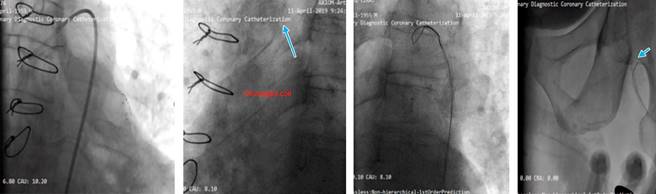
The patient was a 78 years old male with a history of CABG and PCI. His medical history included hypertension, hyperlipidemia and a previous PCI on the SVG to the obtuse marginal (OM) artery (Figure 1).

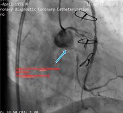
Figure 1: Thrombotic SVG with in-stent restenosis LCx CTO from ostial with no antegrade guide support. (J CTO Score; 4 Long, Calcified, Ambiguous proximal CAP and retry). 

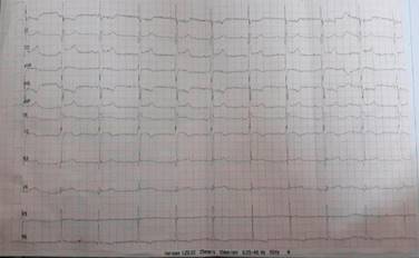
The patient presented with symptoms of acute coronary syndrome, including chest pain and shortness of breath. The ECG was in sinus rhythm and q wave was seen in D2, D3 and AVF leads. (Figure 2) His blood tests showed no significant values. He was referred to our center for tertiary care after coronary angiography performed at another hospital revealed a thrombotic SVG with in stent restenosis and CTO of the left circumflex artery (LCx) (Figure 3).

Figure 2: Sinus rhythm and q wave in D2, D3 and AVF leads. 

Figure 3: Attempt for CTO PCI using SVG as retrograde conduit. 

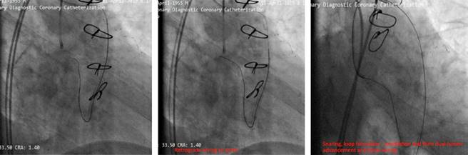
Our aim was to use the SVG as retrograde conduit for CTO PCI. During the procedure stenting was done but the stents balloon ruptured and made a huge dissection which caused a hematoma (Figure 4). As a bailout solution we decided to stent from the ostial LCx (Figure 5). To maintain a sufficient coronary perfusion, we decided to coil occlude due to Thrombolysis in Myocardial Infarction Score (TIMI) flow over 2 in this stenotic and thrombotic SVG so the competitive flow won’t affect the newly opened CTO’s long-term results. While coiling the donor SVG the coil dislodged. During the attempt to snare the dislodged coil with single loop snare, force by this device resulted in fracture in the dislodge coil and made two separated particles. One particle stayed in the descending aorta and the other went to the femoral artery. Both then were snared eventually (Figure 6).

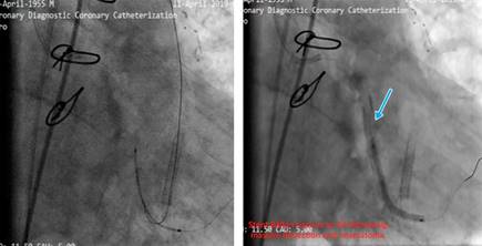
Figure 4: Stenting was done, stent’s balloon ruptured the LCx and created a huge dissection and hematoma. 

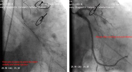
Figure 5: Free wire in aorta to place the stent ostially and avoid injection. Result after stenting and post dilation. 

Figure 6: Dislodged coil, during the attempt to snare the dislodged coil with single loop snare; force by this device resulted in fracture in the dislodged coil and made two separated particles. Dislodged coil particle in descending aorta. Another part of the dislodged coil in the femoral artery. 

Discussion

This case highlights several key challenges in the management of post-CABG patients with PCI to SVGs. First, the management of in-stent restenosis in thrombotic SVGs poses significant risks, particularly when CTO is involved in the native coronary arteries. The decision to use the SVG as a retrograde conduit was justified based on the anatomy;1 but complications such as balloon rupture and SVG dissection underscore the importance of selecting appropriate devices and anticipating potential hazards.2,3

Coil occlusion of SVGs is commonly employed to reduce competitive flow; however, in this case, coil dislodgement led to additional complications. The retrieval of coil fragments from both the descending aorta and femoral artery demonstrates the importance of having a bailout plan in such high-risk interventions.4

Moreover, the fracture of the coil during snaring highlights the need for careful handling of retrieval devices to avoid exacerbating complications.

Prompt and efficient operation is necessary after loop formation; as hemodynamic instability can occur quickly due to the compressive forces of looped wires on the heart.

Balloon rupture leading to dissection and hematoma is a severe complication that necessitates immediate bailout strategies, such as ostial stenting, where techniques like the Szabo technique can be helpful.5

When competitive flow from an SVG is present (TIMI flow ≥ 2), coil occlusion should be considered to ensure long-term patency of the newly opened CTO.6

Conclusions

This case underscores several important technical and procedural considerations for managing complex PCI in post-CABG patients with SVG involvement.

Stenotic or occluded SVGs should be considered the primary retrograde route for CTO PCI when available, as they often lead to fewer complications and greater procedural success compared to other collateral routes. Coil retrieval, when necessary, is best managed with three-looped snares, which are superior to single-loop snares for preventing coil fracture and subsequent embolization. This case highlights the importance of careful procedural planning and the ability to handle unexpected complications during complex coronary interventions.

References

1. Xenogiannis I, Gkargkoulas F, Karmpaliotis D, Krestyaninov O, Khelimskii D, Jaffer FA et al. Retrograde chronic total occlusion percutaneous coronary intervention via saphenous vein graft. JACC Cardiovasc Interv. 2020; 13 (4): 517-526. doi: 10.1016/j.jcin.2019.10.028 [ Links ]

2. Dautov R, Manh Nguyen C, Altisent O, Gibrat C, Rinfret S. Recanalization of chronic total occlusions in patients with previous coronary bypass surgery and consideration of retrograde access via saphenous vein grafts. Circ Cardiovasc Interv. 2016; 9 (7): e003515. doi: 10.1161/CIRCINTERVENTIONS.115.003515 [ Links ]

3. Rinfret S, Dautov R. When SVGs “had enough”: using them as retrograde conduits to resurrect native coronary srteries. JACC Cardiovasc Interv. 2020; 13 (4): 527-529. doi: 10.1016/j.jcin.2019.11.030 [ Links ]

4. Kostantinis S, Simsek B, Karacsonyi J, Alaswad K, Megaly M, Jaffer FA et al. Saphenous vein graft occlusion following native vessel chronic total occlusion percutaneous coronary intervention. J Invasive Cardiol. 2022; 34 (12): E836-E840. doi: 10.25270/jic/22.00221 [ Links ]

5. Simpfendorfer CC, Dimas AP, Zaidi A, Hollman J, Franco I, Knezinek V et al. Balloon rupture during coronary angioplasty. Angiology. 1986; 37 (11): 828-831. doi: 10.1177/000331978603701106 [ Links ]

6. Wilson SJ, Hanratty CG, Spence MS, Owens CG, Rigger J, Spratt JC et al. Saphenous vein graft sacrifice following native vessel PCI is safe and associated with favourable longer-term outcomes. Cardiovasc Revasc Med. 2019; 20 (12): 1048-1052. doi: 10.1016/j.carrev.2019.01.025 [ Links ]

Abbreviations: CABG = Coronary Artery Bypass Graft, CTO = chronic total occlusion, ISR = In-Stent Restenosis, LCx = Left Circumflex Artery, OM = Obtuse Marginal, PCI = Percutaneous Coronary Intervention, SVGs = Saphenous Vein Grafts, TIMI = Thrombolysis in Myocardial Infarction.

2Declaration of patient consent: the patient gave his consent.

3Funding: no financial support was received for this study.

4Conflict of interest: the authors declare no conflict of interest.

Received: November 23, 2024; Accepted: May 29, 2025

*Corresponding author: Melisa Uçar. E-mail: mmelisaucar@icloud.com

Creative Commons License This is an open-access article distributed under the terms of the Creative Commons Attribution License