




 nueva página del texto (beta)
 Inglés (pdf)
 Artículo en XML
 Referencias del artículo
 Traducción automática
 Enviar artículo por email
 Citado por SciELO
  Similares en
SciELO
Similares en
SciELO 
 Permalink

INTRODUCTION
Technological advances have enabled a qualitative leap in the study of organs and anatomical structures, including their function, pathological changes, and mechanisms of injury. The topic in this pictorial essay – the study of bursae – is essential for recognizing the diagnostic accuracy and detailed information provided by high-resolution ultrasound (US) and magnetic resonance imaging (MRI), thereby facilitating appropriate medical and surgical treatment decisions.
In ancient times, bursae were not studied as independent structures but were considered elements associated with joints and body movement. Hippocrates (2nd century BC) and Galen (2nd century AD) described them without assigning a specific name. It is also known that, even in antiquity, thermal baths were recommended for the treatment of inflammation and movement limitations1. The term bursa began to be used during the Middle Ages. It derives from Latin and means “bag” or “pouch,” referring to containers used to store coins or wine. The term was adopted in both anatomical and stock exchange terminology. The formal and systematic study of bursae in medicine is relatively recent2.
Bursae are fluid-filled structures located between the skin and a tendon, or between a tendon and a bone3. Most bursae form during embryonic development; however, some are not present at birth and develop gradually with age as mechanical friction increases. The human body contains approximately 140 bursae4. Their main function is to minimize friction between adjacent moving structures3. They are classified according to their location as subcutaneous, subfascial, subtendinous, and submucosal3. Bursae can be divided into two types: anatomical and adventitial. Anatomical bursae are true synovial-lined fluid-filled sacs located near joints.
In contrast, adventitial bursae lack a synovial lining and may be located farther from the joint; therefore, they are also considered accidental bursae3. A limited number of reports describe the superficial prepatellar and superficial infrapatellar bursae. Most articles focus on anatomical aspects or are case reports of severe bursitis treated with minimally invasive techniques or surgery. This pictorial essay broadens the spectrum of conditions affecting the superficial prepatellar and infrapatellar bursae of the anterior knee and provides a detailed description of their imaging findings.
ANATOMY OF THE SUPERFICIAL PREPATELLAR BURSA
This superficial prepatellar bursa is located anterior to the patella, between the patella, the quadriceps tendon, and the overlying subcutaneous tissue. It is a small superficial structure centered over the patella, although it may project beyond the lateral border by a few millimeters5. It is oriented in the coronal plane in all cases.
Anatomically, the anterior bursae of the knee, particularly the superficial prepatellar bursa, have a complex trilaminar organization, which explains their diverse pathological patterns. The trilaminar configuration of the superficial prepatellar bursa consists of fibrous soft-tissue over the patella6. These structures, from superficial to deep, include (Figure 1):
–. A superficial compartment, known as the subcutaneous prepatellar bursal space, is located between the subcutaneous tissue and the transverse superficial fascia (fascia lata).
–. An intermediate compartment, or subfascial prepatellar bursal space, is situated between the transverse superficial fascia and the intermediate oblique fascia, which is formed by fascial extensions of the vastus lateralis and vastus medialis muscles.
–. The deepest compartment, or subaponeurotic pre patellar bursal space, is located between the intermediate oblique fascia and the deep longitudinal fibers of the rectus femoris tendon, which inserts directly on the patella6.
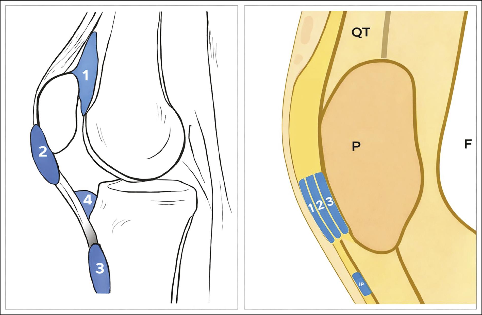
F: femur; QT: quadriceps tendon; PT: patellar tendon; IP: infrapatellar bursa; P: patella.
Figure 1 A: bursae of the anterior compartment of the knee highlighted in blue: (1) suprapatellar, (2) superficial prepatellar, (3) superficial infrapatellar, and (4) deep infrapatellar. B: illustration shows compartmentalization of the superficial prepatellar bursa highlighted in blue: (1) superficial compartment, or prepatellar subcutaneous bursal space, between subcutaneous tissue and transverse superficial fascia; (2) intermediate compartment, or prepatellar subfascial bursal space, between transverse superficial fascia and intermediate oblique fascia; (3) deepest compartment, or prepatellar subaponeurotic bursal space, between intermediate oblique fascia and deep longitudinal fibers of the rectus femoris tendon, which inserts directly into the patella. This illustration serves as an anatomical reference for the pictorial essay. Modified from Aguiar et al.5
This trilaminar arrangement was found in 93% of cases in cadaveric study by Dye et al.6 and in 78% in a study by Aguiar et al.5, who also reported a bilaminar pattern in 22%. There are no reports of a unilaminar or unicameral pattern, as previously suggested in older anatomy textbooks5. The superficial prepatellar bursa normally does not communicate with the knee joint, except in cases of patellar tendon rupture (Figure 2).
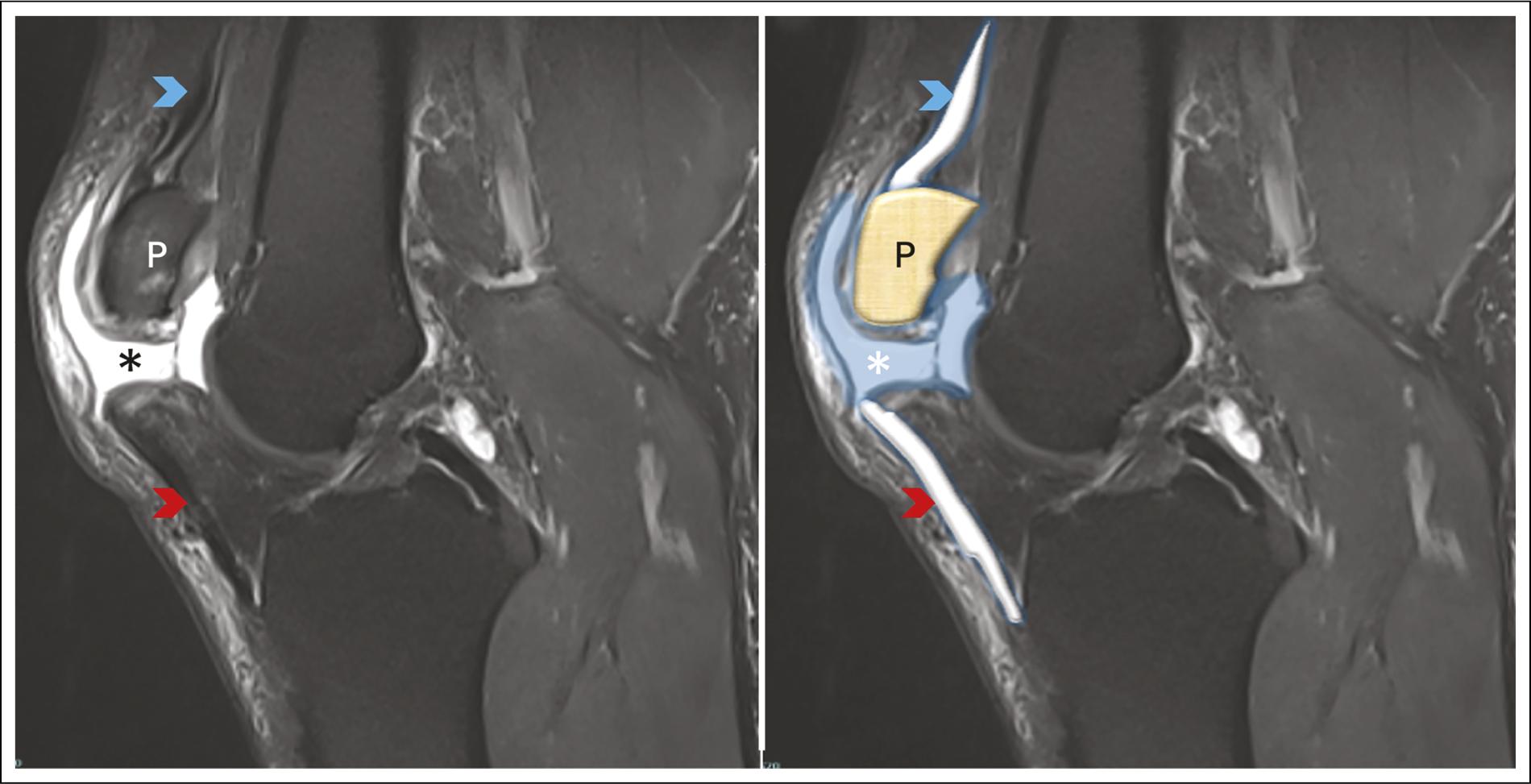
MRI: magnetic resonance imaging; P: patella.
Figure 2 A-B: sagittal proton density-weighted knee MRI with fat suppression in a 42-year-old man shows complete patellar tendon rupture, characterized by fiber discontinuity and marked hyperintense fluid signal at the tear site (red arrowheads). There is abnormal hyperintense fluid communication between the knee joint and the superficial prepatellar bursa (asterisks), highlighted in blue. The patella is shown in brown. The quadriceps tendon is intact (blue arrowheads).
ANATOMY OF THE SUPERFICIAL INFRAPATELLAR BURSA
The superficial infrapatellar bursa, located adjacent to the distal insertion of the patellar tendon, is one of the bursae surrounding the knee7. It has clinical and imaging characteristics similar to those of the superficial prepatellar bursa.
This bursa has both superficial and deep components7. The superficial component lies over the patellar tendon and is most often involved in bursitis. In some pathological situations, the superficial prepatellar and superficial infrapatellar bursae may communicate with each other7.
IMAGING DIAGNOSIS OF BURSITIS
X-ray
The role of X-ray in evaluating superficial bursae of the knee is limited. Plain radiography is primarily used to assess adjacent bone structures and, on anteroposterior and lateral projections, to evaluate soft tissues indirectly. The lateral X-ray projection is the most useful view for evaluating anterior knee soft tissues. Its main usefulness is in identifying associated fractures and detecting soft-tissue swelling or subcutaneous emphysema anterior to the patella. These findings may be relevant in the acute setting, particularly in the context of trauma or infection8.
Plain radiographs may show dystrophic or heterotopic calcifications, which are common in chronic or compli cated bursitis or in some differential diagnoses involving the anterior aspect of the knee. X-ray also serves a com plementary role in postoperative patients, allowing evaluation of surgical hardware position and identifica tion of potential complications related to previous interventions9 (Figures 3 A and B).
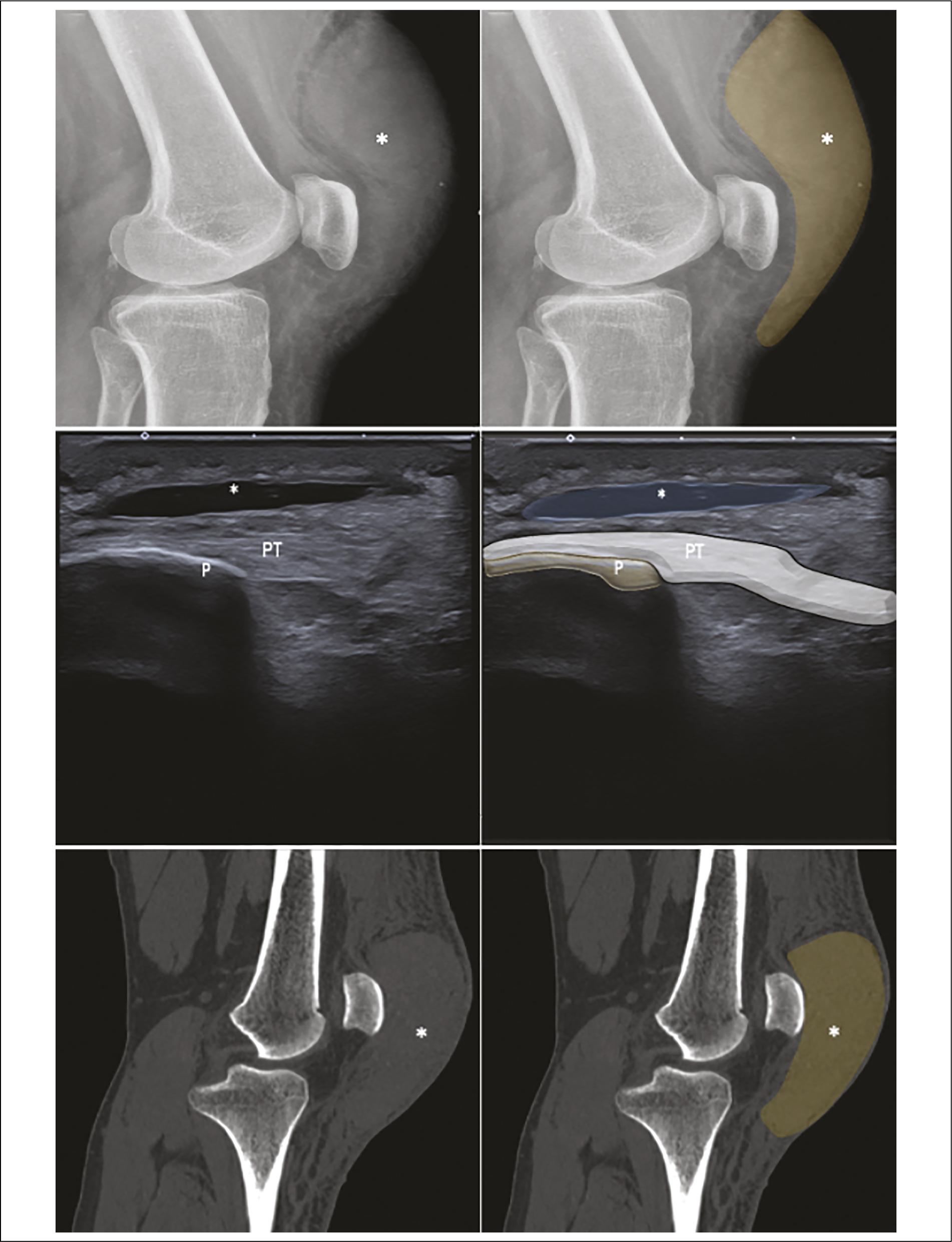
US: ultrasound; CT: computed tomography; P: patella; PT: patellar tendon.
Figure 3 A-B: lateral knee X-rays in a 59-year-old woman show prominent soft-tissue swelling anterior to the patella (asterisks), highlighted in brown, consistent with prepatellar soft-tissue edema. C-D: sagittal US views show prepatellar bursitis, characterized by a well-defined anechoic collection of the prepatellar bursa (asterisks), superficial to the patella, with a thin wall and absence of vascularization, highlighted in blue, suggestive of aseptic (post-traumatic) bursitis. E-F: sagittal CT views show soft-tissue edema anterior to the patella (asterisks), highlighted in brown, with no associated bone abnormalities.
Ultrasound
US is the imaging modality of choice to assess superficial prepatellar and superficial infrapatellar bursae10. It is operator-dependent and, in some cases, considered limited for this reason. The location of the superficial prepatellar bursae allows adequate evalu ation, even by personnel with limited US experience, making it a useful, versatile, cost-effective procedure11. US enables assessment of the subcutaneous tissue and multiple knee structures. Current high-resolution equipment allows the detection of small amounts of fluid.
US allows evaluation of intrinsic bursal characteristics, such as synovial thickening, septations, or internal heterogeneity, findings commonly associated with hemorrhagic, subacute, or chronic processes10,12. US diagnosis of bursitis of the superficial bursae of the anterior aspect of the knee may be based solely on identification of a hypoechoic fluid collection, always in the context of the patient’s clinical presentation12 (Figures 3C and D). Dynamic maneuvers can be performed to assess the functional involvement of the joint. Real-time interaction with the patient during the US examination allows questioning and provides valuable information about the mechanism of injury or the patient´s current clinical status.
A wide range of differential diagnoses can be identified by correlating ultrasound findings with the patient’s clinical presentation. US is the modality of choice for interventional procedures, such as fluid aspiration for diagnosis, therapeutic injections, and placement of per cutaneous catheters10,13.
Comparative studies show the high specificity of US versus MRI for superficial bursitis with good sensitivity in expert hands, although some deep bursae or small collections may go undetected14,15.
Computed tomography
Computed tomography (CT) plays a secondary role in evaluating the superficial bursae of the anterior knee. Nevertheless, it is a useful modality with high resolution for assessing bone structures, particularly for detecting associated fractures16. Its soft-tissue charac terization capability is limited, with performance similar to conventional X-ray; however, it allows identification of findings such as emphysema, calcifications, and edema16 (Figure 3E and F). Dual-energy computed tomography (DECT) is a new tool for soft-tissue evalu ation. It offers emerging opportunities in a field where the utility of CT has traditionally been limited16. However, its scope has not been clearly defined in superficial prepatellar and infrapatellar bursitis.
In postoperative patients, CT allows adequate evalu ation of surgical hardware and identification of potential complications16. However, exposure to ionizing radiation limits its routine use; it should be reserved for selected cases.
Magnetic resonance imaging
MRI offers greater anatomical and tissue resolution and superior characterization of the bursal wall, soft-tissue edema, deep-plane extension, and bone complications (such as osteomyelitis). MRI is more sensitive for detecting secondary findings, such as bone contusions, signal changes in adjacent tendons, and complex intrabursal masses. MRI is useful when clinical findings or US suggest complications or when detailed preoperative mapping is required7,17.
MRI provides advantages, including the evaluation of associated findings and assessment of the internal structures of the knee, as well as the added benefit of avoiding ionizing radiation8,18. MRI exhibits high tissue differentiation, making it the ideal complementary modality to US, particularly in complex cases14,18 (Figure 4).
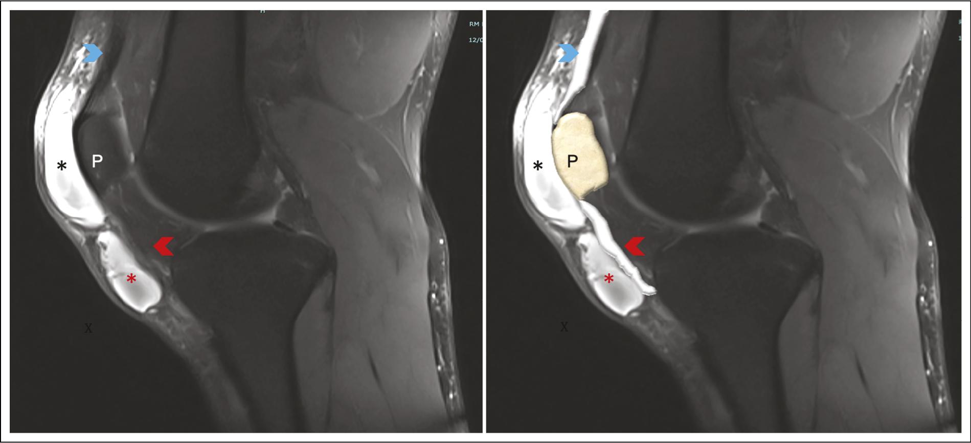
MRI: magnetic resonance imaging; P: patella.
Figure 4 A-B: sagittal proton density-weighted knee MRI with fat suppression in a 29-year-old woman shows a well-defined hyperintense fluid collection involving the superficial prepatellar (black asterisks), and superficial infrapatellar (red asterisks) bursae without imaging evidence of communication between them. The patellar tendon has preserved contour and homogeneous low signal intensity (red arrowheads). The quadriceps tendon is intact, with normal signal intensity (blue arrowheads).
MRI identifies bursal fluid content and soft-tissue edema, making it useful for differentiating simple bursitis from hemorrhagic, infectious, or chronic processes8. It also enables evaluation of synovial thickening, internal septations, debris, and fluid-fluid levels, a characteristic finding related to blood products of different ages and sedimentation effects that can help support the diagnosis to distinguish it from other soft-tissue collections or neoplastic processes19,20 (Figure 5). The use of contrast media allows improved assessment of the bursal wall and tissue behavior in equivocal cases18.
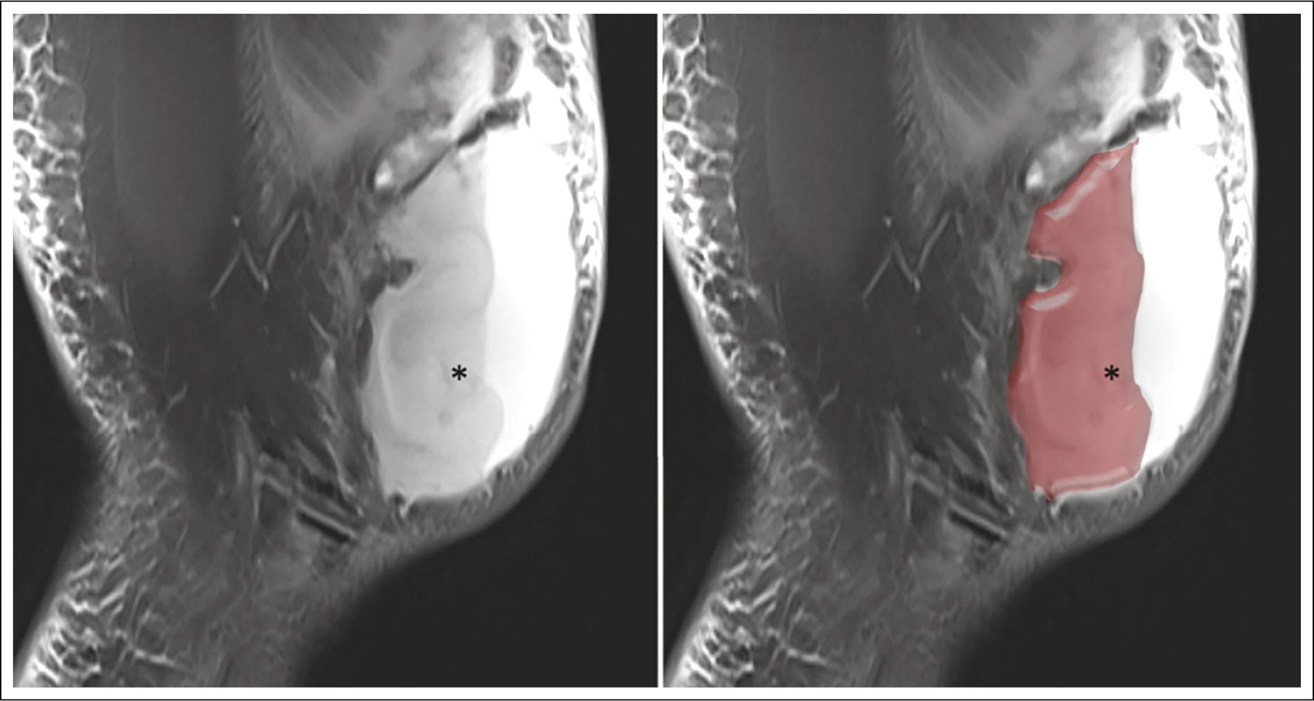
MRI: magnetic resonance imaging.
Figure 5 A-B: sagittal proton density-weighted knee MRI with fat suppression in a 22-year-old man demonstrates a markedly distended superficial prepatellar bursa containing a heterogeneous fluid collection with fluid-fluid levels, showing dependent hypointense sedimented components and nondependent hyperintense fluid signal superiorly (asterisks), consistent with hemorrhagic bursitis. The bursal extent is highlighted in red.
MRI is particularly valuable for assessing involvement of adjacent structures, such as the patellar tendon, Hoffa’s fat pad, subcutaneous tissue, and the patella, allowing exclusion of associated pathologies, such as soft-tissue tumors, fibrocicatricial changes, or intra-articular lesions8.
Despite its advantages, MRI is not usually a first-line procedure due to its higher cost and limited availability compared to US. Therefore, MRI is reserved for cases with suspected complications, lack of treatment res ponse, deep extension, or to refine complex differential diagnoses7,14.
Recommendations for imaging report of bursitis
–. Specify the affected anatomical bursa (superficial prepatellar or superficial infrapatellar) and note any compartmentalization if present7.
–. Describe the contents (anechoic, hyperechoic, heterogeneous), wall thickness, post-contrast enhancement (if MRI), peribursal edema, and adjacent bone findings17,21.
–. Identify findings suggesting infection (circumferential mural enhancement, gas, marked inflammatory edema) and recommend image-guided aspiration when appropriate15,21.
–. Suggest additional imaging modalities (e.g., MRI if US indicates deep extension or bone involvement) and recommend a clinical-microbiological correlation7,14.
–. Emphasize findings that (a) differentiate bursitis from other cystic or soft tissue lesions, (b) recognize signs suggestive of infection or complication, and (c) optimize the imaging algorithm and communication with the clinical or surgical team7,14,17,21.
CLINICAL PATHOLOGIES
The diagnosis of superficial prepatellar and superficial infrapatellar pathology is primarily clinical, but multimodal imaging (US and MRI) provides critical information on location, extent, and complications such as abscesses, extension into adjacent tissues, or osteomyelitis. They also help guide procedures such as needle aspiration or surgery7,14,17,22.
The superficial prepatellar bursa is particularly susceptible to compression and repetitive microtrauma, which are common in professions that require kneeling. Superficial prepatellar bursitis can be acute or chronic with synovial thickening and septations.
Aseptic bursitis
Traumatic superficial prepatellar bursitis can be aseptic or septic. Aseptic superficial bursitis occurs after minor, repetitive trauma in people whose occupations require prolonged kneeling, such as carpet installers and cleaning staff4. Superficial prepatellar bursitis, which involves excessive accumulation of fluid in both superficial anterior knee bursae, is commonly known as “maid’s knee” when it affects the superficial prepatellar bursa (Figure 6) and “cleric’s knee” when the superficial infrapatellar bursa is involved (Figure 7). The appearance of superficial prepatellar and infrapatellar aseptic bursitis is consistent with fluid on US and MRI4.
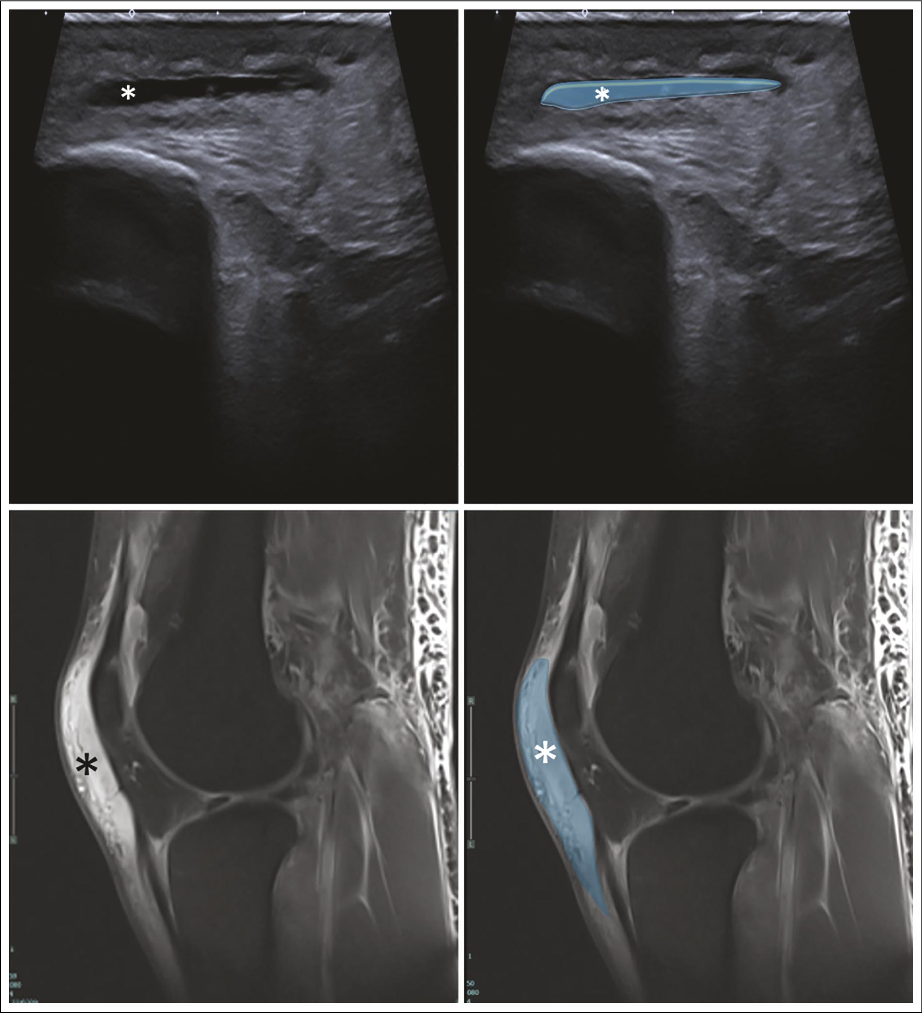
US: ultrasound; MRI: magnetic resonance imaging.
Figure 6 A-B: sagittal knee US in a 30-year-old man shows superficial prepatellar bursitis, characterized by well-defined anechoic fluid collection in the prepatellar bursa (asterisks), highlighted in blue. C-D: sagittal proton density-weighted knee MRI with fat suppression shows a marked hyperintense fluid signal distending the superficial prepatellar bursa (asterisks), highlighted in blue, consistent with superficial prepatellar bursitis (“housemaid’s knee”).
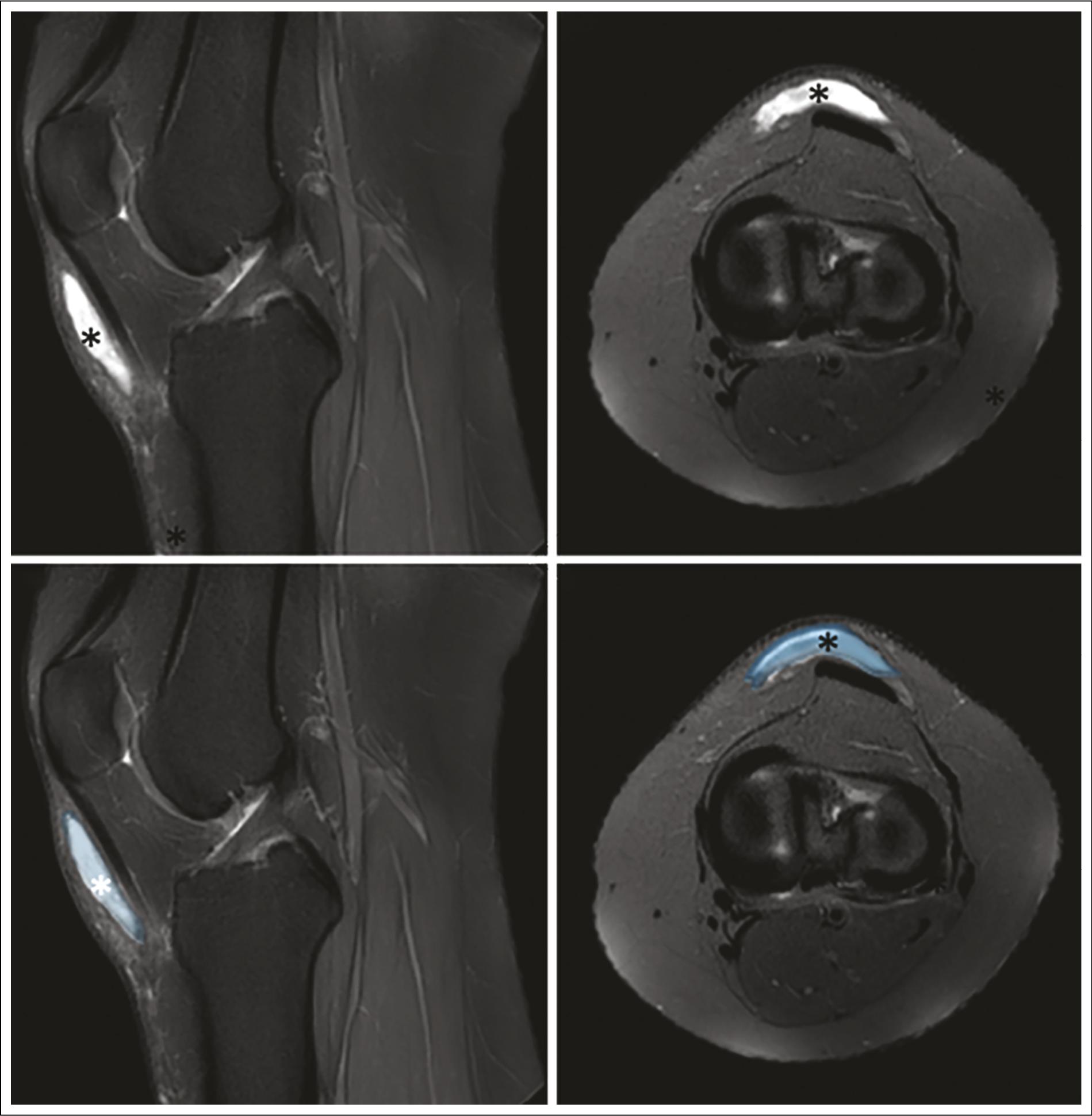
MRI: magnetic resonance imaging.
Figure 7 A, B, C, D: sagittal and axial proton density-weighted knee MRI with fat suppression in a 35-year-old man shows superficial infrapatellar bursitis, characterized by fluid collection in the superficial infrapatellar bursa (asterisks). The bursa is highlighted in blue. This condition is commonly referred to as “clergyman’s knee.”
US is a helpful screening method, with a sensitivity of 87% for detecting fluid in the anterior knee bursae compared with MRI as the gold standard4. With modern high-resolution US, even small amounts of anechoic fluid can be detected. This finding must always be interpreted in the clinical context. Other causes of aseptic prepatellar bursitis include systemic diseases such as gout, rheumatoid arthritis, collagen vascular disorders, and inflammatory arthropathy23.
Septic bursitis
Superficial prepatellar and superficial infrapatellar septic bursitis account for approximately one-third of all bursitis cases and represent between 0.01% and 0.1% of hospital admissions24. The infection is believed to occur through direct inoculation rather than hematogenous spread, likely because of the limited blood supply to the bursa24 (Figure 8).
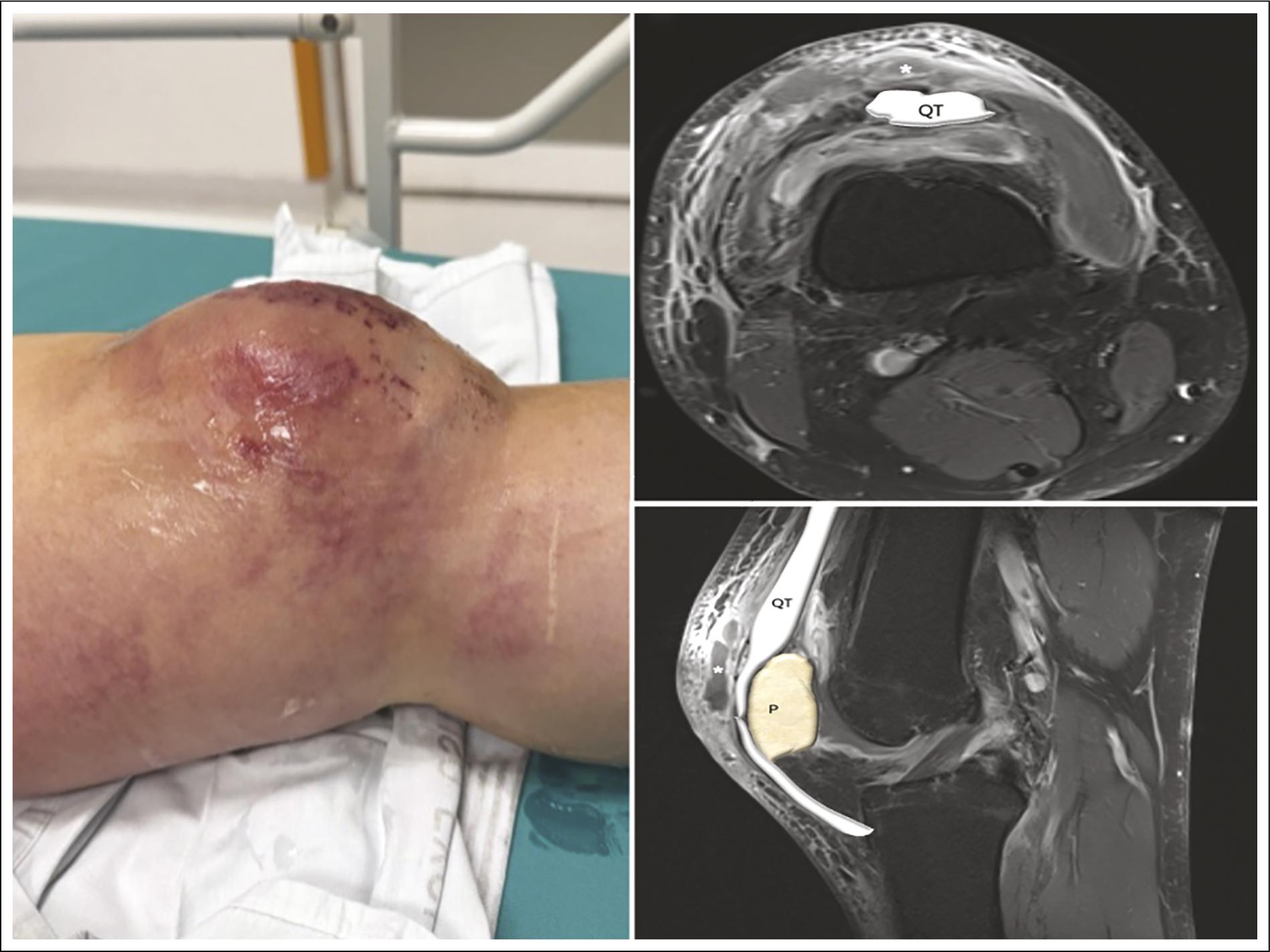
MRI: magnetic resonance imaging; P: patella; QT: quadriceps tendon.
Figure 8 A: clinical photograph of the left knee in a 53-year-old man shows marked edema, erythema, and superficial skin abrasions over the anterior aspect of the knee, suggestive of septic prepatellar bursitis, commonly caused by direct inoculation through skin disruption. B: axial T1-weighted fat-suppressed post-contrast knee MRI shows a hypointense fluid collection in the superficial prepatellar bursa (asterisk) with peripheral bursal wall enhancement and a hyperintense anterior subcutaneous edema. C: axial T1-weighted fat-suppressed post-contrast knee MRI shows a hypointense fluid collection in the superficial prepatellar bursa (asterisk) with peripheral bursal wall enhancement and hyperintense anterior subcutaneous edema.
Diagnosis is primarily clinical. US can help identify hypoechoic fluid accompanied by synovial thickening and hypertrophy24. In some cases, thin septations and increased vascularity on power Doppler US may be observed, which provide useful diagnostic and therapeutic guidance24. Associated soft tissue edema is almost always present. There is a lack of consensus on standardized ultrasound or Doppler parameters that best predict infection10.
The presence of circumferential bursal mural thickening and enhancement on MRI, peripheral enhancement after contrast administration, marked peribursal edema, a heterogeneous collection with debris, and gas in soft tissues increases the likelihood of septic or abscessed bursitis and should prompt urgent aspiration and antibiotic management. However, overlaps are frequent: chronic bursitis with hemorrhage or tophi (gout) can show heterogeneous content and enhancement; therefore, clinical correlation and microbiological/crystal aspiration analysis remain the gold standard for distinguishing etiologies15.
Hemorrhagic bursitis
Hemorrhagic bursitis is commonly post-traumatic, although it can also occur as a postsurgical compli cation19,20. Hemorrhagic bursitis has been extensively described in the literature, largely due to the significant size these lesions can reach. Progressive enlargement can result in visible cosmetic deformity, leading to limited joint mobility and impaired quality of life19.
Its appearance on US is variable and typically cha racterized by predominantly heterogeneous and poorly defined echogenicity, usually without central vascularity on color Doppler imaging. In some cases, hemorrhagic bursitis may show a more complex ultrasonographic appearance, including internal septations, debris, or calcifications, particularly in chronic or organized stages19 (Figure 9). Unlike hematomas in other anatomical regions, hemorrhagic bursae tend to become predominantly organized over time, often developing a fibrous capsule and internal complexity. Several cases requiring surgical management have been reported due to their large size, persistent symptoms, or functional compromise (Figure 10).
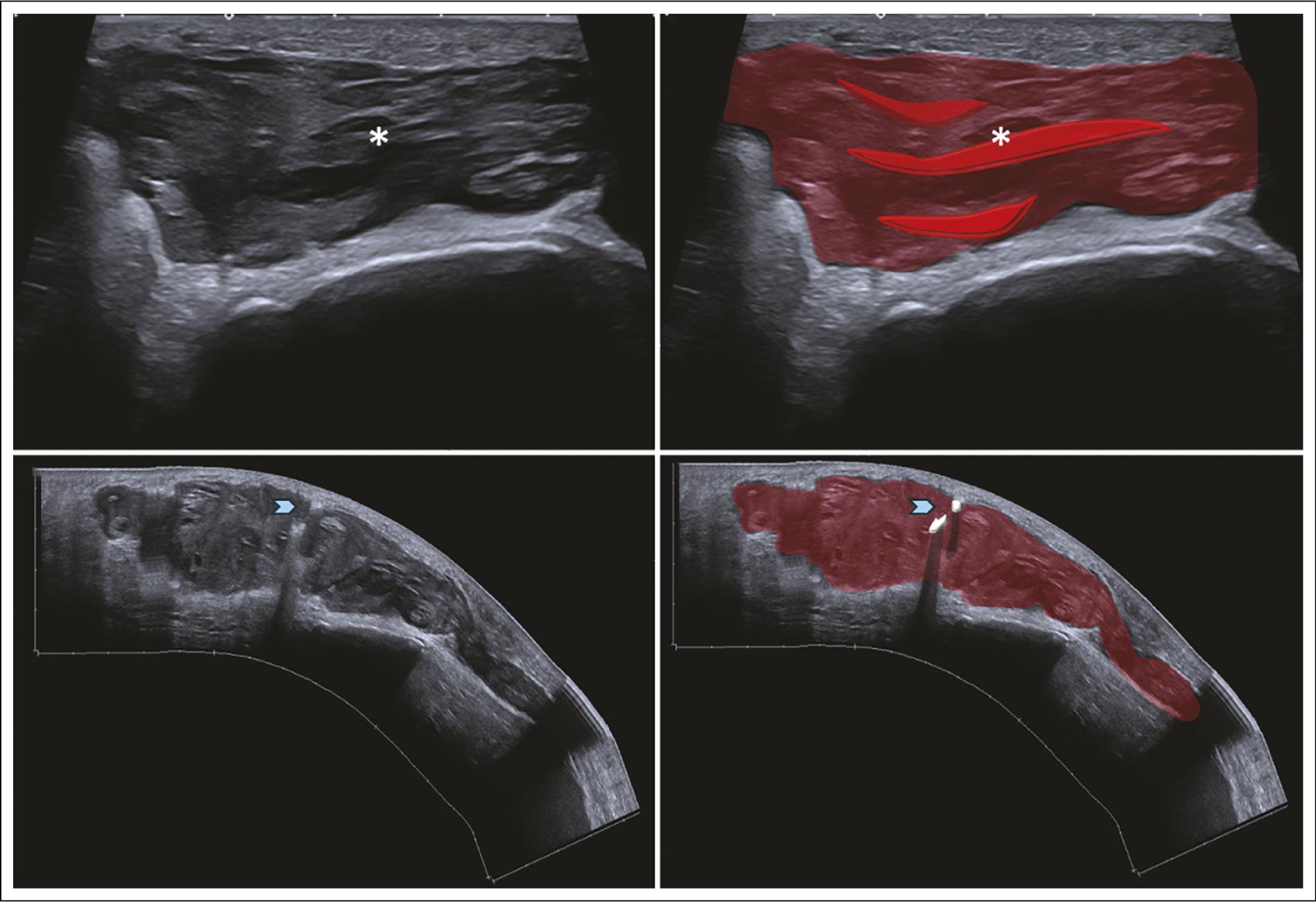
US: ultrasound.
Figure 9 A-B: axial US views of the anterior knee in a 21-year-old woman show hemorrhagic bursitis with an organized hematoma within the bursa (asterisks), highlighted in red. The lesion shows heterogeneous content and echogenic fragments consistent with debris or hemorrhage, one of the most common ultrasonographic features of acute hemorrhagic bursitis. C-D: sagittal US images of the anterior knee in a 43-year-old woman show hemorrhagic bursitis with an organized hematoma highlighted in red, containing internal calcifications highlighted in white (blue arrowheads), findings suggestive of chronicity. In the absence of previous trauma, gout should be considered in the differential diagnosis.
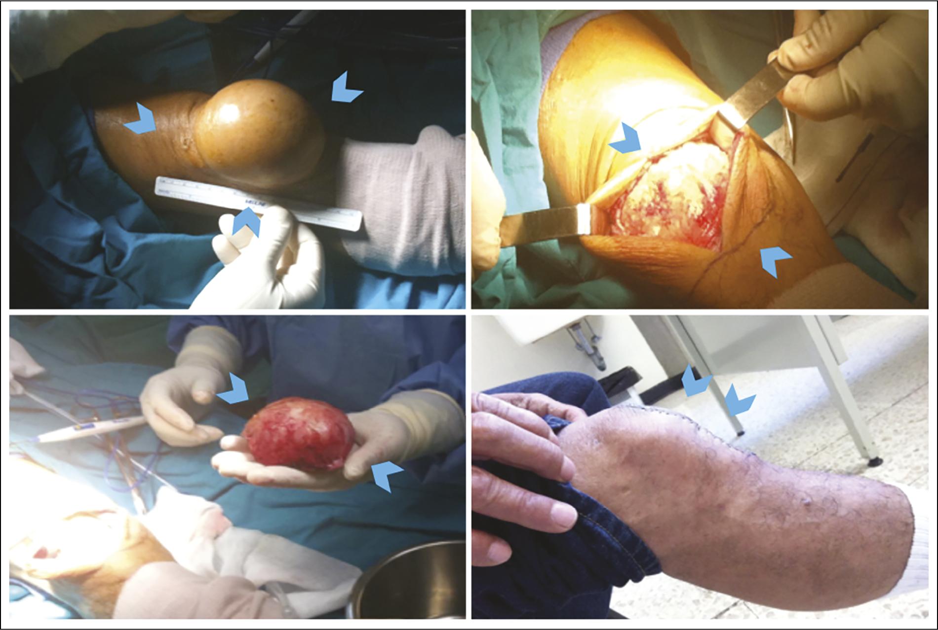
Figure 10 Clinical and intraoperative photographs of a 59-year-old man diagnosed with hemorrhagic superficial prepatellar bursitis. A: marked anterior knee swelling (blue arrowheads). B: intraoperative exposure of a hemorrhagic prepatellar bursal lesion (blue arrowheads). C: surgical specimen showing a large, organized hemorrhagic bursa (blue arrowheads). Post-traumatic hemorrhagic bursae are typically among the largest. D: postoperative clinical appearance showing resolution of anterior knee swelling after surgical excision (blue arrowheads).
Postsurgical and/or iatrogenic bursitis
Patellar surgeries can affect the superficial prepatellar bursa more than the infrapatellar bursa25; therefore, cases of bursitis associated with these procedures may be encountered (Figure 11). However, surgeries involving the patellar tendon may affect the superficial infrapatellar bursa, making bursitis a potential complication of this type of intervention25.
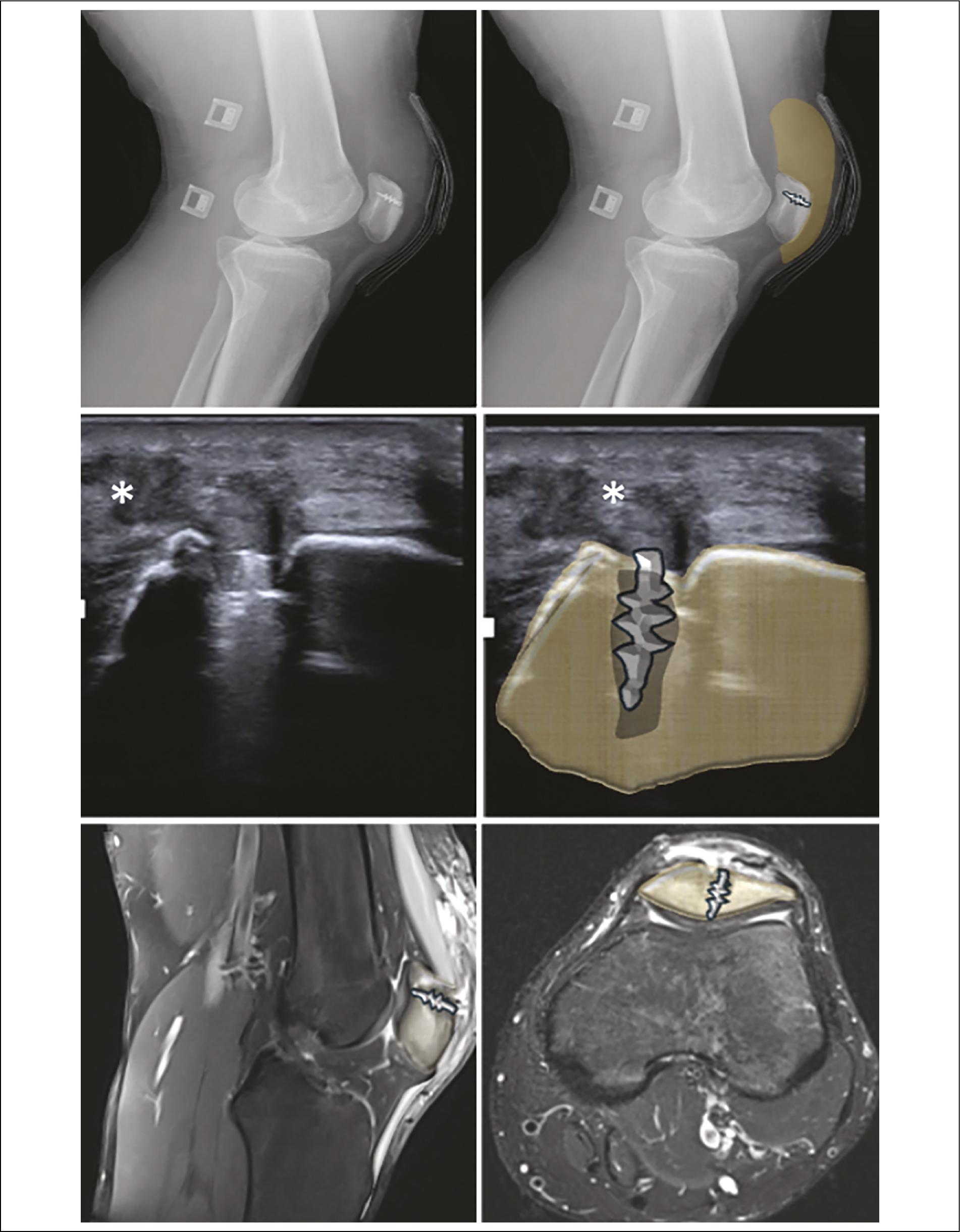
US: ultrasound; MRI: magnetic resonance imaging.
Figure 11 A-B: lateral knee X-rays of a 17-year-old man show the patella with a surgical tendon anchor system and anterior soft-tissue swelling, highlighted in brown. C-D: longitudinal US of the anterior knee shows postoperative soft-tissue edema and fluid collection in the prepatellar bursa (asterisks). The patella is highlighted in brown with the surgical tendon anchor system. E-F: sagittal and axial proton density-weighted knee MRI with fat suppression shows postoperative anterior soft-tissue edema, characterized by heterogeneous signal in the prepatellar soft tissues with hyperintense fluid distension of the superficial prepatellar bursa. The patella is highlighted in brown. Findings are consistent with postsurgical superficial prepatellar bursitis following a patellar procedure.
DIFFERENTIAL DIAGNOSES
Various soft-tissue conditions of the anterior knee are included in the differential diagnoses. Among these, extensive soft tissue hematomas and Morel-Lavallée hematomas are important considerations, as they can mimic intrabursal collections. Therefore, the clinical history (such as trauma or metabolic disease) and characterization by MRI or fluid analysis are essential17,21. These lesions are often associated with previous trauma and tend to occur in regions adjacent to the superficial bursae of the anterior knee (Figures 12 A and B). They result from shearing forces that separate the subcutaneous tissue from the underlying fascia. Due to their characteristic anatomical location, extent, and distribution, differentiation from superficial prepatellar bursitis on US and MRI is usually straightforward.
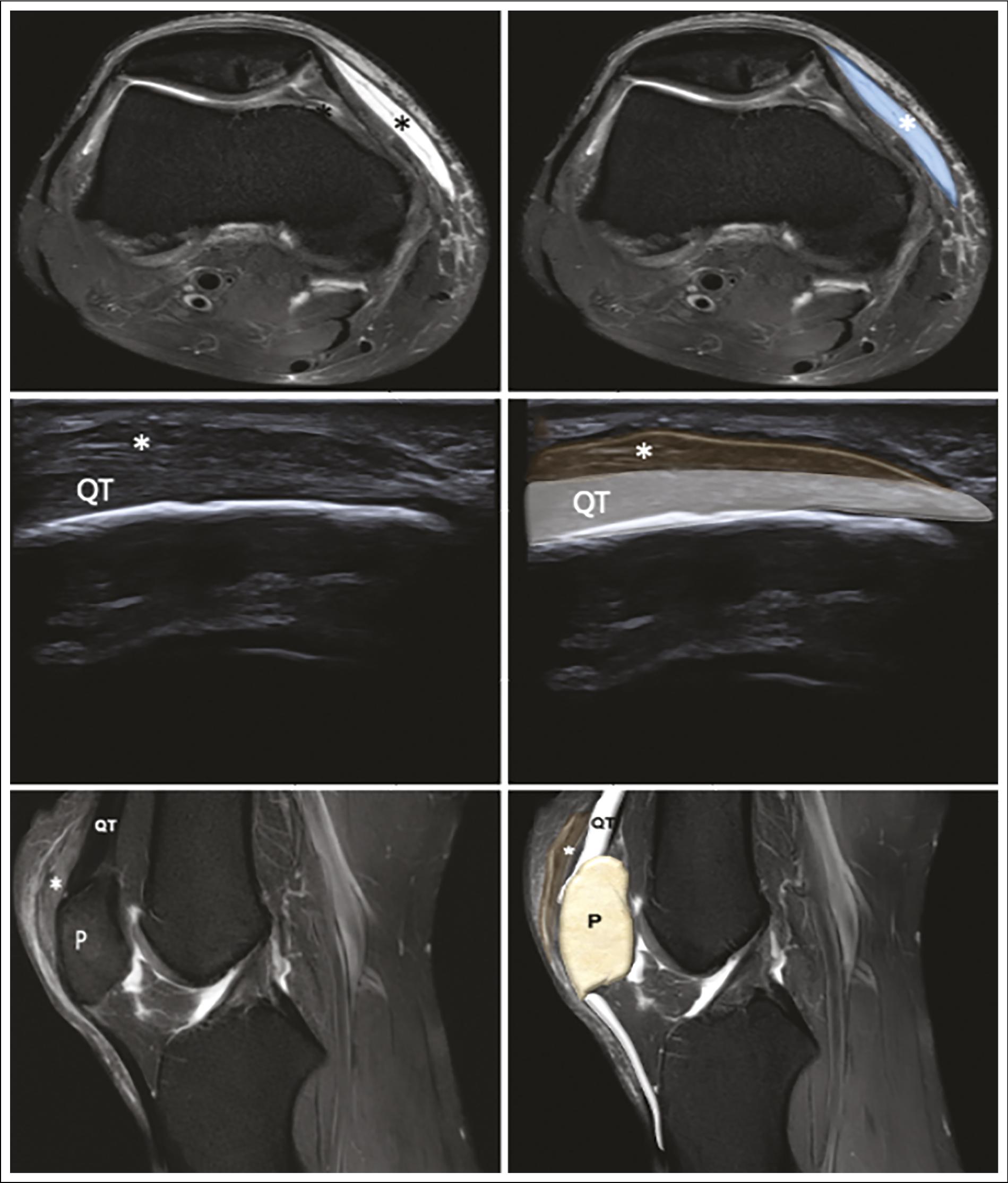
MRI: magnetic resonance imaging; US: ultrasound; P: patella; QT: quadriceps tendon.
Figure 12 A-B: axial proton density-weighted knee MRI with fat suppression in a 17-year-old man shows post-traumatic hematoma along the medial aspect of the knee, located in soft tissue (asterisks) and highlighted in blue. No prepatellar bursa involvement is observed. C-D: longitudinal US view of the anterior knee shows post-traumatic fibrotic thickening of the prepatellar tendon, characterized by focal tendon enlargement with a predominantly hypoechoic echotexture and changes in the normal fibrillar pattern (asterisks). The affected tendon segment is highlighted in dark brown. E-F: sagittal proton density-weighted knee MRI views with fat suppression show post-traumatic fibrotic thickening of the prepatellar tendon (asterisks), characterized by tendon enlargement with predominantly low signal intensity and focal areas of intermediate signal, without fiber discontinuity or hyperintense fluid signal suggesting tendon rupture. The affected tendon is highlighted in dark brown.
Injuries to adjacent supporting structures should also be considered, including post-traumatic scarring and thickening of the prepatellar ligament. These changes are commonly observed after direct trauma and are typically characterized by predominant posterior fibrotic thickening. Both US and MRI are valuable in this setting, with MRI providing crucial information for accurate anatomical assessment and differential diagnosis (Figures 12 C-F). High-energy motorcycle injuries are a frequent cause of these types of injuries.
Soft-tissue masses such as lipomas can mimic bursal pathology. Lipomas generally grow slowly with minimal clinical symptoms. On US, they appear as solid lesions with absent or minimal Doppler vascularity. At the same time, MRI shows distinctive signal intensity characteristics, including high signal intensity on T1-weighted images and signal suppression on fat-suppressed sequences. Importantly, these lesions are located outside the normal bursal topography, facilitating differentiation from true bursitis. Osgood-Schlatter disease should also be included in the differential diagnosis, particularly in cases of superficial infrapatellar bursitis, especially in younger patients.
Postsurgical complications and less common entities within the differential diagnosis, including tendon-related abnormalities and soft-tissue tumors, should not be overlooked. Other less frequent differential diagnoses include tuberculosis, crystal deposition diseases such as gout and calcium pyrophosphate deposition disease (pseudogout), and systemic conditions including rheumatoid arthritis, systemic lupus erythematosus, and uremia4,23.
A high index of suspicion should be maintained for osteomyelitis or intraosseous conditions in pediatric or immunocompromised patients that initially present with prepatellar swelling. MRI should be considered to evaluate bone involvement4.
TREATMENT OF BURSITIS
Imaging identifies the affected bursa and helps determine the treatment plan. The treatment of aseptic superficial prepatellar and superficial infrapatellar bursitis is determined first by the underlying cause and, secondly, the pathological changes within the bursa23. Management of acute bursitis includes PRICE therapy (protection, rest, ice, compression, and elevation) and non-steroidal anti-inflammatory drugs, which generally provide an adequate response4. Patients with septic superficial prepatellar bursitis are managed successfully with non-surgical treatment, including rest, compression, immobilization, aspiration, and antibiotics23.
Surgery is not required in most cases; however, when necessary – particularly in recurrent or treatment-refractory cases – available medical procedures include aspiration and intrabursal injection of an appropriate agent, such as corticosteroids, platelet-rich plasma, autologous blood, sclerosing solutions, and short-term placement of a drainage catheter as a therapeutic option. Excision of a chronically inflamed and thickened bursa is uncommon for superficial prepatellar and superficial infrapatellar bursae23,26.
Collections with signs of suppuration or deep extension require aspiration, culture, and often systemic antibiotics. Persistent or recurrent, and massive or chronic collections with fibrotic tissue may require bursectomy (open or endoscopic). Open bursectomy carries a significant risk of morbidity of the surgical site, including poor wound healing, decreased scar sensation, contracted scarring, atrophic skin changes, accumulation of subcutaneous hematomas, and painful or hypersensitive scarring23. This risk is related to the delicate blood supply of the prepatellar skin and the rich network of anastomoses formed by the descending vertical branches of the anterior divisions of the medial and lateral femoral cutaneous nerves, the intermediate cutaneous nerve, and the infrapatellar branch of the saphenous nerve23. Recent systematic reviews show that endoscopic bursectomy is non-inferior to open bursectomy for prepatellar resection in terms of recurrence and is associated with a shorter hospital stay, supporting fewer invasive options when intervention is necessary4,27.
CONCLUSION
A wide range of pathologies can affect the superficial prepatellar and superficial infrapatellar bursae. Advances in contemporary imaging techniques, particularly high-resolution US and MRI, have significantly improved diagnostic accuracy, enabling more precise characterization of these conditions and facilitating optimal therapeutic decisions. The pathology involving these bursae is wide-ranging, and their anatomy is more complex than traditionally appreciated. Trauma predominates, encompassing low- and high-energy mechanisms. Accurate imaging diagnosis and appropriate management depend on identifying the underlying cause and understanding the biological behavior of the bursae. US is the primary imaging modality for puncture guidance, while MRI is used for detailed evaluation of complications or complex differential diagnoses.

