


 nueva página del texto (beta)
 Inglés (pdf)
 Artículo en XML
 Referencias del artículo
 Enviar artículo por email
 Citado por SciELO
  Similares en
SciELO
Similares en
SciELO 
 Permalink
Introduction
Breast development during adolescence is an important factor in the transition to adulthood1. Breast overgrowth in adolescents was first described in 1910 by Henry Albert, who names this pathology as juvenile hypertrophy or virginal breast hypertrophy2. It is a rare, benign, and sporadic condition that affects adolescents, mainly during puberty.
There are different terms that describe this entity in the medical literature, such as virginal hypertrophy, juvenile gigantomastia (JG), or juvenile macromastia3.
Within juvenile infant breast pathology, virginal breast hypertrophy accounts for 12.5% of all breast diseases in adolescents, while gigantomastia has a prevalence of 1 in 25,000 women and affects only 3.5/1000 adolescents3. The etiology is unknown; however, in the case of patients without comorbidities, with normal hormone levels, it is believed that it is hypersensitivity of the breast tissue to estrogen, resulting in diffuse breast growth4.
Under this hormonal hypothesis, the use of drugs such as tamoxifen, danazol, or bromocriptine is justified, however, the safety and efficacy in the short and long term is unknown5,6. For this reason, the most recommended treatment in most cases is reduction mammoplasty, which is the option with the lowest recurrence rates3.
The most challenging aspect in the management of JM, or juvenile breast hypertrophy, is the effectiveness of definitive management, as its challenging natural history and refractory nature to surgery are well documented7. We report the case of a 12-year-old girl with bilateral juvenile breast hypertrophy of large dimensions, which was recurrent to the initial surgical management.
Case report
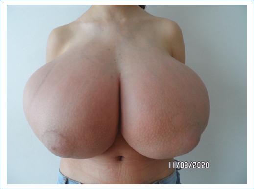
A 12-year-old female patient presented with JG with massive and progressive bilateral breast growth, with a 6-month evolution (Fig. 1). She presents local manifestations of breast overweight such as mastodynia. In addition, added symptoms such as neck pain and severe low back pain limit their interpersonal relationships, causing social distancing. The only important antecedent was pubarche at 11 years of age, without menarche. No other history of relevance to the condition, and he does not take medication on a regular basis.
On physical examination, an ectomorphic patient was found, weighing 45.7 kg and height of 1.52 m with a BMI of 19.8 kg/m2. Measurement of bilateral nipple-to-nipple fork distance of 32 and 33 cm, nipple-to-inframammary fold distance of 15 cm. Disproportionately large, asymmetrical breasts, with Grade 4 ptosis, expanded, dilated subcutaneous veins, diffuse erythema, hard and firm consistency, no palpable masses, no nipple secretions or axillary lymphadenopathy.
Luteinizing hormone, follicle-stimulating hormone, prolactin, thyroid function tests, and cortisol within normal parameters. Breast ultrasound results without masses, only interstitial edema. We performed a bilateral mammoplasty surgery for the reduction of the supermedial pedicle, with a total breast resection of 6167 g (2906 g of right breast tissue and 3261 g of left breast tissue), integrating this resection to the equivalent of 13.3% of your total body weight (Figs. 2 and 3). The pathological report reported diffuse proliferation of the mammary stroma with abundant deposits of collagen, lymphocyte infiltrate, mast cells and extravasated erythrocytes. Dilated capillaries and ducts were identified, with a decrease in breast adipose tissue and the epithelial component. There were no morphological data of malignancy, confirming a diagnostic suspicion of virginal breast hypertrophy.
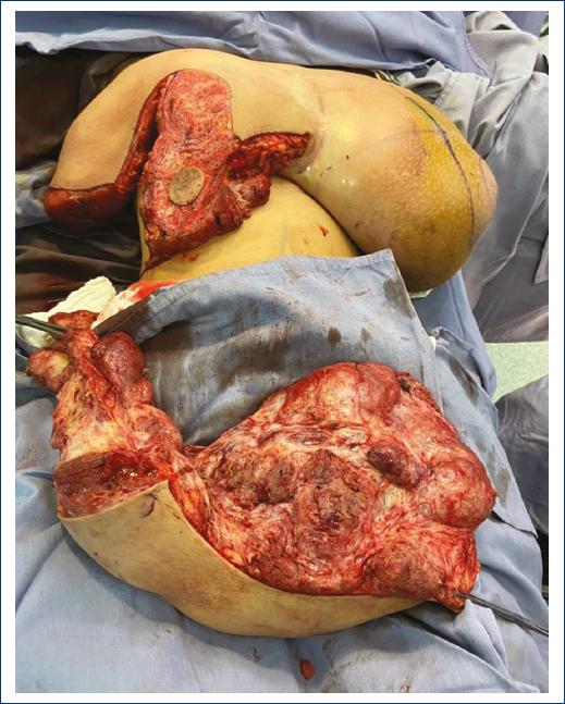
Figure 2 Surgical management with superomedial pedicle reduction mammoplasty. Right resection of 2906 g.
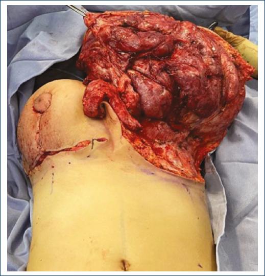
Figure 3 Surgical management with superomedial pedicle reduction mammoplasty. Left resection of 3261 g.
Her post-operative evolution was characterized by progressive breast growth, reaching gigantomastia in just 3 months after the previous breast reduction surgery.
Therefore, it was decided to perform a bilateral subcutaneous mastectomy, with immediate breast reconstruction with subpectoral breast implants, and the use of a dermofatty flap to support and cover the breast implants in the lower breast pole. In this second mastectomy surgery, 2910 g was resected on the right side and 2530 g on the left side, which is equivalent to a growth of 90% and 70% with respect to the initial pre-operative volume before the surgeries, despite having performed a first breast reduction (Fig. 4).

Discussion
GJ is also known as virginal breast hypertrophy, juvenile hypertrophy, or juvenile macromastia. Clinical manifestations include a sudden and continuous growth of breast tissue, usually accompanying the onset of puberty. There is usually a 6-month period of extreme growth, followed by a slower but sustained period8,9.
The definition of gigantomastia varies depending on the author: excessive growth representing 3% or more of the patient’s total weight or more than 1500 cc in volume4,3. It causes physical dysfunction, postural pain, deviation in the spine and dermal alterations, mainly hyperemia, orange peel and even necrosis. Dilation of subcutaneous veins and intertrigo can be observed in inframammary folds; with an impact on the psychosocial development of the patient, eating disorders, social distancing, inability to perform physical activity, esthetic non-conformity with body image distortion and alterations in habitual behavior may also occur10,11.
JG is a diagnosis of exclusion and during the patient’s approach, it is vitally important to rule out all differential diagnoses, which include: breast hypertrophy secondary to the use of medications, pseudo-gigantomastia associated with obesity, fibroepithelial tumors (breast fibroadenoma, phyllodes tumor), fibrocystic disease, endocrinopathies, hypertrophy associated with pregnancy, infection, tumors of benign origin (hemangiomas and lymphangiomas) and tumors of malignant origin (lymphoma, sarcoma). The definitive diagnosis is obtained with the anatomopathological study2.
The most recommended treatment is surgical resection, as GJ is an absolute indication for a breast reduction or resection procedure in its entirety12. Other alternatives to consider are reduction with upper pedicle, lower pedicle, bipedicled, and free nipple grafts13. The other surgical option, used in a smaller proportion, as was the case in this case, is subcutaneous mastectomy, with reconstruction based on prostheses. Normally, it is reserved for cases with suspected malignancy and recurrences, as it is a management with less satisfactory esthetic results than reduction mammoplasty7.
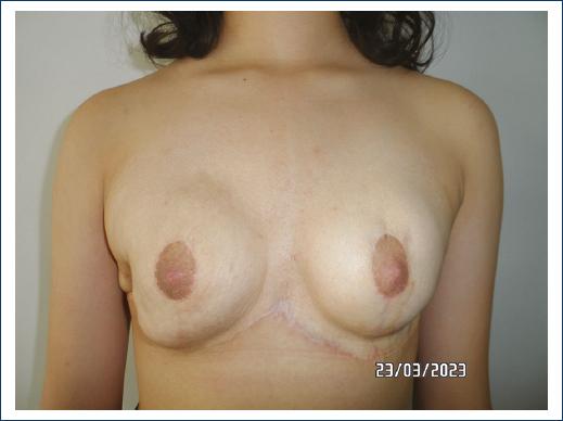
For patients such as the one presented here, the recurrence that reached 70-90% of the initial volume must be treated with radical surgery. In this case, subcutaneous mastectomy and reconstruction with bilateral breast implants, with the aim of minimizing residual breast tissue and obtaining a favorable result for health and esthetics. The surgical technique of subcutaneous mastectomy has the lowest recurrence rate and ensures a reliable and definitive final result. At present, the patient remains under follow-up, undergoing 2 years of evolution without recurrent breast growth, and very satisfied with her result (Fig. 5). If necessary, and especially when the patient reaches an older physical and mental age, subsequent breast surgeries for esthetic purposes may be considered.
Conclusion
Any case of JG merits multidisciplinary management, involving specialties such as endocrinology, psychology, pediatrics, and plastic and reconstructive surgery. To design a complete treatment plan, rule out possible etiologies, differential diagnoses, and obtain favorable results to improve the quality of life of our patients. Now, there are no evidence-based treatment guidelines, only case reports, due to the low incidence of the pathology. Further research is required to define etiopathogenesis, natural history, and response to medical and surgical treatment. However, both for the symptoms and for the patient’s self-esteem and lifestyle, so far, the surgical approach with or without hormonal treatment is indicated.

