




 nueva página del texto (beta)
 Inglés (pdf)
 Artículo en XML
 Referencias del artículo
 Traducción automática
 Enviar artículo por email
 Citado por SciELO
  Similares en
SciELO
Similares en
SciELO 
 Permalink
Introduction
Ureterocele is defined as congenital cystic dilatation of the intravesical part of the distal ureter1. It is frequently left-sided and is 60-80% ectopic located. Eighty percentages of ureteroceles occur with duplicated collecting systems, 10% with a single system, and 10% bilaterally1. The most common method for diagnosing ureteroceles during pregnancy and after birth is ultrasonography (US)2-5. The alternative procedures include voiding cystourethrography (VCUG), intravenous urography (IVU), and cystoscopy.
Although urinary tract infection (UTI) is the most common symptom, other conditions such as fever, poor growth, and post-natal sepsis may also occur5,6. Endoscopic decompression (ED) is the most preferred management method for ureteroceles in recent years despite the lack of a standard treatment protocol. However, for convenient patients, conservative follow-up is advised7. Reconstruction and partial/total nephrectomy are the other options (excision of ureterocele and reimplantation of lower pole ureter).
We wanted to share our institution's 8 years of experience with ureterocele follow-up and treatment.
Method
A retrospective analysis was performed on 52 patients' records who had ureterocele diagnoses and were treated at the Pediatric Surgery Clinic of the Medical School of Dicle University between January 2009 and December 2017. The patients' records were examined for age, gender, diagnostic method, age of diagnosis, and location of the ureterocele, as well as for the presence of a UTI, vesicoureteral reflux (VUR), and procedure of management. After receiving approval from the Ethical Council of Medical School of Dicle University, the trial began (permission code: 18, 05, 2018-163). Patients who had < 6 months of follow-up were disqualified.
The data were analyzed using the IBM SPSS-22 system. X2 and Fisher Exact tests were applied. Statistical significance was defined as p < 0.05.
Results
Of the 52 patients, 45% were male and 55% female. Thirty-six patients had left-sided ureterocele, 12 had right-sided ureterocele, and four had bilateral ureterocele. There was a duplicated collecting system for 27 (52%) of the patients. Thirty-three ureteroceles (63.4%) were intravesical, and 19 (36.6%) were ectopically located. Forty-four patients (84.6%) had hydronephrosis noticeable.
Twelve (23%) of the patients had ureterocele diagnosed prenatally, 14 (26.9%) between the ages of 1 and 6 months, 4 (7.6%) between the ages of 6 and 12 months, and 22 (42.3%) after the age of 1 year. The average age of diagnosis was 20 months, and 7 months was the median age at the time of diagnosis.
Forty patients (76%) ureterocele was detected by US, 4 (7.6%) by VCUG, 2 (3.8%) by IVU, and 6 (11.5%) by cystoscopy.
There were no significant physical examination findings or significant pathological laboratory values, with the exception of the vulvar swelling in three patients.
UTI was significantly more frequent in patients diagnosed after 6 months than those diagnosed before 6 months (p = 0.04). In 18 (54.5%) of 33 intravesical ureteroceles and 7 (36.8%) of 19 ectopic-located ureteroceles, various grades of VUR were observed. The renal scar was reported in 14 of 19 ectopic ureterocele patients and 19 of 33 intravesical patients, and UTI was confirmed in 15 of 19 patients with ectopic ureterocele and 18 of 33 intravesical patients. VUR, UTI, and renal scar were all significantly higher in patients with the duplex system (Table 1).
Table 1 VUR, UTI, and renal scar rates for ureteroceles on single collecting system and duplicated system
| Renal collecting system type | Single collecting system | Duplicated collecting system | p | ||
|---|---|---|---|---|---|
| Present (%) | None (%) | Present (%) | None (%) | ||
| VUR | 7 (33.3) | 14 (66.7) | 17 (63) | 10 (37) | 0.042 |
| UTI | 9 (42.9) | 12 (57.1) | 23 (85.2) | 4 (14.8) | 0.02 |
| Renal parenchymal scarring | 9 (42.9), | 12 (57.1) | 23 (85.2) | 4 (14.8) | 0.03 |
VUR: vesicoureteral reflux; UTI: urinary tract infection.
Every patient was individually treated under the guidance of our algorithm according to their clinical condition and socio-economical-cultural features (Fig. 1).
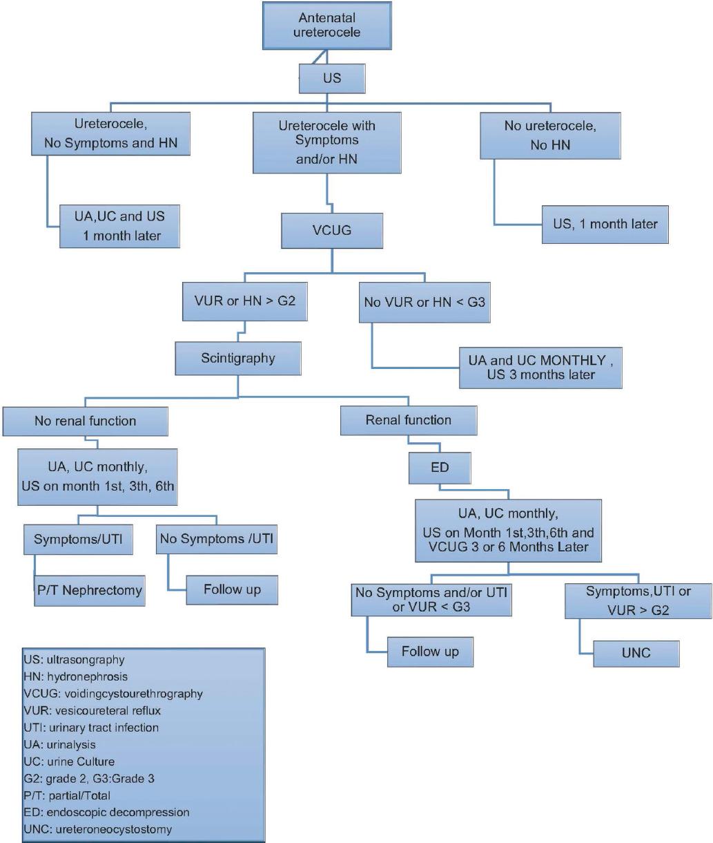
For the initial intervention, ED was performed on 23 patients using the electrocoat (Bugbee electrode), 17 patients using laser electrodes, eight patients using resectoscope knives, and three patients undergoing open surgery. One of the three patients who underwent open surgery had a ureterocele with calculi; as a result, the ureterocele was removed, and a ureteroneocystostomy (UNC) was performed. The other patient underwent upper pole heminephrectomy and ectopic ureterocele surgery due to recurrent UTI and a non-functioning upper pole. The third patient, who was 1.5 years old, underwent UNC and ureterocele excision due to severe hydronephrosis. A patient made a cautious follow-up.
At the summary of the initial intervention, the lasers decompression rate was 82% and the Bugbee's was 91%. Twenty-three patients who underwent ED and two who underwent open surgery required secondary surgery. Eleven patients underwent UNC, four sub-ureteric injections, seven partial nephrectomies, and three total nephrectomies.
Following secondary surgery, three patients required additional surgery. These three patients underwent sub-ureteric injection for persistent VUR, double j catheterization due to hydroureteronephrosis, and cyst excision of a remnant cyst on a previous partial nephrectomy zone, respectively. The average follow-up period was 29 (6-137) months. The management and follow-up of 23 patients are still ongoing.
Discussion
The primary management goals for ureterocele are to prevent UTI and renal damage and to maintain continence7,8.
Due to the high risk of infection, antibiotic prophylaxis is advised; some authors recommend giving it to children up to the age of three whereas others advocate waiting until they can control their micturition9,10. In our clinic, we advise stopping prophylaxis after the 1st year if there are no symptoms after decompression and surgical procedures, or if there is no progressive renal scar and no urinary infection.
With the widespread use in the US, 75% of ureteroceles can be diagnosed antenatally or in early infantile ages1,11. The majority of patients were diagnosed by the US in Chowdharys trial, and Adorisio et al. found 76% of ureteroceles with the US12,13. In our study, the US usage rate was 76%, which was comparable to research findings. The other diagnostic methods used in our clinic were VCUG (7.6%), IVU (3.8%), and cystoscopy (11.5%).
VCUG is crucial for the diagnosis of ureterocele1,14. By virtue of VCUG which shows ureterocele with associated bladder abnormalities and VUR surgery can efficiently be planned. All of our patients with ureterocele and hydroureteronephrosis underwent VCUG, which we advise doing for that patient population. Several authors assert that ureterocele can be diagnosed using computed tomography (CT) and magnetic resonance imaging (MRI)15. Patients with anatomic abnormalities can undergo CT, MRI, and IVU procedures16,17. However, these techniques are not preferred due to high radiation exposure in CT scans and high MRI costs, as well as challenges with imaging in children and occasionally the need for anesthesia. Therefore, for any of our patients, we did not use MRI or CT.
Hodhod et al. found 39 (78%) ureteroceles on duplex systems out of 51 patients with ureteroceles in their study18. In our study, 40% of patients had a single system, and 52% had a duplex system. For these variable rates, we believe that more clinic data sharing is necessary. According to Hodhod et al., UTI was discovered in 41% of duplex systems and 15% of single systems18. In the duplex system, we also discovered notably high rates of UTI, VUR, and renal scar.
The 41-patient trial by Visuri et al. revealed high rates of UTI in ureterocele and also demonstrated decreased rates of UTI with early surgery18. According to Hodhod et al.18, early ureterocele decompression and diagnosis reduce UTI and the need for secondary surgery19. It was discovered that delayed diagnosis and decompression led to an increased UTI. UTI was significantly more common in patients diagnosed after 6 months of birth20,21 than in those diagnosed before 6 months22,23. The most popular ureterocele surgery methods are ED procedures. The primary factors determining the need for secondary surgery are the presence of VUR and UTI1,24.
In a trial comparing ED techniques, there was no discernible difference between laser puncture and electrocautery (Bugbee) incision for decompression success, but the laser puncture group had a lower VUR rate12,13,24-27. According to Ilic et al., there is no discernible difference between laser and electrocautery for decompression success and complications, but electrocautery-incised groups have more VUR25. For ED, we primarily used the Bugbee electrode (45%) and laser probe (33%). Decompression success rates were 91.3% in the Bugbee and 83% in the laser, with no discernible difference. Later, decompression VUR was discovered to be 43.5% and 52.9% for laser and Bugbee, respectively. Both procedures were effective for decompression, but there were no advantages to the development of VUR. To determine the best method, we believe that more information is required.
Patients who have healthy kidney function can be treated permanently with ED as the initial procedure, with no need for additional surgery7,26. According to Di Renzo et al., 24 out of 45 patients did not require surgery after ED14,27. In our study, 52.9% of patients did not require additional surgery after ED, which is consistent with research findings.
Conclusion
Due to the high risk of UTI, VUR, and renal scarring, ureterocele continues to pose a difficult problem for patients, families, and doctors. The risk of urinary infection, VUR, and renal scarring is higher in ureteroceles in a duplex system. Each patient must be managed with an event based on their unique circumstances and clinical setting. UTI and renal scarring could be decreased with early detection and treatment. ED is still the most preferable surgical procedure, and also, we believe that conservative follow-up may be an option for convenient patients.

