


 nueva página del texto (beta)
 Inglés (pdf)
 Artículo en XML
 Referencias del artículo
 Enviar artículo por email
 Citado por SciELO
  Similares en
SciELO
Similares en
SciELO 
 Permalink
Introduction
Sickle cell disease is an inherited blood disorder characterized by the production of erythrocytes that are sickled or shaped like crescents. In sickle cell disease, the body produces hemoglobin S, which is considered abnormal and affects the transport of oxygen around the body. Generally, it is caused by a mutation in the amino acid located at position 6 of the β-globin gene [1]. The amino acid valine replaces glutamic acid in the 6th position of the β-globin chain located on chromosome 11. These series of events lead to the formation of the abnormal red blood cell with hemoglobin S, which displays a sickled shape [2]. This abnormality reduces the efficiency of the sickled hemoglobin to transport oxygen around the body, decreasing the solubility of blood in the deoxygenated form. Moreso, when the partial pressure of oxygen decreases, the abnormal hemoglobin becomes less soluble, forming polymers with other sickle hemoglobin [3]. The inefficiency of abnormal hemoglobin (hemoglobin S) to transport oxygen around the body, coupled with its ability to form polymers that can block blood flow routes, leads to the development of secondary conditions such as oxidative stress, inflammation, pain, cardiovascular diseases, vaso-occlusive crises, organ malfunctioning, and bone degeneration accompanying this disease [4].
This disease condition currently has no known cure, but medications are available to manage the condition and also alleviate the symptoms of the secondary complications associated with it. Furthermore, the use of nutrients such as vitamins, amino acids, and fatty acids in diets for managing the disease condition has long been documented in the literature [5-9]. Several drugs used in the management of this disease condition, such as hydroxyurea, piracetam, calcium antagonists, and several painkillers, are not without side effects and toxicities to body organs, which are attributed to their long-term use [3,10]. These drugs when available in developing countries are very expensive to afford, driving the search for alternatives from plants, which are widely available, relatively safe, and cheap [4].
Numerous studies have reported the application of plant materials for the management of sickle cell disease [4,11]. The plant M. flagellipes, which belongs to the family Leguminosae, is a plant localized mainly in Africa but present in other parts of the world [12]. The high nutritional profile and minerals of the leaves of Mucuna flagellipes have been reported by Ihedioha and Okoye [13]. The phytochemistry profile and mineral estimation of the seeds have shown a high content of flavonoids, vitamin C, zinc, and iron [14]. Furthermore, Uchegbu et al. [15] reported the presence of 9-octadecenoic acid, ethyl ester linoleic acid, ethyl ester, 9-octadecanoic acid, hexadecanoic acid, ethyl hexadecanoate, ethyl octadecenoate, eicosanoic acid, ethyl icosanoate, 3-hydroxypropyl-9-oate, and 9-methylbicyclo (3,3,1) nonane in the plant extract. Ajayi et al. [16] reported the presence of stearic, palmitic, linoleic, and oleic as major fatty acids in the seed oil of M. flagellipes. Recently, animal studies by Abireh et al. [17] reported the potential of M. flagellipes leaf extract to alleviate the bone marrow suppression, anemia, and leukemia effects following cyclophosphamide treatment of malignancies. The extract significantly improved packed cell volume, white blood cell counts, and the number of proliferating cells in the bone marrow. The ethanol extract of the seeds has been reported to possess anti-hypertensive potentials in salt-induced hypertension in rats via its ability to reduce blood pressure parameters [18]. In traditional systems in Eastern Nigeria, plant material is widely employed for the treatment of various ailments by traditional medical practitioners, specifically for the reduction of hypertension and blood-related issues.
Interest is growing in the use of seed oils rich in polyunsaturated and omega-3 fatty acids for managing sickle cell disease [19]. Polyunsaturated and omega-3 fatty acids have been reported to possess antioxidant activities and membrane protection potentials, which are strongly required in sickle cell disease fatty acids and omega-3 fatty acids for managing various diseases [9,20]. Furthermore, the identification of new sources of zinc in the diet of sickle cell patients is a new focus in managing the abnormalities associated with the disease.
Currently, there is no information on the in vitro anti-sickling potential of the seed oil extract of M. flagellipes. Therefore, the study aimed to assess the in vitro anti-sickling potentials of M. flagellipes seed oil (MFSO) by looking at its effects on inhibition and reversal of sickling, inhibition of hemoglobin polymerization, and the Fe2+/Fe3+ ratio. The minerals (zinc and iron), vitamins (A, E, and K), and fatty acid profile were determined using spectrophotometric techniques. Nutritional calculations were employed to determine the safety potential of consuming the seed oil.
Experimental
Methods Chemicals used
All chemicals used for this study were of analytical grade and were obtained from Merck, Germany.
Plant material procurement
The seeds of M. flagellipes were purchased in July 2021 from Orie Ntigha market in Isiala Ngwa North Local Government Area of Abia State, Nigeria. A sample of the seed was identified as M. flagellipes seed by Dr. Magnus C. Nwoko, a taxonomist at the Department of Biology, Federal University of Technology, Owerri, Imo State, Nigeria. A voucher specimen was assigned to the plant material (AEE/21/0155), and the specimen was deposited in the herbarium of the Department of Biology, Federal University of Technology, Owerri, Imo State, Nigeria.
Plant materials preparation and oil extraction
The casing of the seeds was cracked open to obtain the seed cotyledons. The seed cotyledons were washed in running tap water to remove sand and debris and then dried under shade for two weeks to attain an unformed weight. The dried seeds were milled into powder form with a mechanical blender and then stored in an airtight container. Extraction was performed using a Soxhlet extractor and n-hexane as a solvent. Briefly, 50 g of seeds of M. flagellipes powder were extracted and soaked in 500 mL of n-hexane (1:10 w/v) with the temperature set at 35 oC throughout the extraction process. To ensure complete extraction of oil in the sample, the set-up was allowed to reflux continuously till a clear liquid was seen [21]. The extract was evaporated using a rotary evaporator and stored in a sterile brown and amber bottle in the dark prior to further analysis.
Phytochemical profile GC-MS profiling and fatty acid analysis of seed oil extract
GC-MS analysis was performed using the method described by Ariyike et al. [22] at the University of Lagos, Lagos State, Nigeria. The composition of M. flagellipes seed oil (MFSO) was analyzed using a gas chromatograph coupled with a mass spectrometer GC-MS-QP 2010 Plus Shimadzu system. Operating condition of the GC-MS was Column Elite-1 fused silica capillary column (30 m × 0.25 mm 1D × µl df, composed of 100 % dimethyl polysiloxane). The carrier gas was ultra-pure helium (99.999 %) at a flow rate of 1.0 mL/min and an injection volume of 2 µL was employed (split ratio of 10:1). The injector temperature was set at 250 °C, ion source temperature 280 °C. The oven temperature was programmed 110°C (Isothermal for 2 min) with an increase of 10 °C /min to 200 °C then 5 °C/min to 280 °C/min, ending with a 9 min isothermal at 280 °C. Mass spectra were taken at 70 eV; a scan interval of 0.5 s and fragments from 40 to 550 Da. Total GC running time was 22 min and the compounds were identified by direct comparison of the retention times (RT) and mass fragmentation pattern with those from the National Institute of Standards and Technology (NIST) Library [23-24].
Vitamin estimation of MFSO
Vitamin A and E concentrations were determined in the oil sample using spectrophotometric methods as described by De’Leenheer et al. [25]. Briefly, exactly 0.1 g of the sample was dissolved in an acetone/hexane mixture (70:30 v/v), and the total carotenoid and vitamin E determinations were carried out by measuring absorbance at 470 nm and 270 nm, respectively, against a blank sample (solvent). Vitamin K was determined spectrophotometrically, as described by Gul et al. [26].
Mineral estimation of MFSO
The estimation of iron and zinc was performed using the wet digestion method described by AOAC [27]. Briefly, 0.5 g of the oil sample was boiled at 100 °C with 5 mL of concentrated nitric acid and 5 mL of a 30 % hydrogen peroxide solution for about 2 hours in an electric heating mantle until a clear solution was obtained. The solution was cooled and filtered through the Whatman No. 45 filter paper and then through 0.45 Millipore filter paper. The obtained filtrate was made up to 25 mL of the volumetric flask with distilled water and then analyzed for the individual minerals using an atomic absorption spectrophotometer (Bulk Scientific AAS model 210, equipped with a single slot burner and an air acetylene flame). Working standard solutions of zinc and iron were prepared, and the calibration and measurement of absorbance against a blank were performed using an AAS. The absorbance of each element in the filtrate was read at its wavelength from the spectrophotometer, and its concentration in the sample was extrapolated from the standard curve produced from the absorbance of the standard elements.
Nutritional indices of oil sample (MFSO) Fatty acids ratios
The ratio between polyunsaturated fatty acid (PUFA) and saturated fatty acid (SFA) [PUFA/SFA] also regarded as the polyene index was estimated as described by Chen and Liu [28] and Eke et al. [29]. The ratios between monounsaturated fatty acids (MUFA) and SFA [MUFA/SFA], PUFA and MUFA [PUFA/MUFA], and [(PUFA+MUFA)/SFA] were calculated to indicate the nutritional potentials of SOPA.
Oxidizability
The oxidative stability which is also regarded as the oxidizability was determined using the formula described by McCormick et al. [30]
Index of atherogenicity (IA)
The index of atherogenicity was calculated as described by Chen and Liu [28].
where C12:0 is lauric fatty acid; C14:0 is myristic fatty acid and C16:0 is palmitic fatty acids. UFA is summation of unsaturated fatty acids in oil samples.
Index of thrombogenicity (IT)
The thrombogenicity index was calculated as Chen and Liu described [28].
where C18:0 is stearic fatty acid; C14:0 is myristic fatty acid and C16:0 is palmitic fatty acids. MUFA is summation of monounsaturated fatty acids in oil samples.
Hypocholesterolemic/ hypercholesterolemic ratio
The hypocholesterolemic/ hypercholesterolemic ratio was calculated as described by Chen and Liu [28]
Unsaturation index (UI)
The UI which shows the level of unsaturation in oils was determined as described by Chen and Liu [28]
In vitro anti-sickling assay Collection of blood sample and ethical approval
Sickle cell patients attending the Clifford University Medical Centre, Ihie, Isiala Ngwa North LGA, Abia State, Nigeria, provided blood samples for the research after getting their consent and approval. Ethical clearance for the study was obtained from the Clifford University Research Ethics Committee, and ethical clearance number CLU/ETC/21/103 was assigned. Six milliliters (6 mL) of blood were used the same day for analyses.
Sickling inhibition test
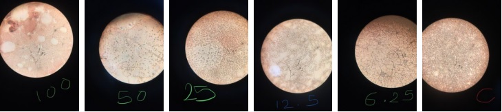
The in vitro sickling inhibition assay was performed using the method described by Egba et al. [31]. Briefly, 2 drops of HbSS blood, 2 drops of freshly prepared 2 % sodium metabisulphite and 2 drops of MFSO were mixed and covered. The solution was incubated for 30 mins at 37 °C and with the aid of a pipette, a drop was put on a slide, covered with a cover slip, and then viewed using a microscope at x 10 and x 40 magnifications. One hundred (100) cells were counted each from three randomly selected fields to evaluate the effect of MFSO on the HbSS erythrocytes. The test was performed in replicate, and the average reading was taking. The sickling inhibition (%) was determined using the equation below:
The control experiment was set up and did not contain MFSO, while p-hydroxybenzoic acid (5 % PABA) was used as standard agent.
Sickling reversal test
The sickling reversal test was performed as described by Ekeke et al. [32]. Briefly, 2 drops of HbSS blood were mixed with 2 drops of freshly prepared sodium metabisulphite and the mixture was covered. The mixture was incubated for 30 min during which sickling was induced. This was followed by the addition of 2 drops of buffered extract and the resulting mixture was incubated for another 30 min. Buffer was used in place of the extract for the control, and a drop of each was placed on a slide and viewed at × 40 magnification. The percentage reversal was calculated as:
Polymerization studies
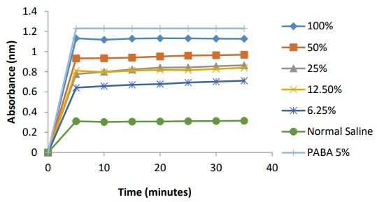
The polymerization study was performed as described by Noguchi and Schechter [33]. Briefly, 0.25 cm3 of normal saline (0.9 % w/v) was added to each test tubes containing 0.5 cm3 of various dilutions of the MFSO respectively. Freshly prepared aqueous sodium metabisulphite (2.2 cm3) and HbSS erythrocytes (0.05 cm3) were added into the test tubes simultaneously. The contents were mixed, and the absorbance taken at 700 nm using a spectrophotometer after standardizing it with distilled water as blank. Readings were taken at 5- min interval for a period of 35 min and appropriate control experiment was set up without the extracts.
Determination of the Fe2+/Fe3+ ratio in sickle cell blood
The method of Tietz [34] was adopted for the determination of Fe2+/Fe3+ ratio in sickle cell blood. This method works on the principle that hemoglobin and methemoglobin absorbs light at 540 nm and 630 nm respectively. Exactly 5.0 cm3 distilled water and 0.02 cm3 sickle cell blood was placed in a test-tube. Exactly 0.02 cm3 saline solution was introduced into the test tube and this served as the control. The test tube was kept for an hour at an ambient temperature and a spectrophotometer was used to determine its absorbance at 540 nm and 630 nm wavelengths. To determine the effect of MFSO on Fe2+/Fe3+ ratio, the process described above was repeated replacing saline solution with different dilutions of the extract. The percentage hemoglobin and methemoglobin were determined thus:
The ratio of Fe2+/Fe3+ was determined by dividing the values of each % Hb with the corresponding values of % MHb (before approximation).
Results
Quantification of MFSO fatty acids and phytocompounds
Table 1 Identified compounds present in M. flagellipes seed oil (MFSO).
| Peak No. | Name of Compound | MW (g) | Fatty acid class | RT (min) | Molecular formular | Concentration (%) |
| 1 | 2-hexanamine, 4-methyl | 115.22 | - | 6.54 | C7H17N | 0.52 |
| 2 | Resorcinol | 110.11 | - | 7.73 | C6H6O2 | 1.37 |
| 3 | Cathinone | 149.19 | - | 8.83 | C9H11NO | 1.33 |
| 4 | Guanidine, N,N-dimethyl | 87.12 | - | 11.84 | C3H9N3 | 4.00 |
| 6 | Pentadecanoic acid** (pentadecyclic acid) | 242.39 | SFA | 14.55 | C15H30O2 | 1.04 |
| 7 | n-Hexadecanoic acid** (palmitic fatty acid) | 256.40 | SFA | 14.82 | C16H32O2 | 5.91 |
| 8 | 9,12-Octadecadienoic acid** (linoleic fatty acid) | 280.44 | PUFA-6 | 15.92 | C18H32O2 | 18.30 |
| 9 | 9-Octadecenoic acid** (oleic acid) | 282.47 | MUFA | 15.99 | C18H34O2 | 31.87 |
| 12 | Octadecanoic acid** (stearic acid) | 284.48 | SFA | 16.46 | C18H36O2 | 1.85 |
| 13 | Phenylephrine | 167.20 | - | 17.76 | C9H13NO2 | 1.87 |
| 14 | I-Guanidinosuccinimide | 141.13 | - | 17.96 | C5H7N3O2 | 0.69 |
| 16 | Propanamide | 73.09 | - | 19.13 | C3H7NO | 1.03 |
| 17 | N-Desmethyltapentadol | 207.31 | - | 19.58 | C13H21NO | 0.79 |
| 19 | Alanyl-beta-alanine | 344.40 | - | 20.03 | C6H12N2O3 | 1.65 |
| 20 | 1,8-Octanediamine, N,N'-dimethyl- | 172.31 | - | 20.09 | C10H24N2 | 1.85 |
| 21 | 3-Propoxamphetamine | 193.28 | - | 20.21 | C12H19N0 | 0.94 |
| 22 | 1-Phenanthrene carboxylic acid | 222.24 | - | 20.29 | C15H10O2 | 10.61 |
| 24 | 2-Heptenoic acid, 4-cyclopropyl-5-methylene-, methyl ester, (E)- | 194.27 | - | 20.43 | C12H18O2 | 9.22 |
| 25 | 3-Keto-isosteviol | 332.40 | - | 20.59 | C20H28O4 | 4.42 |
RT: Retention time (min); MW: Molecular weight (g); ** indicates fatty acids; SFA: Saturated fatty acids; PUFA: Polyunsaturated fatty acids; MUFA: Monounsaturated fatty acids
Vitamin and mineral content of MFSO
Table 2 The vitamin and mineral content of MFSO.
| Vitamin | Concentration (µg/100mg) | RDA(mg/adult/day) |
| A | 220 ± 1.60 (2.2 mg/kg) | 0.9 |
| E | 370 ± 2.20 (3.7 mg/kg) | 15 |
| K | 197 ± 0.23 (1.9 mg/kg) | 0.12 |
| Minerals | ||
| Fe | 170 ± 1.30 (1.7 mg/kg) | 8 |
| Zn | 780 ± 2.50 (7.8 mg/kg) | 7-9 |
Data are mean ± SD, n = 3; MFSO- M. flagellipes seed oil; RDA: recommended dietary allowance
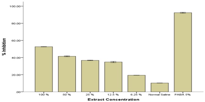
Effects of MFSO extracts on sickling inhibition
The MFSO significantly (p < 0.05) decreased sickling in human erythrocytes when compared to the negative control (normal saline) at all concentrations tested in which sodium metabisulphite was used to exacerbate sickling. The highest inhibition (52.66 ± 0.18) was observed at the highest concentration, 100 %. However, this activity was low compared to the standard agent used p-hydroxybenzoic acid (PABA) used as the positive control.
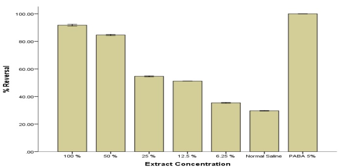
Effect of MFSO on sickling reversal
The MFSO significantly (p < 0.05) reversed sickling in human erythrocytes when compared to the negative control (normal saline) at all concentrations tested in which sodium metabisulphite was used to exacerbate sickling. The highest inhibition (91.76 ± 0.65) was observed at the highest concentration, 100 %. However, this activity was low compared to the standard agent used p-hydroxybenzoic acid (PABA) used as the positive control.
Effect of MFSO on polymerization of sickle cell blood
The MFSO which shows a dose dependent activity, significantly decreased (p < 0.05) polymerization of sickle cell at all studied concentration compared to negative control (normal saline). The observed activity for MFSO was however lower than the standard agent used. This indicates the ability of MFSO to inhibit polymer formation.
Effect of MFSO on % Hb, % MHb and Fe2+/Fe3+ ratio in sickle cell blood
The MFSO inhibited the formation of methemoglobin formation, and this activity was dose dependent. At the highest concentration, the percentage methemoglobin formation has the lowest value of 10.8 ± 2.3 % and the ratio of Fe2+/Fe3+ is 8.2. This activity was however lower than the standard agent used.
Table 5 Showing percentage hemoglobin, percentage methemoglobin and Fe2+/Fe3+ ratio in sickled blood treated with MFSO.
| Conc. (%) | %Hb | %MHb | Fe2+/Fe3+ ratio |
| 100 | 89.2±2.3 | 10.8±2.3 | 8.2 |
| 50 | 88.2±0.7 | 11.8±0.7 | 7.5 |
| 25 | 76.6±1.9 | 23.4±1.9 | 3.3 |
| 12.5 | 67.9±1.2 | 32.1±1.2 | 2.1 |
| 6.25 5% PABA | 65.4±0.5 95.0±0.08 | 34.6±0.5 4.0±0.32 | 1.9 23.8 |
| NS | 64.3±1.4 | 35.7±1.4 | 1.8 |
Discussion
The use of natural products of plant origin in managing and treating disease is on the rise. More specifically, plant materials are strongly employed in managing sickle cell disease, especially in traditional systems in underdeveloped and developing countries. GC-MS quantification of fatty acids in the seed oil presented in Table 1 showed the presence of five (5) fatty acids, such as pentadecyclic acid (1.04 %), palmitic acid (5.91 %), linoleic acid (18.30 %), oleic acid (31.87 %), and stearic acid (1.85 %). The major saturated fatty acid was palmitic acid, while oleic fatty acid was detected as the major unsaturated fatty acid in the seed oil. The result for fatty acid analysis agrees with the report of Ajayi et al. [16], who reported the presence of oleic, linoleic, palmitic, and stearic acids as the major fatty acids in M. flagellipes seed oil (MFSO). Furthermore, the concentration of oleic acid in this study was similar to the value obtained by Ajayi et al. [16] for the seed oil of M. flagellipes. The application of essential fatty acids in managing pain and other abnormalities associated with sickle cell disease has been properly documented [5,9,20]. These fatty acids are also known to promote cell membrane activity and prevent vaso-occlusive crises. The high amount of oleic acid and linoleic acid, which are known polyunsaturated fatty acids, in the oil sample may contribute to phospholipid requirements in sickle cell disease to keep membrane integrity intact. The fatty acid n-hexadecenoic acid (palmitic fatty acid) has been reported to exert its action on sickle red blood cells via stabilization of the sickle cell membrane against hemolysis and formation of HbSS polymers [35]. Furthermore, linoleic acid has been linked to analgesic, antibacterial, antioxidant, antipyretic, antispasmodic, and sedative activities. These activities are highly needed in the sickle cell disease crisis. Thus, this suggests that the seed oil of M. flagellipes may offer many therapeutic potentials in sickle cell disease [3]. Fratianni et al. [36] also reported that oleic acid has antioxidant activity. In sickle cell disease, where abnormal hemoglobin thrives, the hemoglobin stabilizing capacity is negatively affected, thereby making the red blood cells more susceptible to damage from oxidative stress. This may overwhelm the antioxidant defense system. It has been shown that sickle red blood cells produce more quantities of superoxide radicals, hydrogen peroxide (H2O2), and hydroxyl radicals than normal erythrocytes [37].
The result for vitamins and minerals is presented in Table 2. The results showed a great number of fat-soluble vitamins; vitamin A (220 ± 1.60 µg/100mg), vitamin E (370 ± 2.20 µg/100mg) and vitamin K (197 ± 0.23 µg/100mg), which was within the RDA for the vitamins. The values reported for vitamin A and E in the seed oil were lower than the values reported by Nwajagu et al. [38] in the processed and unprocessed seeds of M. flagellipes and its flour product. Deficiencies of antioxidant vitamins such as vitamin A and E have been reported in anemic conditions [39], and due to the nature of sickled cells, there is a constant need to furnish them with minerals and vitamins so as to keep them stable. Vitamins A and E, which are strong antioxidant vitamins detected in the seed oil, could contribute to the overall antioxidant status of the sick patient, and reduce symptoms. These antioxidant vitamins also play a role in the fight against infection, fight oxidative stress, and enhance iron absorption [7]. Vitamin K, a soluble vitamin, is useful in preventing excessive bleeding, which can lead to iron deficiency anemia. Mineral estimation showed an appreciable concentration of Fe (170 ± 1.30 µg/100 mg) and Zn (780 ± 2.50 µg/100 mg) in the seed oil. Interesting, the concentration of zinc was higher than iron in the sample and was within the recommended dietary allowance. The result reported for zinc and iron in the seed oil for this study was lower than the value reported for these minerals in the seed flour of M. flagellipes (32.60 ± 0.14 and 6.40 ± 0.03 mg/100g respectively) [40] and leaves (57.08 ± 8.44 and 5.83 ± 3.14 respectively) as reported by Ihedioha and Okoye [13]. This difference can be attributed to various factors ranging from the source of minerals to planting season, soil type, climate, and genetic and environmental factors [41-42]. Sickle cell anemia has been linked with zinc deficiency, making zinc supplementation in the diet helpful to manage or prevent some clinical manifestations [43]. Similarly, Datta et al. [8] reported the ability of zinc to prevent infection in sickle cell anemia, and reduced zinc bioavailability in sickle cell patients has been reported to lead to the development of several complications, ranging from wound healing problems to delayed or slow growth and vaso-occlusive crises [44]. The activity of zinc in reversing the functional state of sickle erythrocytes to their original biconcave structure, coupled with increasing their oxygen affinity, is established. Considering the value obtained for zinc in the seed oil, this plant material can contribute to the zinc bioavailability of sickle cell patients when consumed. Iron overload has been reported in patients with sickle cell anemia [45-46]. Therefore, it is important that iron supplementation be minimal for sickle cell patients, unless there is a proven case of iron deficiency. The result obtained for iron in this study, which was small compared to zinc, further justifies the use of seed oil in managing anemic conditions as it may not contribute to the overall iron burden of the sickle cell patient.
Sickle cell anemia is an inherited genetic disorder characterized by the inability of the red blood cells (erythrocytes) to carry enough oxygen around the body, leading to anemia and occlusion of small blood vessels. This series of activities leads to the development of excruciating body pains, crises, complications, and other manifestations [47]. The percentage activity of the seed oil on sickling inhibition and reversal is presented in figures 1 and 2. The seed oil at all studied concentrations significantly inhibited sickling and promoted reversal. This activity was however lower compared to 5 % p-hydroxybenzoic acid (PABA), the standard agent used. The anti-sickling potential observed in this study by the seed oil can be linked to the presence of zinc and some key vitamins present in the sample. The mechanism employed by zinc in inhibiting and reversing sickling has been reported [48]. Zinc binding to hemoglobin in sickle cells increases oxygen affinity and increases cell membrane filterability [49]. Zinc also decreases the quantity of hemoglobin linked with the red blood cell membrane, reducing the calcium effect in causing hemoglobin retention by the membrane and thus reducing the clumping that is observed in sickle cells. Zinc inhibits the role played by oxidative stress and proteins involved in inflammation (cytokines) in promoting sickle cell disease, and this is facilitated by its ability to promote and increase anti-inflammatory proteins [50].
The seed oil of M. flagellipes significantly inhibited HbS polymerization compared to the negative control, as seen in figure 3. The ability to inhibit HbS polymerization is a key area when looking for compounds with anti-anemic potential. The role played by minerals and vitamins in the prevention of HbS polymerization has been properly documented [6]. Zinc has been reported to stimulate the synthesis of metallothionein, a protein that binds and traps copper within the intestinal cells, inhibiting copper absorption [51]. The ability to keep copper trapped and not released is a key mechanism employed by zinc in inhibiting HbS polymerization. Copper has been reported to increase HbS polymerization by 33.13 % [6]. The high zinc content of the seed oil of M. flagellipes may contribute to this effect, as seen in this study. A key mechanism for inhibiting HbS polymerization is to increase oxygen affinity. Zinc plays a major role in increasing oxygen affinity in HbS conditions [52]. Experimental data has shown that the effect of oxygen on the inhibition of polymerization could be likened to the 2-state allosteric MWC (Monod-Wyman-Changeux) model of enzymes [53-54]. This suggests that an equilibrium exists between the low oxygen affinity of fully deoxygenated hemoglobin, which is labeled the T quaternary structure, and the high affinity arrangement of fully oxygenated hemoglobin, called the R quaternary structure. Increased oxygen affinity effectively controls polymerization via a shift from the T-R equilibrium towards the R equilibrium and is considered an effective treatment approach in anemic conditions [55]. Also, the treatment of sickled blood cells with plant extracts has been suggested to inhibit the Ca2+-activated K+ channel [56]. The inhibition of this channel prevents the loss of water and potassium ions from sickled blood cells, preventing dehydration. This facilitates a decrease in the intracellular concentration of hemoglobin S and further prevents polymerization and sickling of erythrocytes. This could therefore have been another mechanism employed by the seed oil of M. flagellipes in preventing polymer formation, as seen in this study. The inhibition of HbS polymerization in this study can also be attributed to the presence of vitamins A and E and zinc, as recorded in this study. Studies by Djote et al. [19] attributed the anti-sickling potentials of Azadirachta indica seed oil to the presence of linoleic fatty acids and vitamin E.
The result for the effect of the seed on % Hb, % MHb, and the Fe2+/Fe3+ ratio is presented in Table 4. The oil extracted from the seeds of M. flagellipes showed potential to increase the Fe2+/Fe3+ ratio. This occurs by decreasing the methamoglobin (MHb) concentration that is produced from the conversion of the iron of deoxy-hemoglobin (Fe2+, reduced state) to the ferric state (Fe3+, oxidized state) [57]. The ratio of ferrous and ferric iron indicates the level of oxygen affinity for red blood cell hemoglobin [58]. The high Fe2+/Fe3+ ratio observed in this study, as shown by the seed oil-treated group, corroborates the activity of the extract in improving oxygen affinity in sickled erythrocytes, which can be attributed to the presence of zinc, antioxidant vitamins, and polyunsaturated fatty acids present in the sample. Furthermore, the increased Fe2+/Fe3+ ratio as revealed by the seed oil confirms the sickling reversal, indicating the seed oil's ability to convert deoxy-HbS to oxy-Hbs.
The nutritional index values of the oil extracted from M. flagellipes are presented in Table 3. Nutritional indices of oils are strongly employed to determine how fit an oil sample is for consumption. The PUFA/SFA ratio, also called the polyene index [29], is used to ascertain the impact of edible oils on the cardiovascular system. Saturated fatty acids (SFA) have been reported to promote high serum cholesterol levels, while polyunsaturated fatty acids (PUFAs) are established to reduce cholesterol levels. This has made the PUFA/SFA ratio a useful guide to determining if an oil sample will have positive effects on the body. Generally, a high PUFA/SFA ratio indicates a positive effect of the oil sample on overall cardiovascular health [28]. In this study, the PUFA/SFA ratio for MFSO was 2.07. This value was lower compared to the value report for soya bean seed oil (3.03-3.76) [59]. This suggests a great nutritional potential of MFSO, which is strongly linked to the high amount of PUFAs in the oil sample. The MUFA/SFA, PUFA/MUFA, and (PUFA+MUFA)/SFA values were 3.62, 0.57, and 5.70, respectively, and this corroborated the polyene index of MFSO and can be attributed to the rich amount of unsaturated fats in the oil sample. Consumption of oil rich in unsaturated fats suggests a low potential to develop cardiovascular diseases. The oxidizability value of the oil was 0.18, and this value was lower than the value reported for soya bean seed oil (7.51-7.63) [59]. Generally, oil samples with low oxidizability values are known to be more stable than their counterparts. The index of atherogenicity (IA) and index of thrombogenicity (IT) are employed to characterize the atherogenicity and thrombogenicity potentials of oil samples. While the IA shows the link between SFAs and the sum of unsaturated fats, the IT suggests the potential of the oil sample to form clots in blood vessels. The consumption of fats with low IT and IA has been linked to reducing cholesterol levels and contributing significantly to improved cardiovascular health. The IA value of 0.11 in this study was similar to the value reported for sunflower oil (0.09-0.11) [60]. The low value of IA in this study further suggests a good nutritional property of MFSO. The values reported for IT (0.07) in this study suggest the ability of the oil to prevent or not initiate thrombogenesis. In this study, we ascertained the hypocholesterolemic/hypercholesterolemic (HH) [61] ratio to determine the effect of the oil on the composition of cholesterol. It shows the relationship between hypocholesterolemic fatty acids (cis-C18:1 and PUFA) and hypercholesterolemic FA. A high HH value indicates a reasonable nutritional index of the oil sample. As seen in this study, the HH value was 3.09, which is low compared to Camelina oil (11.2-15.0) [60]. The UI value shown in this study (68.47) was low when compared to soybean oil (148-155) [62]. Oils with high UI values have been reported to maintain membrane fluidity under stress-associated conditions [63].
Conclusions
Despite the myriad uses of this plant in traditional systems, its seed oil's anti-sickling properties have not been investigated. The results obtained from this study have shown that the seed oil has great potential to serve as an anti-sickling agent. The high zinc and vitamin content of the seed oil suggests it can replenish nutrients lost in sickle cell disease. The fatty acid profile of the seed oil shows a high level of unsaturated fatty acids, which are cardio-friendly and thus may not pose a threat when consumed. Further studies will be required to isolate and identify the specific anti-sickling agents in the seed oil. Furthermore, studies to determine the best way of processing and preserving the seed oil are warranted.

