




 nueva página del texto (beta)
 Inglés (pdf)
 Artículo en XML
 Referencias del artículo

 Traducción automática
 Enviar artículo por email
 Citado por SciELO
 Similares en
SciELO

 Permalink

Introduction
Binge eating disorder (BED) is characterized by recurrent episodes of excessive intake, within a limited time, of an amount of food that is larger than what most individuals would consume in a comparable period. These episodes are accompanied by a sense of loss of control over food intake, leading to significant distress regarding the binge eating. These episodes occur on average at least once a week for 3 months and are associated with at least three of the following symptoms and signs: eating much more rapidly than normal, eating until feeling uncomfortably full, consuming large amounts of food when not physically hungry, eating alone due to feeling ashamed about the amount of food being eaten, and feeling disgusted with oneself, depressed, or very ashamed following the binge eating1.
Changes in eating behavior may be related to variations in neurochemical components. Several studies suggest that the neurobiological mechanisms involved in binge eating episodes are primarily associated with the dopaminergic system2-4. However, the literature presents contradictory and ambiguous findings. Some studies, conducted in both humans and animals, report hyperactivation of the system5-11 whereas others observe hypoactivation7,12-21. One possible explanation is that these hyperdopaminergic and hypodopaminergic states are not mutually exclusive but may occur at different stages of binge eating. Dopamine elevations could contribute to the initiation of binge eating behavior, followed by downregulation after a sustained pattern of overconsumption is established22. Taken together, these results suggest that binge eating episodes would generate overstimulation and, ultimately, desensitization of the mesocorticolimbic dopamine system as the main reward circuit3,23,24. Consequently, the overconsumption of appetitive stimuli could reflect the need to reactivate a hypofunctional reward circuit11,25. In this regard, treatments with dopaminergic agonists, such as lisdexamfetamine, improve dopaminergic and noradrenergic neurotransmission26,27 and reduce episodes of overconsumption in both rats28,29 and humans30.
On the other hand, the hypofunction of the dopaminergic system in advanced stages of BED could depend on increased activation of the GABAergic system. Dopamine release activates GABAergic pathways, which act as inhibitors by reducing dopamine release. This regulation by the GABAergic system allows for maintaining a balance in the reward circuit and preventing dopamine overload31,32.
Gamma-aminobutyric acid (GABA) is the principal inhibitory neurotransmitter of the central nervous system and plays multiple roles in both neuronal and non-neuronal tissues. Its synthesis primarily derives from glucose, which is metabolized into glutamate through enzymes of the tricarboxylic acid cycle. The enzyme glutamic acid decarboxylase (GAD), predominantly expressed in GABAergic neurons, catalyzes the decarboxylation of glutamate to produce GABA. Once synthesized, GABA is transported into synaptic vesicles through the vesicular GABA transporter. Subsequently, calcium influx into the presynaptic terminal triggers exocytosis. This cellular process involves the fusion of intracellular vesicle membranes with the plasma membrane, releasing their contents into the extracellular space. It is an energy-consuming active transport pathway utilized by cells to release GABA into the synaptic cleft33.
Their function is modulated by ionotropic (GABAA and GABAC) and metabotropic (GABAB) receptors located on the postsynaptic neuron. Ionotropic receptors are coupled to chloride channels, implying synaptic transmission with lower metabolic expenditure and faster kinetics. In contrast, metabotropic receptors entail additional energy expenditure to perform their function. These receptors are coupled to a G protein (GPCR) and are separated from the ion channel, enabling them to modulate inhibitory transmission that is slower but also more prolonged34,35.
On completion of GABA action on receptors, it is cleared from the synaptic cleft. The mechanism of GABA clearance begins with its reuptake through GABA transporters (GAT), membrane proteins located in presynaptic terminals. The enzyme GABA transaminase (GABA-T) catalyzes the conversion of GABA to succinic semialdehyde. Subsequently, succinic semialdehyde is converted to succinate by the enzyme succinic semialdehyde dehydrogenase. However, not all GABA is converted to succinate; a portion is also recycled for the synthesis of new GABA33.
At the presynaptic terminal, metabotropic receptors CB1, also known as type 1 cannabinoid receptors, are located. These receptors interact with anandamide, an endocannabinoid released by the postsynaptic neuron on depolarization. This interaction leads to the modulation of neuronal activity and causes a decrease in neurotransmitter release36.
Furthermore, GABA plays a fundamental role in neuronal function by regulating neuron excitability and dopamine release. The latter is closely linked to various functions such as motor activity regulation, attention, learning, wanting, and the reward system, among other functions37.
Dopamine synthesis begins with the action of tyrosine hydroxylase, a key enzyme in the biosynthesis of neurotransmitters dopamine, noradrenaline, and adrenaline. This enzyme catalyzes the conversion of the amino acid L-tyrosine into 3,4-dihydroxyphenylalanine (L-DOPA). Subsequently, L-DOPA is transformed into dopamine through the action of the enzyme dopa decarboxylase. Once released into the synaptic cleft, dopamine exerts its effects primarily by stimulating metabotropic dopamine receptors. These receptors are subdivided into different subtypes, including D1-like receptors (D1 and D5) and D2-like receptors (D2, D3, and D4), among others. After fulfilling its function, dopamine is reuptaken by the presynaptic neuron through dopamine transporters known as DAT. Once taken back up, dopamine is metabolized by the enzyme monoamine oxidase in a degradation process38. It is crucial to note that the information provided above is a general outline of the synthesis, action, and elimination processes of dopamine and GABA. However, it is important to highlight that there are other factors and pathways also involved in the metabolism and elimination of these neurotransmitters.
The general objective was to conduct a systematic review to gather research conducted from 2013 to 2023 on the functioning of the GABAergic system in eating behavior and binge eating episodes. It is important to acknowledge that, although validated animal models of BED are available, preclinical studies specifically examining the GABAergic system in BED remain relatively scarce. Consequently, most available studies have focused on general modulation of food intake or overeating behaviors, which may not fully capture the core behavioral phenotype characteristic of BED. This limitation affects the breadth and translational relevance of current evidence regarding the role of the GABAergic system in BED. Therefore, while this review includes studies addressing excessive eating behavior broadly, caution is warranted in extrapolating findings directly to BED.
The review will be exclusively limited to studies using animal models (i.e., rats or mice), as these are a valuable tool for addressing hypotheses about the neurobiological mechanisms of ingestion behavior.
General methodology
Eligibility criteria
The review is based on the guidelines proposed by the PRISMA 2020 methodology for reporting systematic reviews39. This methodology involves establishing the selection criteria for the studies to be included. Research published between 2013 and 2023, reported in articles published in English-language journals, was considered.
We included preclinical studies using rodent models (rats or mice) that assessed the GABAergic system in the context of excessive eating behavior. Eligible studies did not have to model BED explicitly, but were required to examine behaviors operationally defined as binge-like eating or overeating, such as the consumption of large quantities of palatable food in a short period, independent of food deprivation status. Studies had to provide neurobiological data related to the GABAergic system (e.g., receptor expression, pharmacological modulation, or GABA-related gene expression).
Sources of information
The databases ScienceDirect and PubMed were utilized, incorporating the combination of the following keywords in English: (((binge-like eating episodes) OR (binge-eating episodes) OR (overeating)) AND (GABAergic system) AND (rat OR mice)). To ensure literature saturation, forward search strategies (searching for articles citing the included study) and backward search strategies (reviewing the reference lists of included studies) were applied. In addition, the reference lists of systematic reviews and meta-analyses on the topic were reviewed.
General procedure
IDENTIFICATION
The initial search was conducted on March 22, 2023. This identified 488 records across the two databases used: ScienceDirect (n = 480) and PubMed (n = 8; Fig. 1).
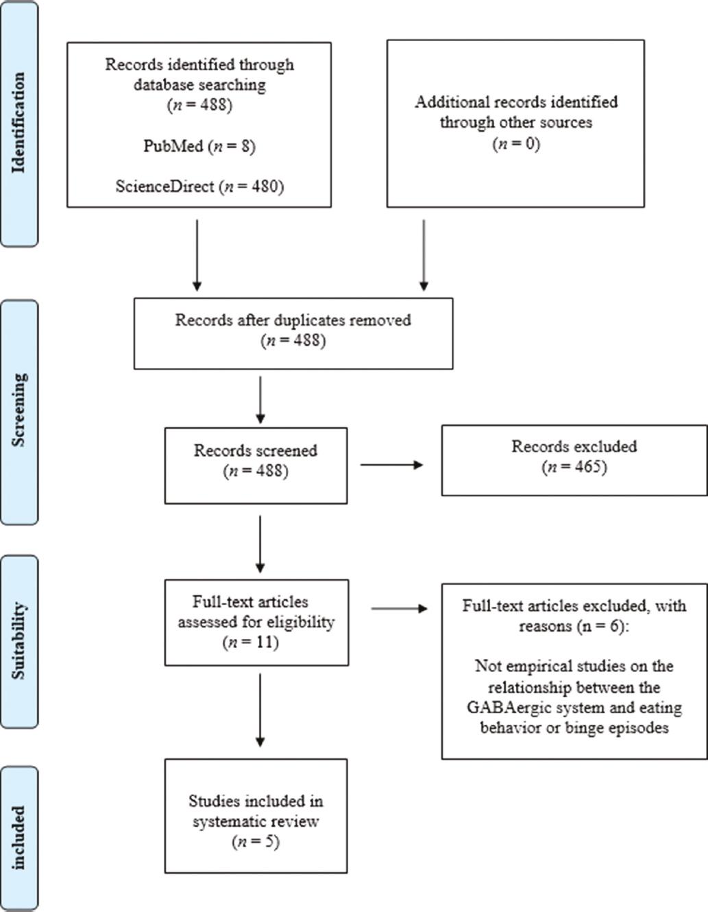
SCREENING
During screening, the number of records was reduced to 23. Articles were excluded (n = 465) based on title screening, as they did not study the relationship between the GABAergic system and feeding regulation.
SUITABILITY
The process of reviewing abstracts of selected articles was conducted. Out of the 23 articles initially considered for eligibility, retrieving 11 articles was requested. On suitability analysis, the number of articles reduced to 5 as they met all inclusion criteria: being published between 2013 and 2023, using animal models of rats or mice, and being empirical research on the relationship between the GABAergic system and binge-eating episodes.
Risk of bias (RoB) assessment
The methodological quality of the included preclinical studies was evaluated using the SYRCLE's RoB tool, which is specifically adapted for animal research. This tool assesses ten key domains:
1. Random sequence generation
2. Baseline group similarity
3. Allocation concealment (housing randomization)
4. Blinding of caregivers
5. Blinding during intervention administration
6. Blinding of outcome assessment
7. Randomization in data/sample analysis
8. Incomplete outcome data (attrition)
9. Selective outcome reporting
10. Other sources of bias (e.g., ethics and conflicts of interest).
Two independent reviewers assessed each domain and classified it as: low risk (clear adherence to the criterion), high risk (criterion not met), and unclear risk (insufficient information). Discrepancies (< 5% of cases) were resolved by consensus or through consultation with a third reviewer.
Assessment results
Eighty percentages of the studies showed low RoB in initial randomization (domain 1), and all studies adequately reported outcome data (domain 8) and declared no conflicts of interest (domain 10), which represent notable methodological strengths. However, all studies presented high risk in the domain related to blinding during intervention administration (domain 5), and 60% showed unclear risk regarding housing randomization (domain 3). These findings suggest that, while the included studies maintained acceptable standards in randomization and transparency, the consistent lack of blinding in intervention delivery may have introduced performance bias. This common limitation in preclinical research highlights the need for cautious interpretation of the observed effects.
Results
The initial search identified 488 results, of which 480 were found in the ScienceDirect database and eight in PubMed. During screening, the number was reduced to 23 articles, of which only five were deemed suitable for this study (Fig. 1). Of the five reviewed studies, 60% (n = 3) were conducted in mice and 40% (n = 2) in rats. In 80% (n = 4) of the studies, only male animals were used, and in the remaining 20% (n = 1), both males and females were used, with no studies exclusively focusing on females. Regarding age, 80% (n = 4) of the articles used adult animals, and 20% (n = 1) did not specify the age (Table 1).
Table 1 Studies reporting GABAergic alterations.
| Authors | Study objective | Methodology | Main results |
|---|---|---|---|
| Hao et al.40 | To evaluate the role of GABAergic cells in the anterior ventrolateral periaqueductal gray (vlPAG) on the regulation of feeding behavior. | Adult male mice (2-4 months old) from the Gad2-ires-Cre and C57BL/6J strains were utilized for the study. The intervention involved both pharmacological and optogenetic techniques. The first technique entailed the administration of muscimol (n = 7) or saline (n = 7) into the anterior vlPAG of C57BL/6J mice without food deprivation. The second technique involved the incorporation of halorhodopsin (NpHR; n = 8) and Channelrhodopsin (ChR2; n = 7) into GABAergic neurons of the same region in Gad2-ires-Cre mice, both with and without food deprivation. Dependent variables included food intake (g) over 30 or 60 min, feeding latency (s), number of approaches to food, and total feeding time (s) over 10 min | The results demonstrate that the suppression of activity of GABAergic cells in the anterior vlPAG, both directly and through their inputs from the lateral hypothalamus (LH) and bed nucleus of the stria terminalis, promotes feeding in satiated mice. Conversely, activation of these cells disrupts food intake in food-deprived mice. |
| Marino et al.41 | Characterize the neural circuits involved in compulsive eating. | Male and female transgenic mice expressing ChR2 in GABAergic projections from the LH to the ventral tegmental area (VTA) were used. The mice (n = 8) were food-satiated and aged between 10 and 20 weeks at the time of surgery. The dependent variable was the feeding time (s) under optogenetic inhibition or stimulation (5, 10, 20, and 40 Hz) for 1 min. | The data indicate that peripheral GABA neurons in the locus coeruleus (LC) are a critical step in the descending circuit for LH neurons, whereas GABA neurons in the VTA are not. Activation of this LH-peri-LC pathway would be necessary and sufficient to induce overeating in satiated mice. |
| Cruz-Martínez et al.43 | Characterize the neurochemical mechanisms related to the hyperphagic effects induced by the activation of CB1 receptors in the paraventricular nucleus of the hypothalamus (PVN) | Male Wistar rats with 21 h of food deprivation before the trial were used. The animals were divided into 4 groups and injected intra-PVN with saline solution (n = 8), ACEA (n = 8), AM 251 (n = 5), and ACEA+AM251 (n = 6). Dependent variables were: food intake in grams (g) and the release of [3H] GABA over a period of 2 h. | The findings suggest that the observed increase in food intake is more strongly associated with GABAergic hyperfunction in the PVN rather than hypofunction. |
| Corwin et al.14 | To evaluate gene expression within the VTA and prefrontal cortex (PFC) in rats experiencing binge-eating episodes (INT) and control rats (D), 20 min before and 20 min after presentation of palatable food. | Male Sprague-Dawley rats (60 days old) were used. One group was exposed to vegetable fat for 1 h on alternate days (INT), while the other group received it daily (D). The intervention procedure on GABAergic pathways consisted of administrations of muscimol/baclofen, which were locally infused into the dorsomedial (n = 18) and ventromedial (n = 14) PFC. Dependent variables were vegetable fat intake (g) over one hour and mRNA expression for GABAA and GABAB receptors. | The findings suggest that initially, binge-eating episodes would lead to an increase in dopaminergic activity, and if this behavior persists over time, it would result in an increase in GABAergic activity to decrease dopaminergic activity. |
| Sato et al.44 | Evaluate the underlying mechanisms of the decrease in body weight generated by the addition of GABA to the supplied food. | In experiment 1, diets containing variable levels of GABA were supplied over a period of 2 weeks: 0% (0 g; n = 8), 0.5% (5 g; n = 8), 2% (20 g; n = 8), and 5% (50 g; n = 8). In experiment 2, subcutaneous injections of vigabatrin, a GABA-degrading enzyme inhibitor (GABA-T), were administered daily to mice receiving diets with 0.5% and 2% GABA. Dependent variables were: intake of balanced food (grams/6 weeks), body weight (grams/6 weeks), and plasma GABA levels (μM) and hippocampal GABA levels (μmol/g). | The findings suggest that increasing GABA levels in the central nervous system can significantly decrease both food intake and body weight. |
Binge eating model
Of the five studies reviewed in this work, only one used a behavioral model to generate binge eating episodes14, while the remaining studies directly intervened in the GABAergic pathways to influence intake. In the study by Corwin et al.14, animals were assigned to one of two conditions, counterbalancing their body weight. One group was exposed to vegetable fat for 1 h on alternate days (INT group), while the other group was exposed to it daily (D group). Both groups were provided with water and balanced food throughout the procedure. The experimental protocol lasted 5-8 weeks. It was observed that binge eating episodes occurred in the intermittent condition during the presentation of highly palatable food.
Intervention procedures in GABA
The experiments conducted by Corwin et al.14 aimed to evaluate gene expression within the ventral tegmental area (VTA) and the prefrontal cortex (PFC) in rats with binge-eating episodes (INT) and control rats (D), 20 min before and 20 min after the presentation of palatable food. The intervention procedure on the GABAergic pathways consisted of administrations of muscimol/baclofen, two agonists that act on GABAergic receptors. Muscimol interacts with both GABAA and GABAB receptors, while baclofen selectively affects GABAB receptors. Both drugs were injected locally into the dorsomedial (n = 18) and ventromedial (n = 14) PFC. The dependent variables were the intake of vegetable fat in grams (g) over 1 h, and the mRNA expression for GABAA and GABAB receptors.
In both the study by Hao et al.40 and Marino et al.41, GABAergic pathways were intervened using optogenetic techniques. This is a genetic engineering technique involving the introduction of opsins into neurons. Opsins are light-sensitive proteins obtained from photosynthetic microorganisms that have the ability to convert light energy into electrical signals within nerve cells. These proteins can function as ion channels or pumps, meaning they allow the flow of ions across the cell membrane in response to light, modulating neuronal activity by either activating or inhibiting it42.
The experiments conducted by Hao et al.40 used adult male mice (2-4 months old) from the Gad2-ires-Cre strain (JAX no 019022) and C57BL/6J strain. The intervention procedure on the GABAergic pathways consisted of pharmacological and optogenetic techniques. The first involved administering an injection of muscimol (n = 7), a selective GABAA receptor agonist, or saline (n = 7) into the anterior ventrolateral periaqueductal gray (vlPAG) of C57BL/6J mice. The dependent variables were food intake in grams (g), the number of approaches to food, and feeding latency in seconds (s). The second technique involved incorporating halorhodopsin (NpHR; n = 8) and channelrhodopsin (ChR2; n = 7) opsins into GABAergic neurons of the same region in Gad2-ires-Cre mice. ChR2 opsin functions as a light-sensitive ion channel, allowing the influx of sodium (Na+) or calcium ions (Ca2+) into the cell, leading to increased activity in GABAergic neural pathways. On the other hand, when NpHR opsin is expressed in GABAergic neurons, exposure to light allows the influx of chloride ions (Cl-) into the cell, resulting in hyperpolarization and consequently reducing neurotransmitter release and GABAergic signal transmission. Finally, a yellow fluorescent protein (eYFP; n = 8), which did not include any opsins, was incorporated into the control condition. The dependent variables were food intake in grams (g), the number of approaches to food, and the total time spent feeding in seconds (s).
In the study by Marino et al.41, male and female transgenic mice expressing ChR2 in GABAergic projections from the lateral hypothalamus (LH) to the VTA were used. The mice (n = 8) were non-food deprived and were between 10 and 20 weeks old at the time of surgery. The dependent variable was the feeding time in seconds (s) in response to optogenetic inhibition or stimulation (5, 10, 20, and 40 Hz) for 1 min.
On the other hand, the experiments by Cruz-Martínez et al.43 intervened in the GABAergic system through the activation of CB1 receptors in the paraventricular nucleus (PVN) of the hypothalamus (PVN). Male Wistar rats were used, with 21 h of food deprivation before the trial.
ACEA (a CB1 agonist) and AM 251 (a CB1 antagonist) were injected into this brain region, and the release of GABA was evaluated. The animals were divided into four groups: intra-PVN injected with ACEA (n = 8), AM 251 (n = 5), a combination of both drugs (n = 6), or vehicle (n = 8). Subsequently, food intake in grams (g) and the release of [3H] GABA were assessed during the following 2 h.
The study conducted by Sato et al.44 aimed to delve into the mechanisms underlying the decrease in body weight induced by dietary GABA administration. In Experiment 1, diets containing varying levels of GABA were provided over a period of 2 weeks: 0% (0 g; n = 8), 0.5% (5 g; n = 8), 2% (20 g; n = 8), and 5% (50 g; n = 8). In Experiment 2, subcutaneous injections of vigabatrin, a GABA transaminase (GABA-T) enzyme inhibitor, were administered daily to mice receiving diets with 0.5% and 2% GABA. The experimental conditions were as follows: 0% + saline (n = 6), 0.5% + saline (n = 6), and 2% + saline (n = 6), 0.5% + vigabatrin (n = 6), and 2% + vigabatrin (n = 6). The dependent variables included intake of balanced food (grams/6 weeks), body weight (grams/6 weeks), and levels of GABA in plasma (μM) and GABA in the hippocampus (μmol/g).
Alterations in the GABAergic system
The pharmacological studies by Hao et al.40 found that muscimol injection led to an exacerbation of food intake and the number of approaches to food, along with a decrease in feeding latency compared to the saline group. On the other hand, in the optogenetic studies, the ChR2 condition showed a decrease in food intake, the number of approaches to food, and the total time spent feeding compared to the control condition eYFP. In the NpHR condition, an increase in food intake, the number of approaches to food, and the total time spent feeding was observed compared to the control condition eYFP.
This suggests that GABAergic cells in the anterior part of the vlPAG play a central role in the regulation of feeding. This structure receives direct inputs from many nuclei related to feeding: the LH, the bed nucleus of the stria terminalis (BNST), the central amygdala nucleus (CeA), the arcuate nucleus (ARC), the PVN of the hypothalamus, and the dorsal raphe nucleus (DR). When examining the LHGABA-vlPAG, BNSTGABA-vlPAG, and CeAGABA-vlPAG pathways, the study found that the suppression of the first two pathways separately was sufficient to induce overeating in sated mice, while optogenetic activation of the CeAGABA-vlPAG pathway did not induce significant behavioral changes. In addition, activation of the LHGABA-vlPAG and BNSTGABA-vlPAG pathways separately resulted in a significant reduction in consumption in food-deprived mice.
In the study by Marino et al.41, a series of experiments were conducted to delineate the neuronal circuitry involved in overeating episodes in satiated mice. The authors hypothesized that stimulation of GABAergic projections from the LH to the VTA could lead to overeating. In addition, they expected strong rostral projections to anterior BNST, the dentate gyrus, and the lateral habenula. Caudal projections extended to and through the VTA and the rostromedial tegmental nucleus, which then branched dorsally toward the locus coeruleus (LC) and ventrally toward the dorsal inferior olive.
However, only photostimulation of VTA projections to the peri-LC induced overeating, while other brain regions either did not produce feeding or their effects did not reach statistical significance. On the other hand, to determine if GABAergic neurons in the LC are linked to overeating induced by LH, GABAergic neurons in the LC and VTA were photoinhibited. The authors observed that photoinhibition of GABAergic neurons in the LC and photostimulation of LH disrupted feeding behavior, while photoinhibition of GABAergic neurons in the VTA and photostimulation of LH led to overeating. These data suggest that GABAergic neurons in the LC are a critical step in the descending circuit for LH neurons, whereas GABAergic neurons in the VTA are not. Activation of this LH-LC pathway would be necessary and sufficient to induce overeating in satiated mice.
Cruz-Martínez et al.43 aimed to characterize the neurochemical mechanisms related to hyperphagic effects induced by CB1 receptor activation in the PVN. Since cannabinoid-induced hyperphagia can be triggered by inhibition of satiety and/or stimulation of orexigenic signals, the hypothesis was raised that CB1 receptor activation in the hypothalamus could induce changes in GABA release.
The results showed that CB1 receptor activation by ACEA increased GABA release and caused a significant increase in food intake compared to animals receiving the vehicle, AM 251, or AM 251 + ACEA. In addition, this effect persisted for a period of 2 h. Finally, animals injected with AM 251 and AM 251 + ACEA exhibited food intake comparable to the vehicle condition. In summary, these findings suggest that increased food intake is primarily associated with GABAergic hyperfunction in the PVN rather than with hypofunction.
Corwin et al.14 found a decrease in the gene expression of GABAB and GABAA receptors in rats with binge eating compared to the control group. Furthermore, the administration of GABAergic agonists in the dorsomedial and ventromedial regions of the PFC increased the binge size in the INT condition, without affecting the intake in the D condition.
In the study by Sato et al.44, it was found that a 5% GABA intake significantly suppressed food intake and body weight gain compared to the other conditions, which did not show significant differences among themselves. In addition, this suppression was associated with significantly elevated concentrations of GABA in plasma.
The reduction in food intake could be modulated by alterations in taste, so that ingesting 5% GABA could have an unpleasant taste compared to 0.5% or 2%. To corroborate this hypothesis, vigabatrin was injected into mice exposed to these latter two conditions. The results indicate that the administration of vigabatrin significantly reduced GABA-T activity in the liver compared to the control groups. This resulted in an increase in circulating and cerebral (cortex and hippocampus) GABA levels, which, in turn, led to a decrease in food intake and body weight.
These findings rule out the explanation that the decrease in intake is due to the unpleasant taste of GABA in the diet. Instead, they suggest that the increase in GABA levels in the central nervous system may decrease food intake and body weight, in addition to its peripheral effects.
Discussion
The general objective of the study was to conduct a literature review on the functioning of the GABAergic system in binge eating episodes. Contradictory results were identified regarding the role of GABA in feeding behavior, as several studies reported variations in opposite directions. Some of these studies showed an increase in intake behaviors with either lower14,40 or higher GABAergic activation,41,43 while others reported the inverse relationship: reduced intake with either higher40,44 or lower activation of the neurochemical circuit41.
Variations in the protocol can lead to the application of procedures with different levels of sensitivity in detecting the phenomenon, which, in turn, could generate contradictory and ambiguous results. In this regard, it is interesting to note that studies conducted by Hao et al.40 and Marino et al.41 have yielded different results despite using similar techniques. This discrepancy could be attributed to differences in dependent variables and the duration of the trial used in their research.
Hao et al.40 recorded both direct behaviors, referring to the amount of food consumed, and indirect behaviors, linked to the time spent on intake as well as the number of approaches to the food, over a period of 30 and 10 min, respectively, while Marino et al.41 only observed indirect behaviors for one minute. Faced with this, it could be argued that an observation period of just one minute may be insufficient to adequately assess the impact of GABA on intake.
This discrepancy in the literature may be attributed not only to procedural differences but also to the specific neuroanatomical structures targeted (Table 2). The studies by Corwin et al.14 and Cruz-Martínez et al.43 reported opposing results following the administration of drugs that enhance GABA activity. These discrepancies could stem from differences in how the neurochemical pathways were manipulated. While Corwin et al.14 directly administered GABAergic agonists into the PFC, Cruz-Martínez et al.43 performed an indirect intervention using cannabinoid agonists targeting the PVN.
Table 2 Summary of the studies included in the systematic review on the GABAergic system and feeding behavior.
| Brain region | GABAergic effect | Outcome on feeding behavior | References |
|---|---|---|---|
| Prefrontal cortex | ↓ GABAB receptor expression | ↑ Binge eating | 14 |
| Paraventricular nucleus | ↑ GABA release (via cannabinoids) | ↑ Hyperphagia | 43 |
| Lateral hypothalamus → Locus coeruleus pathway | ↑ GABAergic activation | ↑ Compulsive eating | 41 |
| Ventrolateral periaqueductal gray | ↓ GABAergic activation | ↑ Intake in food-deprived mice | 40 |
| Hippocampus | ↑ GABA (vigabatrin) | ↓ Food intake | 44 |
Role of the PFC in binge eating and GABAergic modulation
Regarding the PFC, a decrease in the gene expression of the GABAB receptor was found in binge-eating rats compared to the control group. Furthermore, the administration of GABAergic agonists in the dorsomedial and ventromedial regions of the PFC increased the binge size in the experimental condition, without affecting the intake in the control condition. Similarly, the administration of a dopaminergic antagonist of D2-like receptors (i.e., SKF 81297) in the dorsomedial region produced effects similar to inhibition by GABA agonists14.
These findings suggest that D2-like receptors in the PFC are a key component in restraining binge-eating behavior. Binge-eating episodes may generate differential activation patterns within VTA neurons, potentially resulting in increased dopamine release in the PFC14. D2-like receptors located on presynaptic neurons in this cortex would have an inhibitory effect on GABA release in the synaptic cleft. This would increase PFC activity and reduce binge-eating behavior45.
The study by Corwin et al.14 suggests binge-eating episodes serve to restore a hypofunctional reward circuit. For example, rats with binge-eating episodes show alterations in mRNA expression for tyrosine hydroxylase, the dopamine transporter, and the D2-like receptor in the VTA. While binge-eating episodes normalize general aspects of dopaminergic signaling, they do not seem to have the same effect on mRNA expression of the D1-like receptor and the GABAA receptor. There are also no normalizations in mRNA for tyrosine hydroxylase and the GABAB receptor in the PFC. The absence of these normalizations could lead to neuronal adaptations that perpetuate the dysfunctional eating pattern.
These findings suggest that initially, binge-eating episodes would lead to an increase in dopaminergic activity, and if this behavior persists over time, it would result in a subsequent desensitization of this reward system, reducing its activity, causing a hypofunction of the system. GABAergic activity would then be aimed at regulating dopaminergic activity, and this would help explain the differential effect of GABAergic agonists on rats with and without binge-eating episodes14.
In summary, the PFC plays a crucial role in inhibiting binge-eating behavior. When GABA agonists are applied in this region in rats without a previous history of binge-eating episodes (group D), no hyperphagic effects are observed compared to a group that received a saline solution. This effect is only present in the condition with a previous history of binge-eating episodes (group INT), which has been associated with a decrease in GABAergic system function14. Conversely, the administration of Cannabinoid agonists that increase GABA release in the PVN induces hyperphagia in rats without a previous history of binge-eating episodes43. This could be because the PVN may play a central role in regulating satiety. It has been observed in previous studies that lesions in this structure can lead to hyperphagia46. Following this line of reasoning, it could be inferred that increased availability of GABA could inhibit the PVN, resulting in a decrease in the sensation of satiety and, consequently, exacerbating consumption.
Involvement of the PVN and hypothalamus
The modulation of feeding behavior by the hypothalamus exhibits variability depending on various factors, including the hypothalamic region involved in the process, as well as the afferent and efferent signals that this neural structure receives and sends to other anatomical structures. Previous literature suggests that a subset of GABAergic neurons in the LH project to the VTA, inhibiting its activity and promoting dopamine release. Consequently, activation of this GABAergic pathway would have a disinhibitory effect on VTA dopaminergic activity, meaning that LH stimulation can induce compulsive feeding by disinhibiting dopamine neurons47. While these data suggested that GABAergic cells in the VTA played a central role in feeding regulation, the study by Marino et al.41 provided clarifying insights into this relationship. Overeating would be generated by GABAergic projections originating in the LH and terminating in the LC. Peripheral GABAergic neurons in the LC are a critical step in the descending circuit for LH neurons, whereas GABA neurons in the VTA are not. Activation of this pathway would be necessary and sufficient to induce overeating in satiated mice.
Functions of the vlPAG and associated circuits
The study by Hao et al.40 also assigns an important role to the hypothalamus in feeding regulation. The authors found an increase in food intake and body weight on activation of GABAA receptors and on photoinhibition of GABAergic neurons in the anterior vlPAG. In addition, this region is activated by rostral projections to the BNST and connections to the LH. Stimulation of both pathways separately is necessary to induce overeating in food-deprived mice.
Hippocampal GABAergic influence on feeding behavior
Finally, hippocampal GABAergic pathways could play a role in regulating feeding behavior. In the studies by Sato et al.44, a suppression of food intake was found with increased GABA availability. The authors propose three hypotheses regarding the suppressive effects of 5% GABA intake on feeding behavior: alterations in food taste, peripheral effects, and effects on the central nervous system. To test these hypotheses, daily doses of vigabatrin were administered alongside GABA diets that did not show a suppressive effect on intake (0.5% and 2%), resulting in reduced consumption compared to controls. The authors suggest that these data refute the explanation of alterations in taste.
On the other hand, vigabatrin is an inhibitor of the enzyme responsible for degrading GABA (GABA-T), leading to an increase in its availability. The study reported an elevation of GABA levels peripherally, in the cerebral cortex, and the hippocampus44. The authors suggest that the suppression of intake could be explained by effects on the central nervous system: the increased neurotransmitter levels in the hippocampus and cerebral cortex. In addition to its widely reported actions on the hypothalamus47, GABA regulates food intake and body weight by acting on the PFC and the hippocampus, where lower GABA levels are associated with greater body weight gain in rats fed a high-fat diet48.
Limitations and future directions
Finally, certain limitations of this systematic review should be acknowledged. Although the studies included explored the role of the GABAergic system in feeding behavior, only one specifically employed an experimental model of binge eating to investigate BED. This limits the generalizability of the findings to the disorder. Consequently, further empirical research using validated BED models is necessary to clarify the involvement of GABAergic mechanisms and their interaction with the dopaminergic system during episodes of compulsive overeating.
In addition, the RoB assessment revealed some methodological weaknesses across the included studies, particularly related to the lack of blinding during intervention administration and unclear allocation concealment. These limitations, common in preclinical research, may have introduced performance and selection biases, underscoring the need for cautious interpretation of the results and for improved rigor in future animal studies on this topic.
Conclusion
In general, the literature suggests that low levels of GABA in the central nervous system are associated with overeating and obesity49. However, this neurotransmitter operates in various regions of the nervous system, and the underlying mechanisms mediating its influence on feeding behavior are highly complex and have not yet been fully elucidated. While the reviewed studies suggest that GABAergic activity is closely linked to feeding behavior, this relationship may vary depending on the specific neuroanatomical structure in which GABA is present and its connections with other regions of the central nervous system. Specifically, in the anterior vlPAG, it has been observed that suppressing the activity of GABAergic cells promotes feeding in satiated mice, while activating these cells inhibits food intake in food-deprived mice40. On the other hand, a connection has been established between lower GABA activity in the PFC and VTA and increased food consumption, especially in individuals with a history of binge eating episodes14. Conversely, in the hypothalamus, higher GABAergic activity in the PVN43 or LH41 is associated with increased intake. Finally, higher levels of GABA in the hippocampus and cerebral cortex are linked to reduced intake44.
Indeed, these findings highlight the complexity of the mechanisms regulating feeding behavior, suggesting that it largely depends on the inactivation or activation of specific neuroanatomical regions linked to specific feeding functions.

