SciELO - Scientific Electronic Library Online

 
vol.81 número4Caso clínico de síndrome de PI3K-delta activadaLíquidos intravenosos en niños deshidratados con diarrea aguda: la necesidad de evidencias índice de autoresíndice de materiabúsqueda de artículos
Home Pagelista alfabética de revistas  

Servicios Personalizados

Revista

Articulo

Indicadores

Links relacionados

  • No hay artículos similaresSimilares en SciELO

Compartir


Boletín médico del Hospital Infantil de México

versión impresa ISSN 1665-1146

Bol. Med. Hosp. Infant. Mex. vol.81 no.4 México jul./ago. 2024  Epub 04-Mar-2025

https://doi.org/10.24875/bmhim.24000012 

CLINICAL CASES

Cor triatriatum dexter: an uncommon cause of neonatal cyanosis

Cor triatriatum dexter: una causa poco frecuente de cianosis neonatal

Rodrigo Hernández-Benítez1  * 

Horacio L. Reyes-Vázquez2 

1Service of Pediatric Cardiology, Department of Pediatrics. Hospital Español de México

2Department of Pediatrics, Clínica Pigüi, Hospital Español de México. Mexico City, Mexico


Abstract

Background:

Cor triatriatum dexter (CTD) is an extremely rare pathology, with an incidence of < 0.4%. Its main characteristic is a partitioning of the right atrium by the persistence of the embryonic valve of the right sinus venosus.

Clinical case:

In this report, we describe the case of a 7-day-old newborn who presented with persistent cyanosis associated with feeding and crying. The diagnosis of CTD was made after an echocardiogram and confirmed using cardiac magnetic resonance imaging. The patient underwent successful surgery on day 14 with a favorable outcome and without complications.

Conclusion:

The importance of our case lies in the identification of rare heart disease as a cause of cyanosis and desaturation in a neonatal patient in the first days of life who did not present signs of heart failure and whose condition improved with supplemental oxygen. We also demonstrate that early diagnosis with echocardiography and surgical resolution resulted in clear clinical improvement and avoided future complications.

Keywords Cor triatriatum dexter; Congenital heart disease; Cyanosis in neonates; Right valve of the sinus venosus; Case report

Resumen

Introducción:

El cor triatriatum dexter es una cardiopatía muy rara, caracterizada por la división parcial del atrio derecho en dos cavidades por la persistencia de una membrana que embriológicamente representa la valva derecha del seno venoso.

Caso clínico:

En este reporte de caso, presentamos el caso de un neonato en su día 7 de vida que acude a valoración por presentar desaturación persistente con cianosis al llanto. El diagnóstico se realizó con ecocardiograma posterior al cual se decidió la resección quirúrgica de la membrana, procedimiento que fue llevado a cabo el día 14 de vida con éxito sin complicaciones.

Conclusiones:

La importancia de este caso clínico radica en la identificación de una cardiopatía rara como causa de cianosis y desaturación en un paciente en etapa neonatal, el cual no presentaba datos de compromiso hemodinámico. También se muestra como un diagnóstico y tratamiento quirúrgico oportuno permitieron una resolución de los síntomas sin complicaciones futuras.

Palabras clave Cor triatriatum dexter; Cardiopatía congénita; Cianosis en neonatos; Válvula derecha del seno venoso; Reporte de caso

Introduction

In cor triatriatum dexter (CTD), the right valve of the sinus venosus fails to regress, thus separating the right atrium (RA) into two chambers. It represents 0.4% of congenital heart diseases (CHD) in autopsy series and < 0.1% of clinically diagnosed CHD1.

The clinical presentation depends on the degree of chamber separation and flow obstruction. In neonates, the presence of an atrial septal defect (ASD) may lead to cyanosis due to a right-to-left shunt2-4.

This case is significant because it highlights an exceptionally rare diagnosis during the neonatal period. In this scenario, desaturation serves as the primary clinical concern despite the absence of respiratory distress or hemodynamic repercussions. Notably, only a limited number of cases have been documented in the medical literature.

Clinical case

A 7-day-old full-term male newborn was referred to our clinic because of cyanosis and tachypnea associated with feeding and crying, with SpO2 falling as low as 70% during crises. The baby was born by cesarean delivery at 39 weeks of gestation, with good respiratory effort but persistent desaturation of 79% associated with feeding. A cardiology specialist’s opinion was not required, and the baby was discharged home with oxygen.

During the 1st week of life, he presented with tachypnea and desaturation associated with feeding but no fever or hemodynamic instability; therefore, he was referred to our department. We received him in the clinic on day 7 of life, with mild central cyanosis, normal pulse, liver to the right, no heart murmurs, tachypnea during breastfeeding, and no clinical signs of cardiac failure; weight 3.3 kg, height 51.5 cm, blood pressure 90/60 mmHg, heart rate (HR) 125 bpm, SpO2 78%, which increased to 89% after supplemental oxygen (FiO2: 25%).

There was no relevant medical family or psycho-social history.

An electrocardiogram showed a normal sinus rhythm, HR 125 bpm, and no arrhythmia.

Chest X-ray was normal, with no cardiomegaly or abnormal pulmonary overflow.

An echocardiogram showed normal drainage of pulmonary veins, with a membrane in the roof separating the RA into two cavities, the small upper cavity connecting to the left atrium through the patent foramen ovale (Fig. 1). This explained the right-to-left shunt and cyanosis. A diagnosis of ASD and complete CTD was made.

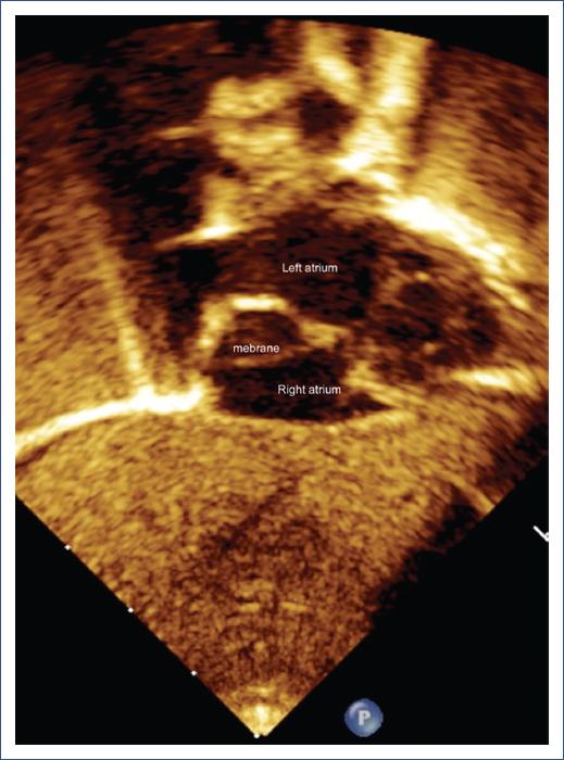
Figure 1 Echocardiography, subcostal view. The arrow shows the membrane (Cor triatriatum dexter) inside the right atrium. 

Cardiac magnetic resonance imaging (MRI) revealed a complete CTD with an ASD and concordant atrioventricular and ventriculoarterial connections (Fig. 2).

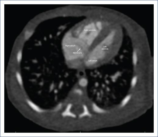
Figure 2 Cardiac magnetic resonance imaging, sagittal plane, showing a membrane (white arrow) in the right atrium. 

Cardiac surgery confirmed the pre-operative diagnosis of complete CTD by the presence of a membrane that separated the RA into two cavities. The membrane was removed, and the patent foramen ovale was closed.

Follow-up and outcomes

After surgery, the patient showed clinical improvement with an increase in saturation to 98% without oxygen supplementation. The patient was discharged 6 days after the procedure with no complications.

Discussion

CTD is a rare CHD characterized by the presence of a membrane that separates the RA into two cavities. This membrane represents the persistence of the venous sinus, causing alterations in venous flow as it passes through the RA, potentially causing a shunt from right to left through defects in the interatrial septum and manifesting clinically with desaturation and cyanosis3,5.

CTD has varying clinical manifestations, depending on the degree of septation of the RA. The symptoms could range from desaturation with mild cyanosis, as in our case, to arrhythmias (supraventricular tachycardias), right-sided heart failure, or obstruction of the tricuspid valve or the right ventricle outflow tract. It is important to alert the pediatrician to immediately request a cardiology evaluation to rule out other severe structural heart diseases such as transposition of the great arteries, pulmonary atresia, or anomalous connection of pulmonary veins2,6,7.

The presence of cyanosis was explained by the ASD, which allowed a right-to-left shunt. As part of the pre-surgical approach, we decided to perform a cardiac MRI to assess the membrane and pulmonary vein anatomy, as recommended in some medical literature8,9.

During the surgical procedure, the membrane was identified, resected, and the foramen ovale closed. The patient’s post-operative course was very good, with a complete resolution of symptoms.

Conclusion

The importance of our case lies in the early identification of rare heart disease as a cause of cyanosis and desaturation in a neonate in the first days of life who did not present signs of heart failure. We recommend that all primary care physicians and pediatricians request a cardiology consultation when detecting any clinical signs of CHD, such as cyanosis, desaturation, tachypnea, increased respiratory effort, murmurs, or hepatomegaly. A simple test, like an echocardiogram, allows for timely diagnosis and treatment of CHD, preventing future complications and increased mortality.

References

1. Talner CN. Report of the New England regional infant cardiac program, by Donald C. Fyler, MD, Pediatrics, 1980;65(suppl):375- 461. Pediatrics. 1998;102:258-9. [ Links ]

2. Hansing CE, Young WP, Rowe GG. Cor triatriatum dexter. Persistent right sinus venosus valve. Am J Cardiol. 1972;30:559-64. [ Links ]

3. Yarrabolu TR, Simpson L, Virani SS, Arora H, Navarijo J, Stainback RF. Cor triatriatum dexter. Tex Heart Inst J. 2007;34:383-5. [ Links ]

4. Mackman CA, Liedel JL, Woods RK, Samyn MM. A case series of patients with cor triatriatum dexter:unique cause of neonatal cyanosis. Pediatr Cardiol. 2015;36:240-3. [ Links ]

5. Battle-Diaz J, Stanley P, Kratz C, Fouron JC, Guerin R, Davignon A. Echocardiographic manifestations of persistent right sinus venosus valve. Am J Cardiol. 1979;124:850-3. [ Links ]

6. Embrey RP. Cor triatriatum, pulmonary vein stenosis, atresia of the common pulmonary vein. In:Mavroudis C, Backer CL, editors. Pediatric Cardiac Surgery. 2nd ed. St Louis, Mo:Mosby-Yearbook;1994. 503-4. [ Links ]

7. Mazzucco A, Bortolotti U, Gallucci V, Del Torso S, Pellegrino P. Successful repair of symptomatic cor triatriatum dexter in infancy. J Thorac Cardiovasc Surg. 1983;85:140-3. [ Links ]

8. Joe BN, Poustchi-Amin M, Woodard PK. Case 56:cor triatriatum dexter. Radiology. 2003;226:701-5. [ Links ]

9. Uvera SG, Guzmán MM, Lamadrid JV. Cor triatriatum dexter:uso de resonancia magne?tica cardiovascular. Case report. Rev Mex Cardiol. 2012;23:12-6. [ Links ]

FundingThe authors declare that they have not received funding.

Ethical disclosures

Protection of human and animal subjects. The authors declare that no experiments were performed on humans or animals for this study.

Confidentiality of data. The authors declare that they have followed the protocols of their work center on the publication of patient data.

Right to privacy and informed consent. The authors have obtained the written informed consent of the patients or subjects mentioned in the article. The corresponding author has this document.

Use of artificial intelligence for generating text. The authors declare that they have not used any type of generative artificial intelligence for the writing of this manuscript nor the creation of images, graphics, tables, or their corresponding captions.

Received: January 24, 2024; Accepted: April 28, 2024

* Correspondence: Rodrigo Hernández-Benítez E-mail: roy210983@gmail.com

Conflicts of interest

The authors declare no conflicts of interest.

Creative Commons License Instituto Nacional de Cardiología Ignacio Chávez. Published by Permanyer. This is an open ccess article under the CC BY-NC-ND license