




 nueva página del texto (beta)
 Inglés (pdf)
 Artículo en XML
 Referencias del artículo
 Traducción automática
 Enviar artículo por email
 Citado por SciELO
  Similares en
SciELO
Similares en
SciELO 
 Permalink
Introduction
Pulmonary embolism (PE) ranks among the most common cardiovascular emergencies and remains a leading cause of morbidity and mortality1. It constitutes a heterogeneous group of clinical presentations, ranging from incidental findings to high-risk PE with shock. The array of therapeutic options has expanded over the last decade, especially catheter-directed therapies2. However, despite these developments, intermediate- and high-risk PE mortality remains high3-6 and robust evidence for comparing each strategy with current standardized guidelines is lacking. The absence of a unified treatment framework for PE and the multispecialty nature of the venous thromboembolism (VTE) spectrum provide a strong rationale for developing a collaborative interdisciplinary team7. A PE response team (PERT) offers a centralized, unique activation process to provide rapid multimodality assessment and risk stratification, formulate an individualized diagnostic and therapeutic approach, and facilitate the implementation of the recommended strategy within a prespecified timeframe in patients with acute PE8.
The Massachusetts General Hospital (MGH) established the first successful rapid-response PERT in 20129. Since then, hospitals worldwide have adopted PERTs to standardize the management of acute PE through a multidisciplinary, multispecialty team of experts. Emerging data highlight the value of PERTs in improving outcomes by changing the care paradigm for acute PE8. In Mexico, we implemented the first PERT at Hospital Zambrano Hellion in 2019 with the VTE Rapid Response Team (PREVENTION) to improve the identification and management of VTE, including deep vein thrombosis (DVT) and PE10. The only other published PERT program in Mexico was launched in 2021 by the General Hospital of Mexico City11, which has cared for 42 patients up until August 202412.
Based on new evidence, PE phenotypes, risk scores, systemic thrombolysis (ST), catheter-directed therapies, extracorporeal membrane oxygenation (ECMO) use, and the need to generate a network with the shock team, we enhanced our previous response team and developed the PERT-PREVENTION protocol with key improvements to further improve the quality of PE patient care, including intermediate-risk patients for whom optimal treatment remains challenging.
Materials and methods
Structure and clinical function of PERT-PREVENTION
The primary objective of PERT-PREVENTION is to provide stratification and diagnostics of PE and standardise PE door-to-management timeframe and therapeutic strategies at the Hospital Zambrano Hellion. PERT-PREVENTION will ensure a fast-track program to initiate specific treatment between 60 and 90 min after code activation, reproducing ST-elevation acute myocardial infarction (MI) and stroke reperfusion programs predominantly for intermediate- and high-risk PE patients. Secondary objectives include (1) increased in-hospital identification of low-risk PE; (2) exploration into the cause of PE by ensuring age-specific cancer-related screening, thrombophilia testing in patients < 40 years with weak triggers, thrombus in unusual sites, or strong family history13; (3) long-term anticoagulation management: election and length of anticoagulation, adherence, bleeding complications, and management; (4) to identify those with a high-risk profile for chronic thromboembolic disease, post-PE syndrome, and chronic thromboembolic pulmonary hypertension; (5) to implement a prospective registry about PE cases at our institution; (6) to provide patient education to improve adherence and reduce recurrence and bleeding complications. According to the PERT MGH stratification9, Hospital Zambrano Hellion has level 1 PERT, signifying that we have the necessary resources to lead a successful program.
Organization
The PERT-PREVENTION structure and organization includes physicians trained in clinical cardiology, vascular surgery, echocardiography, cardiovascular imaging, interventional cardiology, nurses, technicians, cardiology fellows, and students. Effective coordination and communication are mandatory for a successful program. The team must be easily accessible and provide a consistent, rapid, and effective multidisciplinary response in the emergency room, intensive critical care unit, and the in-hospital setting. PERT-PREVENTION is designed to improve PE patient care within our institution. This involves all Zambrano Hellion Hospital’s institutes (cardiology, internal medicine, traumatology and orthopedics, gynecology and obstetrics, neurology, surgery, etc.), strengthening the in-hospital network and allowing for the transfer of care to the PERT-PREVENTION team. If a critically ill PE patient is transferred from an outside institution to Hospital Zambrano Hellion, the emergency department will activate PERT-PREVENTION and the shock team swiftly for the approach and management of the patient.
Activation and execution
The activation of the response team begins with identifying a high clinical suspicion for PE (Table 1). This phenotype comprises common signs and symptoms, including sudden dyspnea at rest or on exertion, pre-syncope or syncope, ischemic chest pain, respiratory distress, hypoxemia, and hypotension14; and relevant patient history information, including active cancer, recent major surgery or infection, active DVT, and pro-inflammatory cardiovascular risk factors. Hospital staff must know the VTE risk factors and how to identify them. Before launching the hospital code, we will conduct educational programs, round table discussions, and case simulations for hospital physicians, nurses, technicians, residents, and students. An activation line will be available 24 h a day, 7 days a week, 365 days a year. The cardiology fellow on call will be responsible for protocol activation, immediate patient evaluation, and obtaining appropriate imaging and laboratory studies. We will complete table 2 by entering information in each box: S signs and symptoms, H history, I image, E electrocardiogram (ECG), L laboratory, D demographics, and B bleeding risk to provide the team with the necessary information to establish a clinical suspicion, diagnosis, bleeding risk, and decision-making (Table 2).
Table 1 Key steps, events, and personnel in the execution of PERT-PREVENTION
| Phase | Key events | Key members |
|---|---|---|
| Pre-activation and activation | PE detection and/or suspicion by referring | Referring to MD |
| MD or a member of the team | Hospital residents | |
| Call placed to the PREVENTION-team line | Nurses | |
| Medical students | ||
| Initial actions | On-call cardiology fellow: | On-call cardiology fellow |
| Calls back referring MD | Referring MD | |
| Gathers case history | ||
| Notifies PREVENTION-Team members of the event and plans an online meeting | ||
| Response | Online meeting | On-call cardiology fellow |
| Case presentation by the on-call fellow with imaging and laboratory results | Referring MD | |
| Consensus treatment | PREVENTION-Team | |
| Treatment recommendation is given to the primary healthcare team in written form | ||
| Transfer | Transfer the patient to the necessary department (ICU, OR, catheterization laboratory) | On-call fellow |
| Nurses and hospital staff | ||
| Execution | Perform the planned treatment | Catheterization laboratory personnel |
| OR personnel | ||
| PREVENTION-Team |
PE: pulmonary embolism; ICU: intensive care unit; OR: operating room.
Table 2 S2HIELDB: Information collected after PERT-PREVENTION activation
| Date: | Activation/initial evaluation time: | Age: | Sex: male/female |
|---|---|---|---|
| Allergies: | Days in the hospital: | Days symptomatic in the hospital/at home: | |
| Signs and symptoms | Patient history | ||
| Signs | Assess PE | VTE: yes/no | Minor surgery recent: |
| Systolic blood pressure: | Dyspnea: yes/no | Obesity: yes/no | yes/no |
| Heart rate: | Ischemic-like chest pain: yes/no | Recent infection: yes/no | Prolonged bed rest/trip: |
| Respiratory rate: | Near or syncope: yes/no | Puerperium recent: yes/no | yes/no |
| O2 saturation: | Respiratory distress: yes/no | Pregnancy: yes/no | Estrogen/OCP use: yes/no |
| Assess DVT | Cardiac arrest: yes/no | Major surgery recent: yes/no | Known active cancer: yes/no |
| Lower limb pain: yes/no | Assess paradoxical embolism | ||
| Swelling: yes/no | Headache: yes/no | ||
| Erythema: yes/no | Back pain: yes/no | ||
| Homans sign: yes/no | Abdominal pain: yes/no | ||
| Ollow sign: yes/no | Paraesthesia: yes/no | ||
| Imaging | |||
| Chest X-ray | Echocardiogram | CT angiogram | Lower limb Doppler US |
| Westermark sign: yes/no | RV: LV > 1: yes/no | Size: | Thrombus: yes/No |
| PA amputation: yes/no | RV hypokinesis: yes/no | Location: | Location: distal/Proximal |
| McConnell’s sign: yes/no | Burden thrombus: | Burden thrombus: floating thrombus: yes/no | |
| TAPSE: | Saddle PE: | ||
| Thrombus-in-transit: yes/no | Pulmonary angiography | ||
| LVOT VTI: | Obstruction site: | ||
| Congested IVC: yes/no | |||
| RVOT mid-systolic notch: | |||
| yes/no | |||
| Paradoxical septal motion: | |||
| yes/no | |||
| 60/60 sign: yes/no | |||
| PASP: | |||
| ECG | Laboratory | ||
| Tachycardia: yes/no | aVR ST elevation: yes/no | Hemoglobin: | High-sensitivity cardiac troponin |
| Atrial fibrillation or flutter: yes/no | V1 qR: yes/no | Platelets: | I: |
| RBBB: yes/no | V1 ST elevation: yes/no | D-dimer: | eGFR: |
| S1Q3T3: yes/no | RV overload: yes/no | BNP: | Lactate: |
| V1-V3 ST dynamic changes: yes/no | NT-proBNP: | ||
| Bleeding risk | |||
| RIETE | Thrombocytopenia: yes/no | Female: yes/no | eGFR < 30 mL: yes/no |
| Recent major bleeding: yes/no | History of stroke: yes/no | BMI < 24 kg/m2: yes/no | Liver/kidney disease: yes/no |
| Creatinine > 1.2 mg/dL: yes/no | Recent major surgery: yes/no | Weight < 50-60 kg: yes/no | Bleeding predisposition: yes/no |
| Anemia: yes/no | Uncontrolled hypertension: | INR > 2.5: yes/no | Alcohol abuse: yes/no |
| Malignancy history: yes/no | yes/no | OAC: yes/no | |
| Clinically overt PE: yes/no | |||
| Age > 75 years: yes/no | |||
| RIETE score: | |||
| PE risk stratification: very low/low/intermediate/high | |||
| Bova score: | |||
DVT: deep venous thrombosis; PE: pulmonary embolism; VTE: venous thromboembolism; OCP: oral contraceptive pills; RV: right ventricle; LV: left ventricle; TAPSE: tricuspid annular plane systolic excursion; LVOT VTI: left ventricular outflow tract velocity time index; PA: pulmonary artery; RV: right ventricle; LV: left ventricle; RBBB: right bundle branch block; BNP: B-type natriuretic peptide; NT-proBNP: N-terminal pro-brain natriuretic peptide; PVD: peripheral vascular disease; MI: myocardial infarction; eGFR: estimated glomerular filtration rate; BMI: body mass index; INR: international normalised ratio; OAC: oral anticoagulation.
Transthoracic echocardiography has emerged as the most practical diagnostic tool for patients with a high clinical suspicion of PE, with or without clinical stability, by detecting signs of right ventricular dysfunction (Supplementary table 1)2,15-24. It represents a crucial instrument for classifying cases by assessing the right ventricular systolic performance in hemodynamically stable patients. When evaluating a patient with PE, the echocardiographer must concentrate on two main objectives. The first is to detect findings that confirm or suggest the diagnosis (e.g., thrombus-in-transit, McConnell’s sign). The second is to assess indicators of right ventricular dysfunction (e.g., tricuspid annular plane systolic excursion, low left ventricular outflow tract velocity index), which could lead to a reclassification of the disease. Transesophageal echocardiography or transthoracic echocardiogram with peripheral intravenous agitated saline bubbles25 will screen for patent foramen oval (PFO) in patients with intermediate- or high-risk PE considered for ST26. Clinicians must rule out PFO in patients undergoing percutaneous thrombectomy to prevent paradoxical embolization during thrombus extraction. The PERT-PREVENTION team must also investigate signs and symptoms suggesting central or systemic embolism in the clinical evaluation of patients with PE. Cerebral magnetic resonance imaging may identify subclinical ischemic strokes related to intracardiac shunting. Hemorrhagic transformation of subclinical ischemic stroke could explain unexpected intracranial hemorrhages following anticoagulation or advanced treatment in PE patients25.
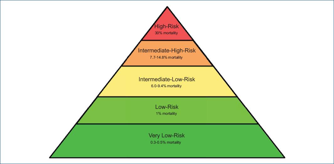
Risk stratification will be driven by the clinical presentation, echocardiogram, and cardiac biomarker findings and will be classified as very low-, low-, intermediate-, and high-risk PE (Fig. 1)3-6,27. Imaging techniques, namely computed tomography angiography (CTA), will prove the final diagnosis. We will perform ECG-synchronized CTA for a triple rule-out of PE, obstructive coronary artery disease, and aortic dissection in cases with diagnostic doubt. Pulmonary angiography can also provide a definitive PE diagnosis in cases of hemodynamic instability in the catheter laboratory. The RIETE score28 will be used to assess and stratify hemorrhage risk during PE treatment. We will also consider other variables associated with a higher bleeding risk.
After protocol activation, the on-call cardiology fellow will contact the PERT-PREVENTION team through an immediate electronic message. The team will communicate through a group chat on a secure messaging platform in < 30 min. At the same time, the fellow will assess and obtain diagnostic studies to confirm the PE diagnosis, quantify the thrombus burden, and evaluate the severity of right ventricular dysfunction. The team will provide a treatment suggestion for intermediate- and high-risk PE patients within 60-90 min. We considered this timeframe to begin management based on (1) our previous experience26,29-34, in which we perform stratification, diagnosis, and ST in the first 90 min after PE patients arrive in the emergency room; (2) the time-dependency of thrombus resistance35, right ventricular ischemia, and MI31; and (3) evidence from mechanical and pharmacological reperfusion in ST-elevation acute MI and ischemic stroke programs24,36-38. In selected cases, we will activate the cardiac catheter laboratory and transesophageal echocardiography units. If the patient has a confirmed DVT, the team will contact a vascular surgeon within 24 h of protocol activation.
Therapeutic approach: anticoagulation
The foundation of PE treatment is anticoagulation, with advanced treatment options considered for patients with impending or clinically unstable PE. Figure 2 shows the clinical decision-making for anticoagulation alone versus advanced treatment in the different phenotypes of PE based on the severity of right ventricular dysfunction. Table 3 presents the recommended anticoagulation therapy in PE treatment’s acute, long-term, and extended phases2,19,24. Long-term anticoagulation is warranted in cases of unprovoked PE, recurrence, active cancer, a proven or strong suspicion of thrombophilia, and a persistently abnormal D-dimer. Direct oral anticoagulants (DOACs) have demonstrated effectiveness and a safer profile than Vitamin K antagonists. Anticoagulation alone is the recommended therapeutic option in very low- and low-risk PE patients (clinical stability, no biomarker expression, without or mild right ventricular dysfunction, and moderate thrombus burden). The treating physician will tailor the anticoagulation type, administration route, and dosing regimen based on clinical judgment and patient needs. We advise enoxaparin in very low- and low-risk PE patients without an intravenous bolus. We will use a dose reduction for elderly patients24. Loading doses of apixaban and rivaroxaban also provide effective and safer management in very low- and low-risk PE patients. In intermediate-risk PE, we advise initial anticoagulation with enoxaparin. However, weight-adjusted unfractionated heparin can be elected over enoxaparin for the first 24-48 h to avoid heparin crossover if clinical status warrants advanced treatment. Patients with high-risk PE will be started on weight-adjusted unfractionated heparin as an adjunctive treatment to advanced treatment with active monitoring of activated partial thromboplastin time. If heparin treatment fails to establish adequate anticoagulation, patients may be switched to enoxaparin, rivaroxaban, or apixaban in-hospital. The regimen of unfractionated heparin as an adjunctive treatment, followed by a switch to enoxaparin, proved effective and safe for Mexican PE patients undergoing ST without intracranial hemorrhage26. Switching to DOACs after 24 or 48 h of unfractionated heparin proved effective and safe for patients who successfully underwent ST39,40. Overall, unfractionated heparin is suitable as an adjunctive treatment in patients with severe renal disease, high-risk bleeding, age > 75 years, hypotension, and impending clinical instability, and as an adjunctive treatment24. In the extended phase, we advise DOACs41,42, while enoxaparin is indicated for patients with active cancer.
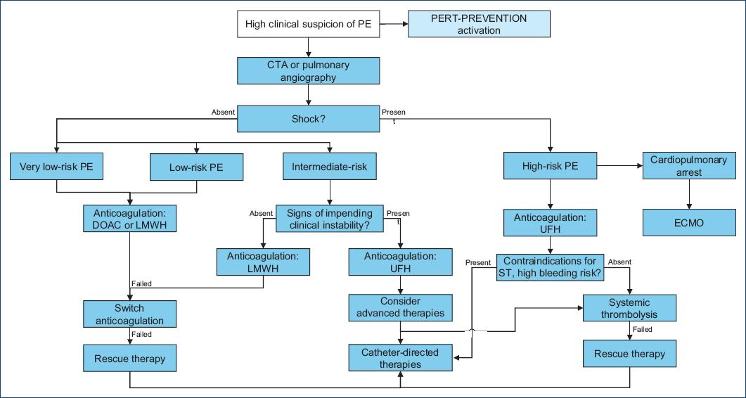
Figure 2 PERT-PREVENTION protocol. PE: pulmonary embolism; CTA: computed tomography angiography; DOAC: direct oral anticoagulant; LMWH: low molecular weight heparin; UFH: unfractionated heparin; ECMO: extracorporeal membrane oxygenation; ST: systemic thrombolysis.
Table 3 Anticoagulation and advanced treatment for pulmonary embolism
| Very low-risk PE | Anticoagulation |
| Low-molecular-weight heparin | |
| Enoxaparin by subcutaneous injection (1 mg/kg BID or 1.5 mg/kg once daily); in patients > 75 years, 0.75 mg/kg BID | |
| DOACs | |
| Apixaban: 10 mg twice daily for 7 days, followed by 5 mg twice daily | |
| Rivaroxaban: 15 mg twice daily for 3 weeks, followed by 20 mg daily | |
| Low-risk PE | Anticoagulation |
| Low-molecular-weight heparin | |
| Enoxaparin by subcutaneous injection (1 mg/kg BID or 1.5 mg/kg once); in patients > 75 years, 0.75 mg/kg BID | |
| DOACs | |
| Apixaban: 10 mg twice daily for 7 days, followed by 5 mg twice daily | |
| Rivaroxaban: 15 mg twice daily for 3 weeks, followed by 20 mg daily | |
| Intermediate-risk PE without impending clinical instability | Anticoagulation |
| Low-molecular-weight heparin | |
| Enoxaparin intravenous bolus 30 mg followed by subcutaneous injection (1 mg/kg BID or 1.5 mg/kg ONCE); in patients > 75 years, no bolus and 0.75 mg/kg BID | |
| Advanced treatment: consider in case of impending clinical instability | |
| High-risk PE or intermediate-risk PE with impending clinical instability | Adjunctive treatment |
| Weight-adjusted unfractionated heparin | |
| 60 U/kg bolus (maximum 4000 U) followed by 12 U/kg infusion (maximum 1000 U)/24 h or 48 h, followed by enoxaparin 1 mg/kg BID or 1.5 mg/kg ONCE/5 days or apixaban or rivaroxaban | |
| Advanced treatment | |
| Sistemic thrombolysis | |
| 25 mg of alteplase in 1-2 h in > 75 years | |
| 50 mg of alteplase in 1-2 h in > 60 years | |
| 100 mg of alteplase in 1-2 h in < 60 years | |
| Weight-adjusted tenecteplase bolus in < 60 years: 30 mg<60 kg, 35 mg 60-70 kg, 40 mg 70-80 kg, 45 mg 80-90 kg, 50 mg > 90 kg | |
| Catheter-directed thrombolysis | |
| 30 ± 10 mg of alteplase | |
| Pharmaco-invasive approach | |
| Thrombus fragmentation with pigtail catheter, 20 mg alteplase infusion in the pulmonary artery, and manual aspiration | |
| Catheter-directed thrombectomy | |
| Thrombus fragmentation and aspiration with FlowTriever® System | |
| Long-term anticoagulation (3-6 months) and extended treatment (> 6 months) | Vitamin K antagonists |
| Warfarin 5 mg daily, overlapped with heparin for the first 5 days until two consecutive INR in therapeutic ranges (2-3) and then dose-adjusted to maintain INR 2-3 | |
| Low-molecular-weight heparin | |
| In patients with active cancer: subcutaneous injection of 40 mg once daily | |
| DOACs | |
| Apixaban: 5 mg or 2.5 mg BID | |
| Rivaroxaban: 20 mg or 15 mg once daily |
PE: pulmonary embolism; DOAC: direct oral anticoagulant; INR: international normalized ratio.
Advanced treatment: PE thrombolysis
International and national guidelines recommend ST and adjunctive treatment with unfractionated heparin in high-risk patients with PE (Table 3)2,19,24,43,44. We will use weight-adjusted unfractionated heparin and ST with 1- or 2-h 100 mg alteplase infusion or tenecteplase in a bolus according to weight over 5-10 s in well-selected high-risk PE patients < 60 years. We prefer 50 mg short-term alteplase infusion for patients over 60 years of age and 25 mg short-term alteplase infusion for patients over 75 years45, considering the higher incidence of intracranial hemorrhage in this population, especially in female patients46. Half-dose short-term alteplase has no evidence of intracranial hemorrhage in the elderly population. We recommend avoiding unnecessary venous and arterial punctures to reduce bleeding complications.
ST in selected intermediate-risk PE patients remains controversial. The PEITHO study showed improved in-hospital outcomes with systematic thrombolysis in this group, although at the expense of an increased risk of major bleeding and stroke29,30,32,33,47. We consider advanced treatment in intermediate-risk PE patients with impending clinical instability as a stage II or III in the Bova score48. The other variables considered are shown in Table 4 based on historically associated factors of impending clinical deterioration. The multidisciplinary team will collaborate to reach a consensus and determine an individualized therapeutic strategy in these cases.
Table 4 Impending clinical instability risk factors in intermediate-risk pulmonary embolism
| Clinical parameters | Oxygen desaturation < 90% |
|---|---|
| Tachycardia > 100 bpm | |
| Respiratory rate > 20 bpm | |
| Systolic blood pressure < 110 mmHg | |
| Comorbidities: chronic heart failure, active neoplasm, active DVT | |
| Laboratory parameters | Lactate > 2 mmoL/L |
| High-sensibility cardiac troponin I > 99th percentile URL | |
| NT-proBNP > 600 pg/mL | |
| BNP > 300 pg/mL | |
| Echocardiographic parameters | TAPSE ≤ 16 mm |
| RV: LV > 1 | |
| Severe global right ventricular hypokinesis | |
| McConnell’s sign | |
| Left ventricular outflow tract velocity-time index ≤ 15 cm | |
| Thrombus-in-transit | |
| Congested IVC | |
| TAPSE/PASP < 0.4 | |
| Electrocardiographic parameters | Advanced right bundle branch block |
| ST-dynamic changes in V1-V3 | |
| ST-segment elevation in aVR | |
| qR in V1 | |
| ST-elevation in V1 | |
| S1Q3T3 | |
| New-onset atrial fibrillation | |
| CT Angiogram parameters | Saddle PE High proximal thrombus burden |
| RV: LV > 1 |
DVT: deep vein thrombosis; URL: upper reference limit; NT-proBNP: N-terminal pro-brain natriuretic peptide; TAPSE: tricuspid annular plane systolic excursion; RV: right ventricle; LV: left ventricle; IVC: inferior vena cava; PASP: pulmonary artery systolic pressure; CT: computed tomography, PE: pulmonary embolism.
Advanced therapy: catheter-directed therapy
We recommend alternative treatment options for patients with contraindications for ST2 (Table 5) or those at intermediate and high bleeding risk24,43 (table 6). Catheter-directed thrombolysis is an adequate option for patients with ST contraindications or persistent hypotension. Based on the evidence, percutaneous thrombectomy is an alternative for patients with major contraindications to ST, high bleeding risk, or unsuccessful systemic or catheter-directed thrombolysis. Both catheter-based therapies may also be used as rescue therapy for patients with hemodynamic deterioration while on anticoagulation and electively for intermediate-risk PE with several impending clinical instability risk factors45,46. In a failed primary treatment, the procedure will be performed 2-4 h after the completion of ST. Percutaneous thrombectomy can be electively proposed in intermediate-risk PE patients with multiple risk factors for impending clinical instability and high thrombus burden in pulmonary CTA, intermediate or high bleeding risk, contraindications of ST, or no clinical, echocardiographic, or biomarker improvement after optimal anticoagulant therapy during 48 h.
Table 5 Contraindications for thrombolysis
| Absolute | History of hemorrhagic stroke or stroke of unknown origin |
|---|---|
| Ischemic stroke in the previous 6 months | |
| Central nervous system neoplasm | |
| Major trauma, surgery, or head injury in the past 3 weeks | |
| Bleeding diathesis | |
| Active bleeding | |
| Relative | Transient ischemic attack in the past 6 months Oral anticoagulation |
| Pregnancy or the first post-partum week | |
| Non-compressible puncture sites | |
| Traumatic resuscitation | |
| Refractory hypertension (systolic blood pressure > 180 mmHg) | |
| Advanced liver disease | |
| Infective endocarditis | |
| Active peptic ulcer |
Table 6 Therapeutic alternatives in patients with high-bleeding risk or ST absolute contraindications
| Percutaneous | Pharmaco-invasive approach |
|---|---|
| Low-dose catheter-directed thrombolysis (alteplase 20-40 mg) | |
| Catheter-directed thrombectomy | |
| Surgical | Surgical embolectomy |
| Other | Vena cava filter |
Percutaneous thrombectomy will be performed in consensus with the PERT team for a trained interventional cardiologist or vascular surgeon. The preferred device for this procedure is the FlowTreiver (INARI), which consolidates expertise with a simple device based on the clinical evidence published about safety and effectiveness (FLASH, FLARE, and FLAME)6,49,50. Secondarily, Indigo-Penumbra can be an option when the primary choice is not immediately available. We will prioritize femoral vein access unless a thrombus obstructs the proximal deep venous system. The jugular vein can serve as an alternative access. Suture closure device systems will be the standard hemostatic strategy in all cases.
Catheter-directed thrombolysis, catheter fragmentation, and manual aspiration have shown efficacy and safety in the Mexican population51. Given the outcomes of standard catheter-directed thrombolysis compared with EKO-assisted catheter-directed thrombolysis48,49, the standard approach will serve as the primary option for advanced percutaneous treatment. Finally, we recommend temporary inferior vena cava filters in patients with absolute contraindications for anticoagulation and thrombolysis and an active DVT, and consider them for selected patients undergoing peripheral thrombectomy24.
Advanced therapy: mechanical circulatory support
ECMO serves as a critical supportive therapy for massive PE, particularly in high-risk patients with hemodynamic instability, impending cardiogenic shock, or cardiac arrest52. Veno-arterial (VA)-ECMO is often used as a bridge to more definitive reperfusion therapies. It can be rapidly deployed to stabilise patients with refractory shock or impending cardiopulmonary arrest, providing time for thrombus resolution through pharmacological or mechanical means53-55. The use of ECMO has been associated with improved hemodynamic stabilization and survival outcomes, mainly when implemented before cardiac arrest occurs52,54-57. However, ECMO is not without risks. It is associated with complications such as bleeding, coagulopathy, and systemic inflammatory response, which must be carefully managed58,59. The decision to use ECMO must be based on a comprehensive assessment of the patient’s condition, including the severity of right ventricular dysfunction and contraindications to thrombolysis55,60. VA-ECMO as a support therapy is most effective when used in conjunction with other reperfusion strategies rather than as a standalone treatment52-60.
Interhospital transfer of critically ill PE patients
For the transfer of patients to our level 1 PERT institution, we propose the following plan. The local hospital or medical center will stabilize the patient and establish the diagnosis of PE, with the appropriate risk stratification. Anticoagulation therapy must be started upon PE diagnosis, regardless of transfer status. For patients with high-risk or intermediate-risk PE with impending clinical instability, anticoagulation should preferably be given with unfractionated heparin, and the local hospital will activate PERT-PREVENTION remotely with the emergency room department at Hospital Zambrano Hellion. Patients will be considered for transfer if they are eligible for advanced PE therapy. The patient will be transferred to our institution by a qualified medic or paramedic with critical care capabilities, limiting patient movement, continuous vital sign monitoring, a diversion plan in case of clinical worsening, closed-loop communication with the receiving centre, and the patient’s medical record61. Upon arrival, the PERT-PREVENTION will receive the patient and stabilize them, if necessary, followed by repeated risk stratification and considering advanced therapies, as per the standard protocol.
Follow-up
The team will follow up on the clinical condition, treatment response, and in-hospital complications to consistently adjust and improve patient care. D-dimer, high-sensibility cardiac troponin (hs-cTn) I, and brain natriuretic peptide (BNP), or N-terminal pro-BNP measurements will be repeated daily during the first 5-7 days of hospitalization. All patient information, including ECG, chest X-ray, cardiac biomarkers, diagnostic studies, therapeutic approach, and complications, will be captured in an online collaborative database with controlled access. Upon discharge, patients will continue to follow up with their cardiologist in the outpatient clinic. The team will repeat an echocardiogram and CTA after 3-6 months of anticoagulation to screen for post-PE syndrome, including chronic thromboembolic pulmonary disease with and without pulmonary hypertension62.
Research and Educational Activities of PERT-PREVENTION
The core team members will be the steering committee for all PERT-PREVENTION team strategies. The secondary objectives include leading research protocols, providing patient education, and expanding our network. The collaborative online database will store patient information for future research and analysis. Primary and secondary prevention to reduce the incidence and recurrence of PE is mandatory. In-hospital strategies, such as thrombosis risk stratification and pharmacologic and non-pharmacologic primary prevention, are currently used to reduce thrombotic events. However, these strategies are lacking in patients at home. As such, patient and family education stimulates early VTE recognition, identifies trigger factors, and allows the implementation of secondary non-pharmacologic prevention, aiming to extend the concept of a thrombosis-free hospital to a thrombosis-free home. Finally, network expansion will allow us to implement our system in other clinical settings, identify areas of opportunity, and improve awareness of PE.
The team will hold regular meetings to review protocol activations, assess the team’s response, troubleshoot and address any emerging system problems, and establish connections and make the PERT-PREVENTION protocol known to other institutes within our hospital. Academic activities, such as clinical presentations and discussions, teaching sessions, and case simulations, will be essential to maintain the program’s quality.
Discussion
Recent evidence indicates that PE mortality rates have risen over the past decade despite technological advancements and the increasing implementation of multidisciplinary PERT care models. The National PERT Consortium Registry, established in 2012, aims to clarify current treatment patterns and outcomes across US centres that provide specialized, multidisciplinary care for patients with PE63. The registry collects data on mortality, treatment complications, major bleeding, and the rapidly evolving therapeutic options for acute PE. Findings from the registry reveal that high-risk PE patients experienced advanced treatment use in 41.9% of cases and a 20.6% mortality rate, compared with 30.2 treatment use and 3.7% mortality in intermediate-high-risk patients. Among high-risk patients experiencing cardiogenic shock or cardiac arrest, the in-hospital mortality rate reached 42.1%. Previous observational studies in similar populations reported between 30% and 50% short-term mortality rates. The registry findings also showed a lower overall mortality rate of 20.5% in all high-risk patients, likely reflecting the impact of PERT activation. In addition, high-risk PE patients more frequently received advanced treatment, including ST, surgical embolectomy, and mechanical circulatory support, compared with their intermediate-risk counterparts.
In contrast, a preliminary analysis of the PREVENTION code10 highlights an increase in PE identification, with 66 cases documented between 2019 and 2024, compared to 24 cases between 2011 and 2018. This analysis also identified a non-significant higher trend in the identification of intermediate-risk PE population (69.7 vs. 62.5%, p = 0.51) and the use of ST (28.79 vs. 20.83%, p = 0.45). However, there was a significant decrease in catheter-directed therapy (10.61 vs. 29.17%, p = 0.04). Mortality rates (10.61 vs. 8.33%, p = 0.75) and major bleeding events (9.09 vs. 4.17%, p = 0.67) showed no significant differences. Likewise, 10-year PERT analysis at the MGH64 revealed improved identification of intermediate-high-risk PE and increased use of catheter-directed therapies (1-14%) and all advanced treatment (9-19%), primarily among patients with intermediate-high-risk PE in the post-PERT cohort. The proportion of patients undergoing ST or surgical thrombectomy did not change before and after PERT implementation, and the 30-day mortality and major bleeding rates showed no significant differences.
PERTs guide each institution’s management of acute PE according to PE risk stratification. Various guidelines are available to stratify PE risk based on the patient’s clinical status. The American Heart Association guidelines categorise PE into three risk levels: massive, submassive, and low risk2. Meanwhile, the ESC guideline divides patients into high-risk, intermediate-high-risk, intermediate-low-risk, and low-risk. Stratification of PE patients using the ESC guidelines directly correlates with the associated mortality risk (Fig. 1). High-risk patients experienced an all-cause mortality of 30.2% in a study involving 782 patients4. The mortality rates for the intermediate-risk group range from 3 to 15%3,5,6. In the intermediate-high-risk group, reported rates range from 7.7 to 14.8%, whereas the intermediate-low-risk group shows rates between 6.0 and 9.6%65. Low-risk PE patients exhibit a mortality rate of approximately 1%. The protocol’s classification will include a new phenotype designated as very low-risk based on a meta-analysis that identified PE patients with normal right ventricular performance by echocardiogram, the absence of hs-cTn and BNP expression, and a mortality rate of 0.3-0.5%27.
Considering that the intermediate-high-risk PE population represents a heterogeneous group with an in-hospital mortality range of 3-15%, optimal treatment remains a significant challenge66. Improving the quality of care for this population will be an essential goal. Several factors linked to clinical deterioration and worse outcomes in patients with intermediate-risk PE may help identify those who could benefit from advanced treatment. Bova et al. developed a prognostic model for intermediate-risk PE based on clinical presentation, right ventricular dysfunction, and myocardial injury assessment in a meta-analysis of 2874 normotensive patients from six studies48. The model classifies patients into three stages to predict the risk of 30-day PE-related complications, including death from PE, hemodynamic collapse, and recurrent nonfatal PE (stage I: 4.4%, stage II: 18%, stage III: 42%). Now termed the Bova score, this model has been validated and assessed in a meta-analysis to confirm its generalizability to effectively discriminate normotensive PE patients with adverse short-term prognoses67-69. Under our PERT protocol, the team will discuss and consider advanced treatment for patients with intermediate-risk PE classified as stage II or III on the Bova score. Additional individual risk factors for developing hemodynamic deterioration and worse outcomes have been studied extensively in intermediate-risk patients. Factors directly associated with right ventricular overload, including cardiac biomarker expression and right ventricular dysfunction by imaging, are historically related to more severe PE and worse outcomes. They may independently aid in identifying intermediate-risk PE with a higher probability of worse early outcomes. Recently, low left ventricular outflow tract velocity index measured through echocardiography has also been associated with death, cardiac arrest, hemodynamic deterioration, or need for reperfusion among intermediate-risk PE patients70,71. Table 4 summarises relevant indicators of higher-risk PE and PE-related death and hemodynamic deterioration2,6,70-76 that we will consider under our PERT to support individualized decision-making for the need for advanced treatment.
PERTs need to assess the bleeding risk to consider in decision-making regarding the appropriate therapy, especially for advanced treatment candidates. Different tools evaluate this risk in patients with PE while on anticoagulation, such as RIETE and VTE-BLEED scores. A study performed from the RIETE registry identified six independent variables associated with the risk of major bleeding: recent major bleeding, creatinine levels > 1.2 mg/dL, anemia, cancer, clinically overt PE, and age > 75 years28. Each variable received a numerical value, and the total score categorized the risk as low (0 points), intermediate (1-4 points), or high (> 4 points). Unlike the VTE-BLEED score77, which estimates the risk of major or clinically relevant bleeding after day 30 of anticoagulation, the RIETE score evaluates the risk of bleeding starting from the day of diagnosis and initiation of anticoagulation therapy. The RIETE score, developed and validated through a multicentre prospective registry involving a robust and diverse population, focuses on the early bleeding risk associated with anticoagulant therapy and is considered the most suitable tool for assessing bleeding risk for this in-hospital response team. The PE-cerebral hemorrhage score was developed to specifically evaluate the risk of intracranial hemorrhage in patients with PE undergoing ST. Scores of 0, 1, 2, and ≥ 5 points are associated with intracranial hemorrhage risks of 1.2, 1.9, 2.4, and 17.8%, respectively78. However, this score has not been extensively validated.
According to the current guidelines, anticoagulation is the mainstay of acute, long-term, and extended PE treatment. Advanced treatment, including systemic thrombolytics, catheter-based approaches, surgical embolectomy, and mechanical circulatory support, is considered for patients with hemodynamic instability or worsening79. Percutaneous management options allow for rapid improvement of right ventricular function and potentially lower the risk of bleeding associated with ST. They may be directly undertaken in high-risk PE patients with absolute contraindications for ST or as rescue therapy in patients with clinical worsening. In severe cases, mechanical circulatory support may be used as a bridge to stabilize high-risk patients before definitive therapy can be undertaken.
Advanced treatment for intermediate-risk PE patients remains controversial. Guideline-directed therapy for this group comprises anticoagulation alone. However, multiple risk factors may identify impending clinical instability in selected patients who may benefit from advanced treatment, as discussed above. Results from the PEITHO study showed a reduction in the composite outcome of early death or hemodynamic decompensation in normotensive patients with intermediate-risk PE who underwent ST but an increase in the risk of major bleeding and stroke when compared with anticoagulation alone, with no difference in mortality. A subgroup analysis shows that those with significant respiratory failure had a better outcome when they received ST compared with anticoagulation alone, demonstrating a high-intermediate risk phenotype of patients who could benefit from advanced pharmacological treatment47. Other evidence has shown decreases in mortality with ST in this group80 and better outcomes with a reduced dose of alteplase46. Catheter-based therapies may especially favor patients with intermediate-risk PE and signs of impending clinical instability by improving right heart function with a lower risk of bleeding. Recently, the PEERLESS trial prospectively compared large-bore mechanical thrombectomy and catheter-directed thrombolysis in intermediate-risk PE patients with right ventricular dilation and clinical variables for impending clinical instability81. The mechanical thrombectomy strategy showed significantly fewer clinical deterioration and bailout episodes, and less post-procedural intensive care unit use and all-cause readmissions. There were no differences in mortality, intracranial hemorrhage, or major bleeding between the two strategies. Ultimately, the PERT-PREVENTION’s multidisciplinary team will collaborate to decide on an appropriate therapeutic strategy and whether to undergo advanced treatment for each individualized patient.
Conclusion
The PERT model has transformed the care of PE patients worldwide, improving identification, treatment, and overall standards. The PERT-PREVENTION protocol builds on these advancements through collegiate decision-making by enhancing the quality of care even further, including intermediate-risk patients for whom optimal treatment remains challenging. Close collaboration with the shock team allows faster and more effective decision-making for patients in the initial stages of cardiogenic shock. It also enables timely clinical decisions about the use of mechanical circulatory support for patients in the compensatory or progressive stages, optimizing outcomes and improving the efficiency of patient care. We are confident that the ongoing re-engineering of PERT-PREVENTION will enhance the care processes, ultimately elevating the quality of care delivered at our center.

