


 nueva página del texto (beta)
 Inglés (pdf)
 Artículo en XML
 Referencias del artículo
 Enviar artículo por email
 Citado por SciELO
  Similares en
SciELO
Similares en
SciELO 
 Permalink
INTRODUCTION
In recent years, technological advances have prompted a change in anatomy education from the traditional way of the last century. Due to the increase in the number of students in medical schools and changes in the anatomy curriculum, the time devoted to this subject has decreased. This situation has led to a lack of anatomy knowledge among new doctors[1]. Anatomy is the basis for clinical examinations, surgery and radiology, so medical and healthcare students must understand the key concepts, structures and their relationships[2].
The human brain is one of the most complex organs in human anatomy, so the subject of neuroanatomy is one of the most difficult and can sometimes discourage students[3]. Traditionally, the study of neuroanatomy is based on 2D images, however, this often does not adequately reflect the complex geometry and spatial arrangement of anatomical structures[4]. On the other hand, neuroanatomical examination using cadaveric dissection or fixed anatomical pieces is another method, and although the experience and realism provided by this option are unquestionable, it is an expensive resource with low availability, accessibility, and high costs for laboratory maintenance[5]. In this way, using neuroanatomical artificial physical models is another traditional study tool, but often with limited accuracy.
Recently, the advent of 3D technologies has improved the way of learning the structural and functional connectivity of the brain for neuroscience applications[6]. Nowadays, there are several virtual modeling tools able to reproduce realistic and easily explorable objects. Many works in literature confirm the evolution of study techniques from traditional resources to digital and 3D[7]. There is evidence that understanding of anatomy can be improved by using visual table images, virtual reality (VR) and augmented reality (AR)[8]. However, medical experts are still unaware of the many advanced 3D-MI visualization techniques that could enhance their ability to analyze data and help them make decisions about specific medical problems[9].
In neuroscience, one of the common challenges is to understand how the brain works and how to study brain disorders. To this end, Virtual Brain (TVB) is a neuroinformatics platform with a brain simulator that incorporates a range of neuronal models and dynamics[10]. In this sense,[11] a realistic virtual model of the human brain is proposed, which could be used in a neurosurgical simulation for both educational and preoperative planning purposes. To address the advantages of visualizing complex 3D anatomies for neuroscience educational and surgical purposes, several virtual reality (VR) and augmented reality (AR) solutions have been proposed[6][12][13][14][15][16][17][18]. However, although there are notable results with AR and VR systems, they have some limitations to overcome due to problems caused by VR headsets and motion sickness.
Although there is significant progress in neuroanatomy 3D visualization, the limitation in using commercially available 3D models remains their absence of a systematic approach to achieving scientific accuracy. In addition, complexity in the creation and interpretation of realistic models is another important element; not only technical skills are required, but also artistic creativity and expert mastery of medical knowledge and teaching[19]. On the other hand, traditional neuroanatomy curricula offer a limited approach to educating a variety of learning styles. Similarly, the fact that visualization technologies exist is not a sufficient consideration to use these resources in teaching, as a focused approach to educational intervention is required[8]. In addition to the previous challenges, in this work, scalability, reproducibility, and usability assessment issues are addressed[20].
Therefore, in this work, we aim to model and assess a 3D web-based interactive human brain using 3D design technologies, WebGL and the improved MEEDERV for neuroanatomy teaching purposes. The research question we have addressed is: How does the proposed methodology allow the design and evaluation of an interactive 3D web-based model for neuroanatomy teaching purposes? The main contribution of this work focuses on the addition of elements to MEDEERV, consisting of the integration of the usability testing stage and a feedback loop from that stage to the functional design stage with the purpose of re-engineering the model for continuous improvement. Finally, the novelty consists of using web-based technology for basic neuroanatomy teaching purposes, as a proof of concept to verify that the 3D model can be usefully implemented in teaching.
MATERIALS AND METHODS
The development of software projects depends on several factors such as the type of project, the size, the development environment and the available resources. Many software development methodologies (SDMs) have been introduced and these are easy to understand and implement[21]; however, in most of the methodologies, the detailed explanation for the elements to develop a virtual environment in a playful and interactive way is not provided. Although in[22], they present a methodology for designing virtual reality applications, they are based on the SDMs approach. For this reason, in this work, we improve the MEEDERV adding the usability testing stage and the feedback loop to the functional design stage as a cyclic process. Figure 1, shows each one of the stages of the improved version of the MEDEERV[23].
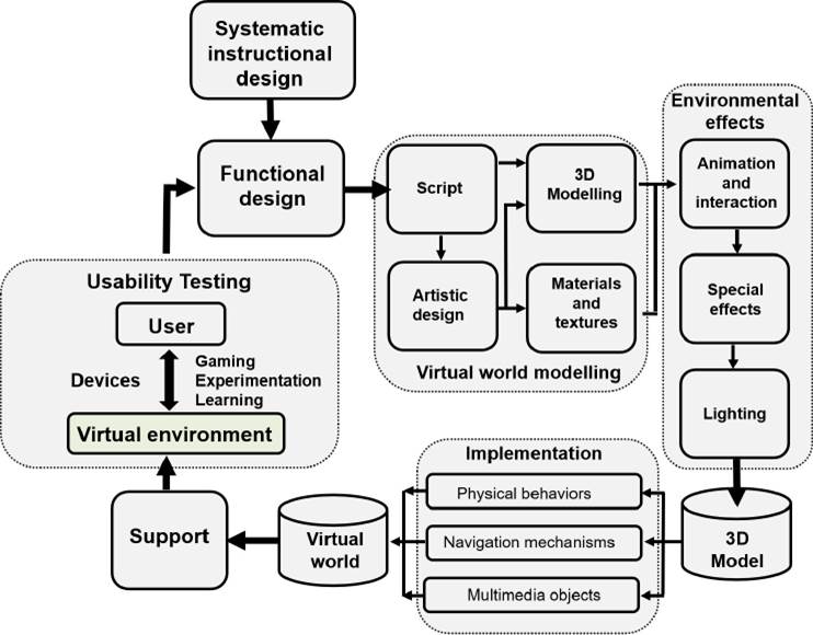
Figure 1 Methodology for the Development of Virtual Reality Educational Environments including usability and feedback. Image adapted from “Metodología para el modelado de sistemas de realidad virtual para el aprendizaje en dispositivos móviles” by Torres-Samperio et al.[23].
The methodology consists of seven main stages, the systematic design, the functional design, the virtual world modeling, the environmental effects, the implementation, the user-virtual environment, and the usability testing; as well as other secondary blocks. Each of these stages contains a series of phases that must be broken down to create an optimal virtual environment. These stages are described below.
Systematic instructional design
Instructional design is the process of creating effective, interactive and engaging learning plans focused on how people learn. In this stage, the competencies and skills that the student should develop depending on the learning area and the target population are identified. In this case, the expected result that undergraduate health science students will have to acquire through interaction with a 3D virtual model of the brain, is learning the basic neuroanatomical structures.
Functional design
In this phase, the requirements and actions that the system will take for its proper functioning are indicated, with the aim of making it intuitive and dynamic with a gamified approach. The aim is that the user can develop skills such as spatial visualization, memory and deductive reasoning by manipulating objects within the 3D brain virtual environment. To create the virtual scenario, the learning objectives that the students are intended to develop when browsing the application must be established. Two objectives that have been proposed with the subordinate skill are presented in Table 1.
Table 1 Objectives and design of functional skill.
| Design of functional skill | |
|---|---|
| Learning Objectives | Subordinate Skill |
| Learn the main structures of the human brain in a detailed and free form. | The user will develop memory to identify the composition of the organic groups of the complete brain system. |
| The user will be able to identify the position occupied by an element within the 3D complex system. | |
| Reinforce the general information of each element that makes up the brain system. | The user will logically and deductively identify the possible structures of the brain. |
Virtual world modelling
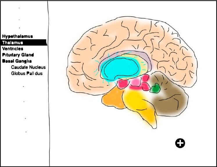
The process of 3D brain modeling through scripting and artistic design represents the first step of the third phase in MEDEERV. In this process, it is necessary to have a script of the system where the actions that the user can perform in it are described step by step. This script identifies the modeling techniques that will be implemented for the creation of the brain. Next, the artistic design is established, where the main models of the virtual world will be identified. The 3D block of the brain that should be embedded in the web page will be managed from a main menu. For example, Figure 2, illustrates an animation perspective of a selected part that is highlighted during the interaction trough the artistic design.
The process of creating the 3D models is a time-consuming activity. Table 2, presents the position in which each element is located, to prioritize the order in which the elements that are part of the brain will be modeled.
Table 2 Names of the brain parts in blueprints.
| Right hemisphere | Middle brain | ||
|---|---|---|---|
| Left hemisphere | Hindbrain | ||
| Frontal lobe | Pons | ||
| Parietal lobe | Cerebellum | ||
| Temporal lobe | Oblongata Medulla | ||
| Occipital lobe | |||
| Hard body | |||
| Limbic system | Diencephalon | ||
| Cingulate Gyrus | Thalamus | ||
| Hippocampus | Hypothalamus | ||
| Griseum Indusium | Pineal Gland | ||
| Septal Nucleus | Optic chiasma | ||
| Toothed twist | Pituitary gland | ||
| Fornix | |||
| Amygdala | |||
| Mammillary Body | |||
| Basal ganglia | Ventricles | ||
| Putamen | Lateral ventricle | ||
| Caudate Nucleus | Third Ventricle | ||
| Side Pale Globe | Fourth ventricle | ||
| Medium Pale Globe | |||
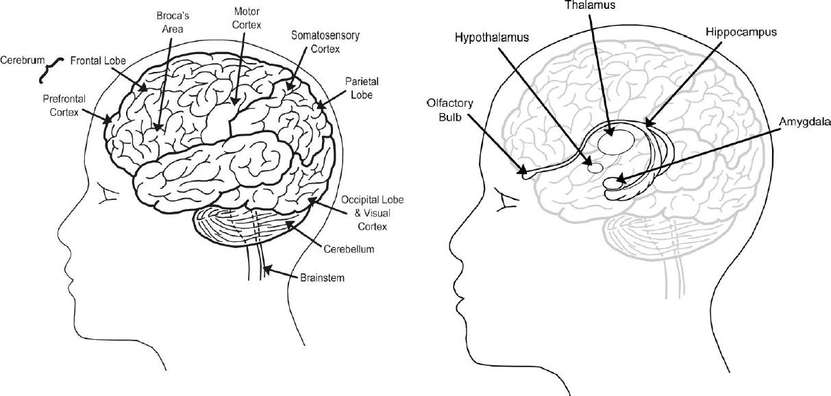
For the creation of each of the brain elements, reference images that allow copying or tracing the geometric shape of the figure to be modeled are necessary. To this end, there are blueprint images, which show orthogonal views of different sides of the object and are based on figures presented in neuroanatomy literature (Figure 3).
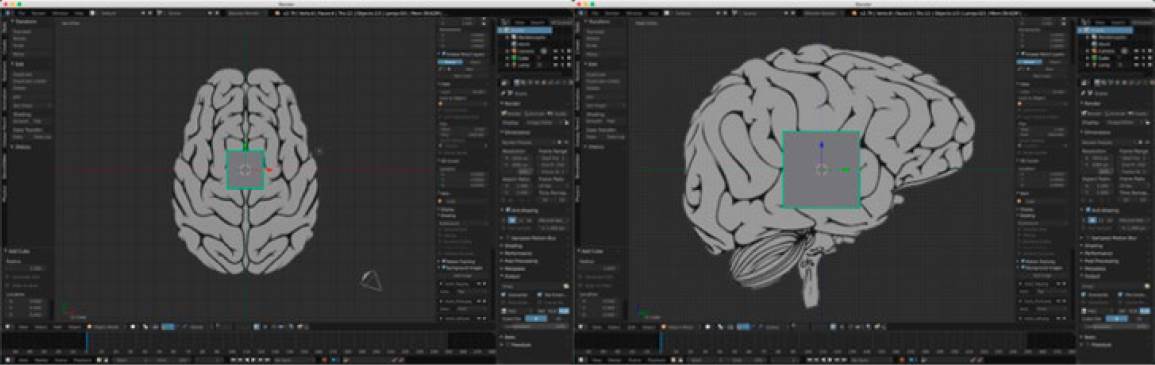
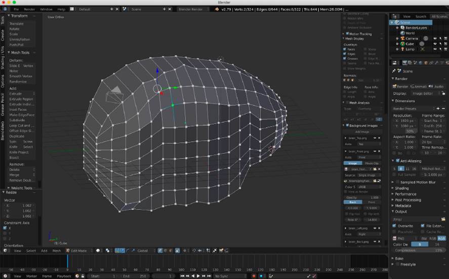
Then, these blueprints are posed in each orthogonal view of the Blender scene, to give a shape from initial single primitive (Figure 4).
Subsequently, using the box modeling technique to model anything from a simple primitive, we get a model volumetrically resembling the cerebral hemispheres as shown in Figure 5.
To obtain a smoothed surface model, the model is modified by subdivision of surfaces. Then, the sculptural modeling technique is used to deform the surface. Figure 6 presents the model obtained up to this stage of the process.
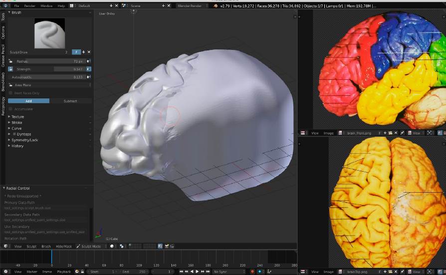
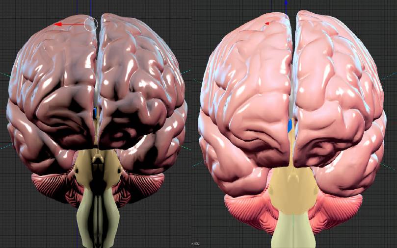
Figure 6 Perspective view of the model resulting from the sculpting process with photographic references.
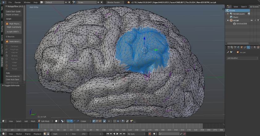
The previous model involves computationally expensive rendering work. Since there are more than twenty parts that compose the complete model of the human brain, it cannot be considered to have detailed models, so it is required to perform retopology of the most complex parts in the mesh. The same process is replicated for each organ to be modeled, some of them are simpler shapes compared to the level of detail of the cerebral hemispheres, therefore, retopology is not necessary. The final result of the retopology process is a less dense mesh as shown in Figure 7.
The materials, colors and textures section, to simulate a more realistic appearance, is based on traditional neuroanatomy teaching and achieved by implementing Oren-Nayar shadows and Toon type reflection. These tools are applied to each component of the brain displaying the complete organ in greater detail. Figure 8 and Figure 9 show the cerebellum and cerebral hemispheres after having assigned Matcap-type material and colors, respectively.
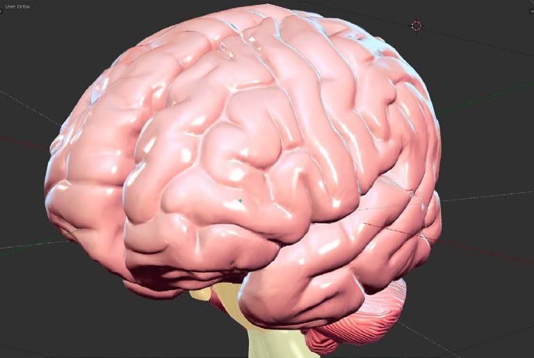
Figure 8 Perspective view of the cerebellum with assigned material and texture. Environmental effects
Environmental effects
Once the models are finished, we proceed to animate the objects with which users will interact in the virtual world. In the same form, the special effects such as particles, fire, among others are included. To complement the stage, depending on the purpose the lighting scene is setup. Lighting settings offer a more realistic experience, taking into account the angle of the objects with the aim of reducing shadows. A scene without lighting results in a lack of depth and 3D appearance. Figure 9, shows a perspective view of two kinds of lighting, sun and ambient.
Implementation
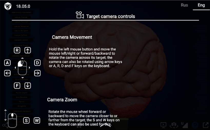
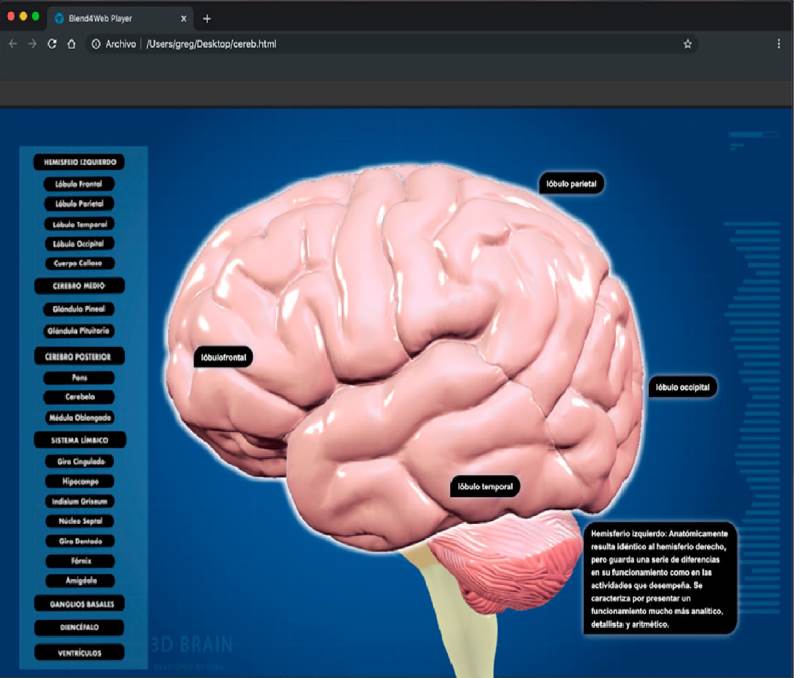
In this stage, the goal is to animate the 3D model by creating the physical behaviors to show, hide or highlight the model elements, when an organ is selected. The animation process is performed by programming parameters in a timeline, using nodes to create a logical tree of decisions. This process is done with the Blend4Web tool, added as a plugin in Blender, useful for creating the logical animation of the scene elements and uploading the model to a web page. Navigation mechanisms are those tools that will be provided to the user to navigate the virtual world. Twentyfour buttons have been added to the scene to access navigation to both organ sets and individual organs. Figure 10, shows the navigation mechanism of 3D brain web-based model.
Finally, in this stage, multimedia objects, for better interaction with the virtual world, videos, audios, music or text can be implemented. There are multimedia objects with information cards so the user can have greater knowledge of the brain, as well as the different organs that make it up. The implementation of the human brain as a completely independent 3D element is a reference as the first step in the process of creating completely 3D-based web pages.
Support
To visualize the 3D brain on the web, the Blender scenario was exported to an HTML file using Blend4Web, which allows navigation in different browsers. The technical requirements to run the model are: latest browser version of Google Chrome, Firefox, Safari or Opera; any 64-bit CPU, graphics card compatible with OpenGL 4.3 or higher and a minimum of 2 GB RAM.
Usability testing
In this section, we just describe some of the essentials to conduct usability testing. Usability testing is the process of learning about users by observing them using a product to accomplish the specific goals of interest. It is crucial to focus on the user and not the product, because you know what works for your users[25]. In that sense, in this work usability is the extent to which the 3D brain can be used by undergraduate students to achieve specific objectives effectively, efficiently and with satisfaction in a neuroanatomy education context.
Among the essential planning steps, after having established the test objectives, it is determined how the product will be tested and how the user groups will be established. On the other hand, there has been a debate about how many participants are needed in a reliable usability test to identify usability issues[26][27] and[28] consider that a majority or about 80 % (given a 30 probability of detection) of usability issues will be observed with the first five participants[29]. In fact, a study can be conducted with 5 users and get excellent results as long as the users are all from the same subgroup. If there is time and budget to test with 10 participants, two or even three subgroups can be identified[25]. In[30] a study was conducted with 12 participants to assess the user experience in virtual upper limb rehabilitation environments. While in[13] SONIA: an immersive customizable virtual reality system for the education and exploration of brain networks was evaluated by 11 subjects, showing attractive visual design and good educational value. Regarding the usability evaluation of Web-Based 3D Medical Image Visualization of the brain, 12 participants evaluated the system, the experimental results show the 3D visualization method improves the educational performance of students[31][32]. Subsequently, we have organized the 3D brain model test using a navigation test and created a post-test questionnaire to get immediate feedback from participants after the virtual trip, whose opinion response options were based on the Likert scale[33]. Table 3 shows a real response sample of the post-test usability questionnaire provided by an undergraduate medicine participant.
Table 3 Real response sample of the post-test usability questionnaire provided by an undergraduate medicine participant.
| Post-test usability questionnaire | |||||
|---|---|---|---|---|---|
| Possible answers | |||||
| Question | Strongly disagree 1 | Disagree 2 | Neutral 3 | Agree 4 | Strongly agree 5 |
| 1. Is the app intuitive? | ✓ | ||||
| 2. Is the application easy to use? | ✓ | ||||
| 3. Does the application give freedom of navigation? | ✓ | ||||
| 4. Did the application present any errors? | ✓ | ||||
| 5. Does the application meet the teaching objective? | ✓ | ||||
| 6. Are the elements of the application model easy to understand? | ✓ | ||||
| 7. Did you find the content of the application structured? | ✓ | ||||
| 8. Is the information displayed in a structured and concrete way? | ✓ | ||||
| 9. Did you find the interface attractive? | ✓ | ||||
| 10. Did the app help reinforce your knowledge? | ✓ | ||||
Experimental setting
Following the guidelines to develop the usability test of our early version, the experimental sample was divided into four subgroups of five participants each. The characteristics of each subgroup were defined depending on the domain knowledge as it relates to the brain neuroanatomy (medicine, dentistry, and gerontology) and the technical skills with 3D computing modelling (computer science). All participants in each stratum were randomly selected among undergraduate students of the final semesters. Before performing the usability test, an informed consent form will be given to users to read and approve, if applicable. The study was developed according to the ethical principles of medical research involving human subjects established in the Declaration of Helsinki.
The technical test consists of using the 3D model brain in the web. The model was run in the browser using WebGL, the library that enhances the graphical capabilities of HTML5 web browsers using JavaScript and OpenGL. EWebGL is included in the Google Chrome, Firefox, and Safari web browsers and specifically in any updated browser. The virtual brain was presented in the Firefox and Google Chrome browsers, since they are the most used by average users. The computers where the test was run were Intel Core i5-4590 CPU @ 3.30 GHz, memory (RAM) 4.0 GB. The 3D web-based human brain based on the 3D web is shown in Figure 11.
After the test, users will answer the usability post-test questionnaire on the Google Forms platform, through the corresponding link and QR code.
RESULTS AND DISCUSSION
The most significant visualization and usability results of the 3D interactive web-based human brain proposed in this research are described below. In 2024, a usability study was conducted on the use of a 3D interactive web-based human brain for the teaching of basic neuroanatomy. The purpose of the study was to determine whether users could easily learn the basis of neuroanatomy using a web-based platform. The web-based 3D interactive human brain was tested by 20 participants (9 women; 11 men, mean age = 22.1 years, SD = 0.70). Each participant spent an average time of 10 minutes to complete the exploration of all modules and then answered the post-test questionnaire described in the previous section. The questions 5, 6, 8 and 10 were oriented to assess the functionality of neuroanatomy learning purpose, while questions 3, 4 and 7 were for operability; and usability was assessed with the questions 1, 2 and 9.
Firstly, to estimate the internal consistency reliability of the post-test questionnaire scores, we calculate the alpha of Cronbach (α)[34]. Where for this case, α=0.93, so it is considered that the set of items is consistent to applied research. Figure 12, shows a bar chart to summarize the answers of the users for the first five questions.
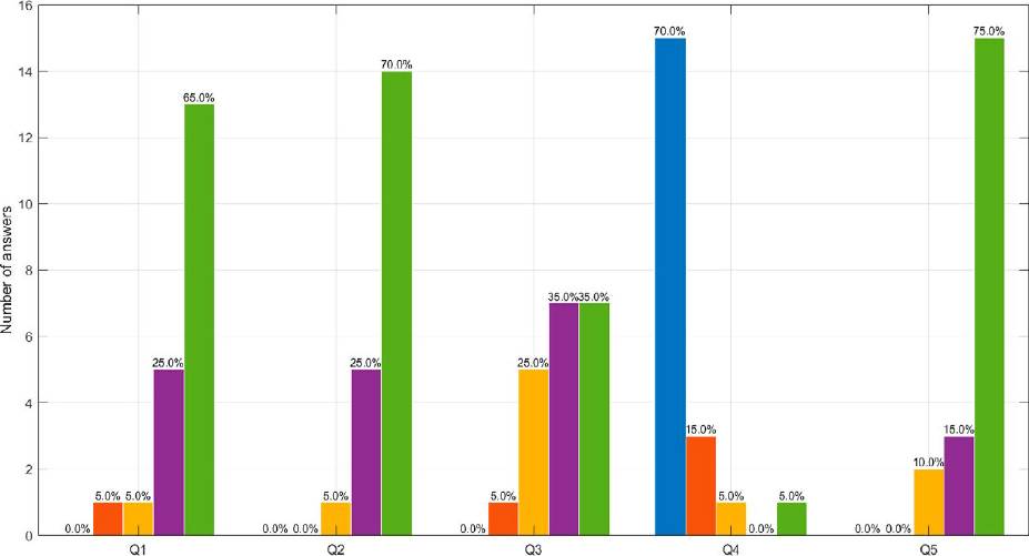
Figure 12 Number of answers to questions 1 to 5 of the acceptance and usability test. Strongly disagree (Pos 1- blue), disagree (Pos 2-orange), neutral (Pos 3-yellow), agree (Pos 4-purple) and strongly agree (Pos 5-green), where position (Pos) is sorted from left to right.
The first question about intuition is related to user interaction with the app without information about its use. 65 % of users totally agree and 25 % agree that the app is intuitive. Question 2 aims to determine the ease of handling the app, for which 70 % completely agree on the ease of handling and 25 % agree. Regarding freedom of navigation in the web environment, an equal amount of 25 % of users totally agreed. Although, in terms of execution errors in the web environment 15 users commented that there were none, some errors were found during the test that required attention. One of the most important objectives of this project is the teaching-learning purpose of neuroanatomy. In that sense, 75 % of the responses lead to the fact that the 3D model and the visualization of the human brain totally agree with this objective. Figure 13, shows the results for questions 6 to 10.
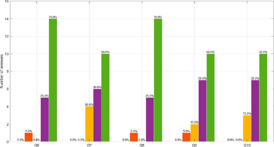
Figure 13 Number of answers to questions 6 to 10 of the acceptance and usability test. Strongly disagree (Pos 1- blue), disagree (Pos 2-orange), neutral (Pos 3-yellow), agree (Pos 4-purple) and strongly agree (Pos 5-green), where position (Pos) is sorted from left to right.
Question 6 refers to an easy understanding of the elements of neuroanatomy in the 3D model, for which 70 % of the users totally agreed and 25 % agreed. 16 participants positively answered about the organization in a logical and coherent way, which is proof that the users had no difficulty identifying the usefulness of each content. Regarding measurement, if the learning elements are excessive or too technical, 70 % of the responses focused on the total agreement of the concrete nature of the information, which implies that the material is accessible to a wider group of users who are not experts in the subject. 50 % respondents totally agree that the app is attractive with respect to colors, text size, and gadgets; and 35 % agree respecting the same aspect. Finally, 17 users answered that the model helped reinforce their knowledge, even when there are health sciences and computer users among them. With the objective of developing a general usability analysis for each subgroup by testing each of the questions, Figure 14 and Figure 15 present the responses to the post-test questionnaire for each subgroup.
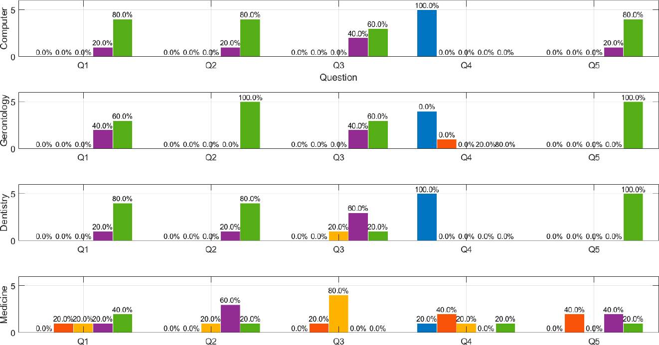
Figure 14 Number of answers to questions 1 to 5 related to acceptance and usability test by stratum. Strongly disagree (Pos 1- blue), disagree (Pos 2-orange), neutral (Pos 3-yellow), agree (Pos 4-purple) and strongly agree (Pos 5-green), where position (Pos) is sorted from left to right.

Figure 15 Number of answers to questions 1 to 5 related to the acceptance and usability test by stratum. Strongly disagree (Pos 1- blue), disagree (Pos 2-orange), neutral (Pos 3-yellow), agree (Pos 4-purple) and strongly agree (Pos 5-green), where position (Pos) is sorted from left to right.
Questions 1, 2, and 9, are related to the usability of the web-based system. In this sense, students of computer science, dentistry, and gerontology, reflected to be in agreement or total agreement in this section, however, students of medicine, who are the most knowledgeable ones in neuroanatomy, had the least favorable responses. With respect to the operability of the system (Q3, Q4), the perception is generally the same among groups as in the case of usability, while in question 7 referring to the structure of the application content, the 4 subgroups have a similar perception tending to be favorable. Finally, regarding the neuroanatomy learning proposal, the computer science, gerontology, and dentistry students (Q5, Q6 and Q8) reported equal favorable perception, while medical students commented that they disagreed or were neutral with the learning objective, ease of understanding and structure of the application content. Regarding whether the app helped to reinforce their knowledge (Q10), gerontology students agreed positively, while medical students were 60 % neutral.
On the other hand, to get a general performance over the usability test, the most frequent response was strongly agreed with 33 apparitions. The mean of the responses was 4.4 and the standard deviation was 0.83. This implies that overall, users responded positively to the evaluation of the brain model. An overview of the results of the usability study appears below, with recommended changes for areas where users found disadvantages and difficulties.
Favorable findings
The users liked the brain model and the fact that it is available on the web.
They all found the drop-down menus and multimedia objects attractive.
Six users annotated the simplicity and clarity of the 3D models as pleasing
Most of the users commented that the neuroanatomy information was easy to find and very informative.
Recommendations
The following is a list of the top user recommendations, some of them were considered to reengineer the early version.
Enlarging the catalog, since when selecting the group of diencephalon organs, it only shows general information of the group and the information pertaining to this group is missing (was considered).
Increasing zoom speed.
Reducing camera speed (was considered), and
Improving the artistic design of the background.
Although we do not use the system usability scale as a measure of usability, there are some works such as the one presented in [14], where they have used the SUS score of the user study with a value 79.8 ± 11.6, which means a positive user-interaction, as presented in this work. While our system in its early version as a proof of concept has interesting results and represents a novel contribution to the teaching of neuroanatomy using web-based 3D technology, it has some limitations. Our small sample may help explain the lack of significant group differences on some question perceptions. On the other hand, the study was developed only among students, so it is necessary to get teachers and professionals of neuroanatomy to participate. In addition, a long-term comparative study of the findings between traditional neuroanatomy teaching methods and the proposed system is also needed.
CONCLUSIONS
In this study, we have presented a 3D interactive web-based human brain modeling process and visualization approach for neuroanatomy education settings, using 3D web technologies and the improved MEDEERV. This methodology allows creating significant learning and establishing best practices to industry standards for generating a more cooperative environment among professionals. The usability capabilities of the web model were evaluated using a post-test questionnaire. The early version as a proof-of-concept of the brain model provides preliminary evidence that the brain model is user-friendly, has an effective user interaction, and is useful to learn about basic neuroanatomy. Then, it is possible to implement the proposed methodology to model and visualize 3D anatomy models in a scalable, reproducible, and standardized way.
The 3D web-based brain visualization provides an advantage towards the learning process in neuroanatomy education settings, which could be expanded to other areas of health and anatomy. The work represents an advance in the integration of interactive 3D technologies in medical education, particularly in the field of neuroanatomy. The web implementation facilitates access and use of this tool, which could positively impact the training of future health professionals and the understanding of brain anatomy by a broader audience. The advantage of interactive visualization of 3D medical objects is focused on improving diagnosis and decision-making in neurological clinical environments. This technology can be used to complement traditional cadaver approaches to better understand complex anatomy such as the brain.
Finally, in this work, the participants who conducted the usability test were undergraduate students in the final semesters of health and computing sciences, as future work we proposed including practicing health professionals and medical educators would provide a broader perspective. Likewise. a simultaneous comparative study with traditional neuroanatomy learning methods is also proposed to determine the success and motivation of medical school students in neuroanatomy courses.

