Artículos científicos
An integrative approach reveals the identity and interoceanic distribution of the diatom Halamphora adumbratoides Stepanek & Kociolek 2018 (Bacillariophyceae: Amphipleuraceae)
Un enfoque integrativo revela la identidad y la distribución interoceánica de la diatomea Halamphora adumbratoides Stepanek & Kociolek 2018 (Bacillariophyceae: Amphipleuraceae)
López-Fuerte, Francisco Omar1
 http://orcid.org/0000-0002-0828-9107
Lora-Vilchis, María Concepción2
http://orcid.org/0000-0003-1839-6974
Murugan, Gopal*2
http://orcid.org/0000-0002-2672-7430
http://orcid.org/0000-0002-0828-9107Lora-Vilchis, María Concepción2
http://orcid.org/0000-0003-1839-6974Murugan, Gopal*2
http://orcid.org/0000-0002-2672-7430
-
Publication dates-
October 22, 2024
Jul-Dec , 2024
- Article in PDF
- Article in XML
- Automatic translation
- Send this article by e-mail
- Share this article +
Abstract
Halamphora adumbratoides was described in 2018 from the Atlantic Ocean (Florida Bay, USA), the type locality of this species. In this report, we present the morphological characteristics and molecular identity of this taxon (strain CIBA 160) collected in the Gulf of California, Mexico (Pacific Ocean region). Cells of the strain CIBA 160 obtained from laboratory cultures showed morphological characteristics inconsistent with the original description, specifically the shape of the valve and the presence of pores at the end of the dorsal raphe ledge at each end of the raphe. Regarding size, the length (4.1-6.6 μm) of the CIBA 160 strain is much shorter than in the original description (10.0-18.0 μm). However, the valve width and number of striae do coincide with the ranges in that description. At the molecular level, DNA sequences of the CIBA 160 strain showed 100, 99.8, and 99.5% similarity, respectively, for SSU, rbcL, and psbC with the H. adumbratoides sequences from the Atlantic Ocean. Our study supports the first case of interoceanic distribution and the second overall recording of this species.
Key words:
first recording, Gulf of California, Pacific Ocean, psbC, rbcL, SSU
Introduction
Diatoms are an important and diverse group of organisms that fix almost 20% of global carbon (Armbrust, 2009; Malviya et al., 2016; D. G. Mann, 1999; Onuma et al., 2017). However, aside from the toxic diatoms, the taxonomy of the specious class of Bacillariophyceae is ‘in a rather poor state’ (Mann et al., 2020). The taxonomic history of the genus Halamphora has been dynamic since its revival by Levkov (2009) produced a considerable number of inclusions via transfers of species new nomenclatural combinations, mostly from the genus Amphora, descriptions of new species, and taxonomic validations. The increased number of taxonomically accepted species and intraspecific names registered in Algaebase is remarkable, from 110 in 2018 to 163 in 2023 (Guiry & Guiry 2023), which reflects the degree of complexity that this genus still represents.
-
Armbrust, 2009The life of diatoms in the world’s oceansNature, 2009
-
Malviya et al., 2016Insights into global diatom distribution and diversity in the world’s oceanProceedings of the National Academy of Sciences of the United States of America, 2016
-
Mann, 1999The species concept in diatomsPhycologia, 1999
-
Onuma et al., 2017Regulation of chloroplast and nucleomorph replication by the cell cycle in the cryptophyte Guillardia thetaScientific Reports, 2017
-
Mann et al., 2020Ripe for reassessment: A synthesis of available molecular data for the speciose diatom family BacillariaceaeMolecular Phylogenetics and Evolution, 2020
-
Levkov (2009)Amphora sensu latoDiatoms of Europe: Diatoms of the Europe inland waters and comparable habitats, 2009
-
Guiry & Guiry 2023AlgaeBase. World-wide electronic publication, 2023
In diatom, monoclonal cultures form a vital part of an integrative taxonomic approach to identifying species using morphological and molecular data (Mohamad et al., 2022). Also, electron microscopy of monoclonal cultures is commonly used to describe new diatom species with ultrastructural details. Stepanek & Kociolek (2018) described H. adumbratoides and complemented their description with molecular tools. Electron microscopy and molecular information have contributed significantly to the determination of new species of diatoms, taxonomic validations, and the transfer of taxa from one genus to another. For example, taxonomic relocations of taxa have been carried out with the closely related genera Amphora, Cymbamphora, and Seminavis (Levkov, 2009; Stepanek & Kociolek, 2013; 2015, 2018, 2019; Van de Vijver et al., 2014).
-
Mohamad et al., 2022Morphological variability and genetic marker stability of 16 monoclonal pennate diatom strains under medium-term cultureDiatom Research, 2022
-
Stepanek & Kociolek (2018)Amphora and Halamphora from coastal and inland waters of the United States and Japan, with the description of 33 new speciesBibliotheca Diatomologica, 2018
-
Levkov, 2009Amphora sensu latoDiatoms of Europe: Diatoms of the Europe inland waters and comparable habitats, 2009
-
Stepanek & Kociolek, 2013Several new species of Amphora and Halamphora from the western USADiatom Research, 2013
-
2015Three new species of the diatom genus Halamphora (Bacillariophyta) from the prairie pothole lakes region of North Dakota, USAPhytotaxa, 2015
-
2018Amphora and Halamphora from coastal and inland waters of the United States and Japan, with the description of 33 new speciesBibliotheca Diatomologica, 2018
-
2019Molecular phylogeny of the diatom genera Amphora and Halamphora (Bacillariophyta) with a focus on morphological and ecological evolutionJournal of Phycology, 2019
-
Van de Vijver et al., 2014Revision of the genus Halamphora (Bacillariophyta) in the Antarctic RegionPlant Ecology and Evolution, 2014
Morphometric similarity between taxa of different genera is rare; however, it is not uncommon among species of closely related genera. For example, the morphology of H. adumbratoides is very similar to that of Amphora adumbrata in terms of the shape of the valve and the fine striae composed of a single elongated areola (Stepanek & Kociolek, 2018). The number of striae was a key characteristic for differentiating these taxa by traditional taxonomy (morphology) (Desianti et al., 2015; Stepanek & Kociolek, 2018). At the molecular level, SSU, rbcL, and psbC markers were used to identify H. adumbratoides (Stepanek & Kociolek, 2018).
-
Stepanek & Kociolek, 2018Amphora and Halamphora from coastal and inland waters of the United States and Japan, with the description of 33 new speciesBibliotheca Diatomologica, 2018
-
Desianti et al., 2015Examination of the type materials of diatoms described by Hohn and Hellerman from the Atlantic Coast of the USADiatom Research, 2015
-
Stepanek & Kociolek, 2018Amphora and Halamphora from coastal and inland waters of the United States and Japan, with the description of 33 new speciesBibliotheca Diatomologica, 2018
-
Stepanek & Kociolek, 2018Amphora and Halamphora from coastal and inland waters of the United States and Japan, with the description of 33 new speciesBibliotheca Diatomologica, 2018
In geographical terms, H. adumbratoides has been recorded only from the coasts of the Atlantic Ocean, in Florida Bay, Cotton Key, Monroe Country, FL, USA. In this publication, we present morphological characteristics and use molecular identity of this taxon (strain CIBA 160) to report its presence in the Pacific Ocean region from the coastal Balandra lagoon (La Paz Bay) in the Gulf of California, on the southern coast of the Baja California Peninsula, Mexico. It is the second recording but the first interoceanic registration of H. adumbratoides. We also register considerable morphological features and morphometry variations in the laboratory-cultured cells.
Materials and methods
Sample collection and morphological observation
Water samples of 50 mL were taken by triplicate from the Balandra lagoon (24°19′9.01′′N 110°19′18.17′′ W) near La Paz, Baja California Sur (Figure 1), in December 2018, and brought to the laboratory. The samples were filtered through a 30-μm Nitex mesh and f/2 medium with silicates added to the filtered samples (Guillard, 1975).
-
Guillard, 1975Culture of Phytoplankton for Feeding Marine InvertebratesCulture of Marine Invertebrate Animals, 1975
Thumbnail
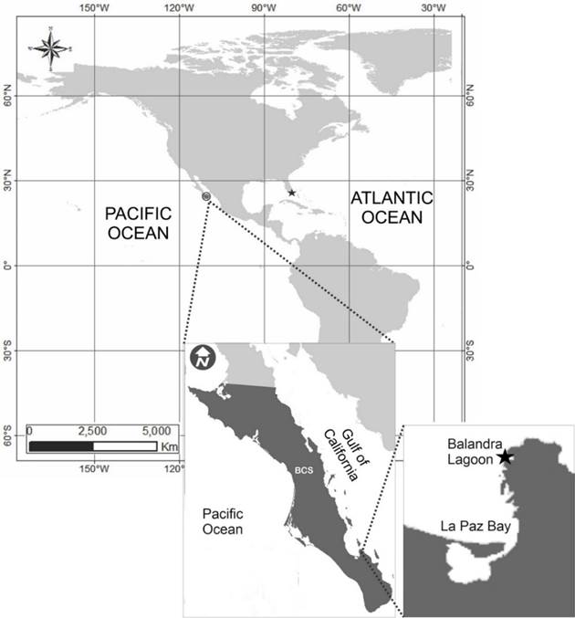
Figure 1
Current recordings of Halamphora adumbratoides in the Pacific and Atlantic Ocean (solid stars). Inset figures show an expanded map of the collection site in the Balandra Lagoon, Mexico.
Current recordings of Halamphora adumbratoides in the Pacific and Atlantic Ocean (solid stars). Inset figures show an expanded map of the collection site in the Balandra Lagoon, Mexico.
After two weeks, microalgal cells were isolated in a 1:50 dilution in f/2 medium and streaked on marine agar plates containing the same medium. Single-cell colonies were isolated under low magnification in a microscope, with a sterile hypodermic needle and then transferred to 24-well plates containing 2 ml of culture medium. Two weeks later, the cells were transferred to test tubes with 5 ml of culture medium, and the unialgal culture of isolated cells was examined under light microscopy. Once confirmed, the monoclonal cultures were deposited in CIBNOR’s Microalgae Collection under catalog number CIBA 160 (https://www.cibnor.gob.mx/investigacion/colecciones-biologicas/coleccion-de-microalgas) after three reseeding periods. We have a collection of three fixed slides with the skeletons of the cells, that remains in the laboratory.
For light and electron microscopy and molecular analyses, cells were cultured in triplicate in 500-mL flasks with 250 mL of medium at 25±1°C, 40 μmol·m-2·s-1, 12:12 h, and 35 PSU. At the exponential stage (on the sixth day), separate samples were taken for each analysis. Live and cleaned cells were observed under a Zeiss Axio Lab A1 microscope, and photographs were taken with a Canon EOS Rebel T5i camera (Figure 2). The cells were prepared for scanning electron microscopy (SEM) as indicated previously by López-Fuerte et al. (2020). Briefly, the organic matter was eliminated by oxidation, heat-assisted with concentrated nitric acid. The cells were then washed in distilled water to reach a neutral pH. SEM images of the cells were taken with a Hitachi SU3500 electron microscope operating at 10 kV and a 6-mm working distance. A coverslip holding the cells was attached to a 32-mm aluminum stub using conductive carbon tape and coated with around 15 nm of gold in a Hummer 6.2 sputtering unit.
-
López-Fuerte et al. (2020)Halamphora siqueirosii (Bacillariophyta), a new diatom species isolated from a hypersaline evaporation pond in Baja California Peninsula, MexicoPhytotaxa, 2020
Thumbnail
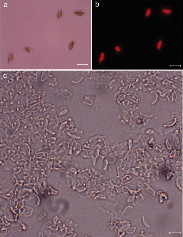
Figure 2
a-c. Halamphora adumbratoides, a. live cell showing plastids; b. fluorescence microscopy showing plastids; c. cleaned specimens. Scale bars represent 5 μm.
a-c. Halamphora adumbratoides, a. live cell showing plastids; b. fluorescence microscopy showing plastids; c. cleaned specimens. Scale bars represent 5 μm.
The key morphological characteristics -length, width, and density of the striae- were measured by light and electron microscopy and compared to those reported for Halamphora adumbratoides (Stepanek & Kociolek, 2018). The validity nomenclatural status of the name was verified on the Algaebase website (Guiry & Guiry 2023).
-
Stepanek & Kociolek, 2018Amphora and Halamphora from coastal and inland waters of the United States and Japan, with the description of 33 new speciesBibliotheca Diatomologica, 2018
-
Guiry & Guiry 2023AlgaeBase. World-wide electronic publication, 2023
DNA extraction, PCR amplification, and sequencing
Cells of the CIBA 160 strain obtained from the exponential stage cultures were utilized to extract genomic DNA by the CTAB method (Doyle, 1991; Herrera et al., 2014). Four regions of DNA were analyzed: fragment of two nuclear genes, the small sub-unit of nuclear gene 18S ribosomal RNA (SSU), and the large subunit of nuclear gene 28S ribosomal RNA (LSU) were amplified following the PCR conditions described in López-Fuerte et al. (2020). A partial sequence of the ribulose bisphosphate carboxylase/oxygenase gene (rbcL) from the chloroplast was amplified according to the published protocols (López-Fuerte et al., 2020). Also, the photosystem II CP43 protein gene (psbC) was amplified with 30 picomoles of the psbC+ and psbC- primers (Alverson et al., 2007) in 40 μL of a PCR reaction mixture containing genomic DNA, 0.3 mM dNTPs, 2 mM MgCl2, 4 μL of 10X PCR buffer, and 2 units of platinum Taq polymerase (InvitrogenTM).
-
Doyle, 1991DNA Protocols for Plants BTMolecular Techniques in Taxonomy, 1991
-
Herrera et al., 2014Are Prorocentrum hoffmannianum and Prorocentrum belizeanum (DINOPHYCEAE, PROROCENTRALES), the same species? An integration of morphological and molecular dataJournal of Phycology, 2014
-
López-Fuerte et al. (2020)Halamphora siqueirosii (Bacillariophyta), a new diatom species isolated from a hypersaline evaporation pond in Baja California Peninsula, MexicoPhytotaxa, 2020
-
López-Fuerte et al., 2020Halamphora siqueirosii (Bacillariophyta), a new diatom species isolated from a hypersaline evaporation pond in Baja California Peninsula, MexicoPhytotaxa, 2020
-
Alverson et al., 2007Bridging the Rubicon: Phylogenetic analysis reveals repeated colonizations of marine and fresh waters by thalassiosiroid diatomsMolecular Phylogenetics and Evolution, 2007
The PCR conditions for psbC amplification were denaturation at 94°C for 3.30 min, then 35 cycles of 30 s at 94 °C, 50 s at 52 °C, and 80 s at 72 °C. The final extension was for 10 min at 72 °C. Both strands of all markers were sequenced with the amplification primers and an internal primer for the SSU and chloroplast genes (18S 962R, rbcL1255, psbC857).
We edited the four sequences (SSU, LSU, psbC, and rbcL) in DNA Baser 4.5 program (http://www.dnabaser.com) and compared the SSU, rbcL, and psbC sequences to those of H. adumbratoides in the GenBank database. The phylogenetic relationship of the CIBA 160 strain was inferred based on the concatenated nucleotide sequences of SSU, LSU, psbC, and rbcL with 23 Halamphora species (Table 1) using the maximum likelihood (ML) method in PAUP 4.0a166 (Swofford, 2002) and Bayesian inference (BI) in MrBayes 3.2.7a (Ronquist et al., 2012). The best fit nucleotide model TIM3+I+G selected according to the BIC criterion in jModeltest 2.1.10 (Darriba et al., 2012; Guindon & Gascuel, 2003) for the concatenated nucleotide sequences was implemented in both analyses. Maximum likelihood analysis was performed with 500 pseudoreplicates under the option of heuristic tree-searching with tree bisection-reconnection branch swapping to generate a majority rule ML consensus tree. Bayesian inference was run for 10 million generations, and a majority rule BI consensus tree was obtained after eliminating 25% of the initial trees using the burn-in option.
-
Swofford, 2002PAUP*. Phylogenetic Analysis Using Parsimony (*and Other Methods), 2002
-
Ronquist et al., 2012MrBayes 3.2: Efficient Bayesian phylogenetic inference and model choice across a large model spaceSystematic Biology, 2012
-
Darriba et al., 2012jModelTest 2: more models, new heuristics and parallel computingNature Methods, 2012
-
Guindon & Gascuel, 2003A Simple, Fast, and Accurate Algorithm to Estimate Large Phylogenies by Maximum LikelihoodSystematic Biology, 2003
Table 1
Halamphora and Amphora sequences used in the phylogenetic analyses. Tabla 1. Secuencias de Halamphora y Amphora utilizadas en los análisis filogenéticos.
Halamphora and Amphora sequences used in the phylogenetic analyses. Tabla 1. Secuencias de Halamphora y Amphora utilizadas en los análisis filogenéticos.
| Taxa | 18S | rbcL | psbC | 28S | Reference |
|---|---|---|---|---|---|
|
Halamphora |
MG027270 | MG027434 | MG027514 | NA | Stepanek & Kociolek, 2019 |
|
Halamphora |
ON714546 | ON736839 | ON736840 | ON714544 | This study |
|
Halamphora |
MG027316 | MG027481 | MG027561 | MG027401 | Stepanek & Kociolek, 2019 |
|
Halamphora |
MG027320 | MG027486 | MG027566 | MG027407 | Stepanek & Kociolek, 2019 |
|
Halamphora |
MG027259 | MG027423 | MG027503 | MG027337 | Stepanek & Kociolek, 2019 |
|
Halamphora |
MG027275 | MG027439 | MG027519 | MG027355 | Stepanek & Kociolek, 2019 |
|
Halamphora |
MG027296 | MG027461 | MG027541 | MG027381 | Stepanek & Kociolek, 2019 |
|
Halamphora |
MG027327 | MG027493 | MG027573 | MG027414 | Stepanek & Kociolek, 2019 |
|
Halamphora |
MG027321 | MG027487 | MG027567 | MG027408 | Stepanek & Kociolek, 2019 |
|
Halamphora |
MG027306 | MG027471 | MG027551 | MG027391 | Stepanek & Kociolek, 2019 |
|
Halamphora |
MG027297 | MG027462 | MG027542 | MG027382 | Stepanek & Kociolek, 2019 |
|
Halamphora |
MG027299 | MG027464 | MG027544 | MG027384 | Stepanek & Kociolek, 2019 |
|
Halamphora |
MG027326 | MG027492 | MG027572 | MG027413 | Stepanek & Kociolek, 2019 |
|
Halamphora |
MG027271 | MG027435 | MG027515 | MG027350 | Stepanek & Kociolek, 2019 |
|
Halamphora |
MG027269 | MG027433 | MG027513 | MG027348 | Stepanek & Kociolek, 2019 |
|
Halamphora |
MG027308 | MG027473 | MG027553 | MG027393 | Stepanek & Kociolek, 2019 |
|
Halamphora |
MG027260 | MG027424 | MG027504 | MG027338 | Stepanek & Kociolek, 2019 |
|
Halamphora |
MG027278 | MG027442 | MG027522 | MG027359 | Stepanek & Kociolek, 2019 |
|
Halamphora |
MG027328 | MG027494 | MG027574 | MG027415 | Stepanek & Kociolek, 2019 |
|
Halamphora |
KJ463451 | KJ463481 | KJ463511 | KP229528 | Stepanek & Kociolek, 2014; Stephanek et al. 2015 (28S) |
|
Halamphora |
KJ463452 | KJ463482 | KJ463512 | KP229530 | Stepanek & Kociolek, 2014; Stephanek et al. 2015 (28S) |
|
Halamphora |
KJ463453 | KJ463483 | KJ463513 | NA | Stepanek & Kociolek, 2014, 2018 |
|
Halamphora |
KJ463450 | KJ463480 | KJ463510 | KP229529 | Stepanek & Kociolek, 2014; Stephanek et al. 2015 (28S) |
|
Amphora |
MG027314 | MG027479 | MG027559 | MG027399 | Stepanek & Kociolek, 2019 |
|
Amphora |
KP229526 | KP229547 | KP229549 | KP229545 | Stepanek et al., 2015 |
-
Stepanek & Kociolek, 2019Molecular phylogeny of the diatom genera Amphora and Halamphora (Bacillariophyta) with a focus on morphological and ecological evolutionJournal of Phycology, 2019
-
Stepanek & Kociolek, 2019Molecular phylogeny of the diatom genera Amphora and Halamphora (Bacillariophyta) with a focus on morphological and ecological evolutionJournal of Phycology, 2019
-
Stepanek & Kociolek, 2019Molecular phylogeny of the diatom genera Amphora and Halamphora (Bacillariophyta) with a focus on morphological and ecological evolutionJournal of Phycology, 2019
-
Stepanek & Kociolek, 2019Molecular phylogeny of the diatom genera Amphora and Halamphora (Bacillariophyta) with a focus on morphological and ecological evolutionJournal of Phycology, 2019
-
Stepanek & Kociolek, 2019Molecular phylogeny of the diatom genera Amphora and Halamphora (Bacillariophyta) with a focus on morphological and ecological evolutionJournal of Phycology, 2019
-
Stepanek & Kociolek, 2019Molecular phylogeny of the diatom genera Amphora and Halamphora (Bacillariophyta) with a focus on morphological and ecological evolutionJournal of Phycology, 2019
-
Stepanek & Kociolek, 2019Molecular phylogeny of the diatom genera Amphora and Halamphora (Bacillariophyta) with a focus on morphological and ecological evolutionJournal of Phycology, 2019
-
Stepanek & Kociolek, 2019Molecular phylogeny of the diatom genera Amphora and Halamphora (Bacillariophyta) with a focus on morphological and ecological evolutionJournal of Phycology, 2019
-
Stepanek & Kociolek, 2019Molecular phylogeny of the diatom genera Amphora and Halamphora (Bacillariophyta) with a focus on morphological and ecological evolutionJournal of Phycology, 2019
-
Stepanek & Kociolek, 2019Molecular phylogeny of the diatom genera Amphora and Halamphora (Bacillariophyta) with a focus on morphological and ecological evolutionJournal of Phycology, 2019
-
Stepanek & Kociolek, 2019Molecular phylogeny of the diatom genera Amphora and Halamphora (Bacillariophyta) with a focus on morphological and ecological evolutionJournal of Phycology, 2019
-
Stepanek & Kociolek, 2019Molecular phylogeny of the diatom genera Amphora and Halamphora (Bacillariophyta) with a focus on morphological and ecological evolutionJournal of Phycology, 2019
-
Stepanek & Kociolek, 2019Molecular phylogeny of the diatom genera Amphora and Halamphora (Bacillariophyta) with a focus on morphological and ecological evolutionJournal of Phycology, 2019
-
Stepanek & Kociolek, 2019Molecular phylogeny of the diatom genera Amphora and Halamphora (Bacillariophyta) with a focus on morphological and ecological evolutionJournal of Phycology, 2019
-
Stepanek & Kociolek, 2019Molecular phylogeny of the diatom genera Amphora and Halamphora (Bacillariophyta) with a focus on morphological and ecological evolutionJournal of Phycology, 2019
-
Stepanek & Kociolek, 2019Molecular phylogeny of the diatom genera Amphora and Halamphora (Bacillariophyta) with a focus on morphological and ecological evolutionJournal of Phycology, 2019
-
Stepanek & Kociolek, 2019Molecular phylogeny of the diatom genera Amphora and Halamphora (Bacillariophyta) with a focus on morphological and ecological evolutionJournal of Phycology, 2019
-
Stepanek & Kociolek, 2019Molecular phylogeny of the diatom genera Amphora and Halamphora (Bacillariophyta) with a focus on morphological and ecological evolutionJournal of Phycology, 2019
-
Stepanek & Kociolek, 2014Molecular phylogeny of Amphora sensu lato (Bacillariophyta): an investigation into the monophyly and classification of the amphoroid diatomsProtist, 2014
-
Stephanek et al. 2015Description and phylogenetic position of Amphora aliformis (Bacillariophyta), a new species from Tokyo BayPhycologia, 2015
-
Stepanek & Kociolek, 2014Molecular phylogeny of Amphora sensu lato (Bacillariophyta): an investigation into the monophyly and classification of the amphoroid diatomsProtist, 2014
-
2018Amphora and Halamphora from coastal and inland waters of the United States and Japan, with the description of 33 new speciesBibliotheca Diatomologica, 2018
-
Stepanek & Kociolek, 2014Molecular phylogeny of Amphora sensu lato (Bacillariophyta): an investigation into the monophyly and classification of the amphoroid diatomsProtist, 2014
-
Stephanek et al. 2015Description and phylogenetic position of Amphora aliformis (Bacillariophyta), a new species from Tokyo BayPhycologia, 2015
-
Stepanek & Kociolek, 2019Molecular phylogeny of the diatom genera Amphora and Halamphora (Bacillariophyta) with a focus on morphological and ecological evolutionJournal of Phycology, 2019
-
Stepanek et al., 2015Description and phylogenetic position of Amphora aliformis (Bacillariophyta), a new species from Tokyo BayPhycologia, 2015
Results
The length and width recorded for the valves of the laboratory cultured cells of the strain CIBA 160 (Halamphora adumbratoides) are 4.1-6.6 and 1.6-2.4 μm, respectively. The number of dorsal and ventral striae for the CIBA 160 strain are 57-60 and 60-70 in 10 μm, respectively. The valve shape is semi-elliptical, and the ends are broadly rounded (Figure 3a -j). The raphe is straight with straight proximal ends, while the distal raphe ends are deflected to hooked dorsally (Figure 3a-b). Externally, two pores are visible at the end of the dorsal raphe ledge at each end of the raphe (Figure 3a-d black arrow), or they may appear fused (Figure 3d white arrow), while internally two, only a single pore is observed (Figure 3e-g, j). Internally, the proximal raphe ends terminated in a broad, fused central helictoglossa (Figure 3e-f, h, white asterisk). The dorsal raphe ledge is nearly absent, especially through the valve center (Figure 3a-d). A reduced dorsal raphe ledge is present, more accentuated towards the distal end of the valve (Figure 3a-b, black asterisk). The dorsal and ventral striae are continuous through the valve center, slightly radiate (Figure 3a-d), and not resolved in the LM (Figure 2b). Figure 3e-j shows internal views of the deformed valves and other deformed structures, like the raphe, which is in a different position than in the undeformed valves.
Thumbnail
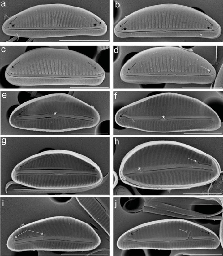
Figure 3
a-d. External view of the valve. a-d showing two distal pores (black arrows). The external and internal proximal raphe ends are not deflected, but the external distal raphe ends are dorsally deflected, a-d, e. Externally, the dorsal and ventral raphe ledges are small but continuous along the length of the valve, e-j. Internal view of the valve. e, f, h. Internally, the proximal raphe ends terminate in a broad, fused central helictoglossa (white asterisks), e. f, g, h, j. One distal pore is visible (black arrows). f, g, h, i, j. White arrows show the abnormal dorsal raphe ledge. Scale bars correspondence: a, 3 μm, b-j, 2 μm.
a-d. External view of the valve. a-d showing two distal pores (black arrows). The external and internal proximal raphe ends are not deflected, but the external distal raphe ends are dorsally deflected, a-d, e. Externally, the dorsal and ventral raphe ledges are small but continuous along the length of the valve, e-j. Internal view of the valve. e, f, h. Internally, the proximal raphe ends terminate in a broad, fused central helictoglossa (white asterisks), e. f, g, h, j. One distal pore is visible (black arrows). f, g, h, i, j. White arrows show the abnormal dorsal raphe ledge. Scale bars correspondence: a, 3 μm, b-j, 2 μm.
Molecular analysis
We obtained 4,004 bp nucleotide sequences (SSU 1375 bp, rbcL 1444 bp, psbC 1185 bp; GenBank accession numbers: ON714546, ON736839, ON736840) for the CIBA 160 strain (H. adumbratoides) from the Pacific Ocean. Comparison of our sequences to those of the type species from the Atlantic Ocean in GenBank showed 100, 99.8, and 99.5% similarity for SSU, rbcL, and psbC, respectively. The BI and ML phylogenetic analyses placed the H. adumbratoides strains in one clade with H. pellicula as a sister clade (Figure 4). This node was supported strongly by the BI (100%) and ML (92%) analyses.
Thumbnail
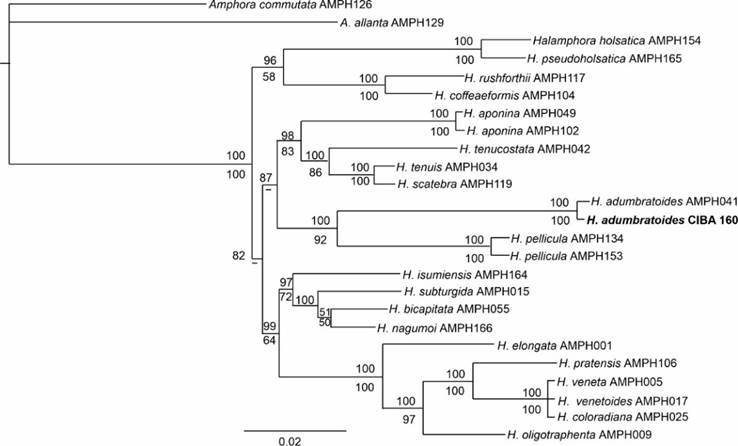
Figure 4
Bayesian tree of the Halamphora species based on the partial sequences of SSU, LSU, psbC, and rbcL. Bootstrap values of maximum likelihood analysis are given below, and posterior probabilities of Bayesian inference are given above the nodes. Nodes supported by less than 50% of Bootstrap values are indicated with the symbol ˋ-ˊ
Bayesian tree of the Halamphora species based on the partial sequences of SSU, LSU, psbC, and rbcL. Bootstrap values of maximum likelihood analysis are given below, and posterior probabilities of Bayesian inference are given above the nodes. Nodes supported by less than 50% of Bootstrap values are indicated with the symbol ˋ-ˊ
Discussion
Accurate identification of a species depends, in principle, on the methods' reliability. Historically, and even today, the identification of a diatom species is based on morphometric aspects observed under light microscopy, which requires extensive taxonomic experience (Mora et al., 2019). The characterization of certain diatom species is impossible only with this tool because of their small size and inconspicuous diagnostic structures (Li et al., 2018; D. Mann et al., 2020). Fortunately, this issue can be resolved with electron microscopy. However, not all diatom species can be delineated using morphology, especially species with subtle morphological differences (Beszteri et al., 2007; Evans et al., 2007; Malviya et al., 2016). To aid in determining species boundaries among diatoms, molecular tools, such as DNA sequences are applied (An et al., 2017; Beszteri et al., 2007; Evans et al., 2007; Guillard, 2005; D. Mann et al., 2010, 2020; Medlin, 1991). However, this methodology has limitations as it may be unable to resolve the species identity (Malviya et al., 2016) without conspecific and/or closely related species sequences in the nucleotide database. Therefore, morphological, and molecular methods are important in diatoms and should be considered as elements of a synergistic approach to species identification challenges.
-
Mora et al., 2019Morphology and metabarcoding: a test with stream diatoms from Mexico highlights the complementarity of identification methodsFreshwater Science, 2019
-
Li et al., 2018The morphology and molecular phylogenetics of some marine diatom taxa within the Fragilariaceae, including twenty undescribed species and their relationship to Nanofrustulum, Opephora and PseudostaurosiraPhytotaxa, 2018
-
D. Mann et al., 2020Ripe for reassessment: A synthesis of available molecular data for the speciose diatom family BacillariaceaeMolecular Phylogenetics and Evolution, 2020
-
Beszteri et al., 2007An assessment of cryptic genetic diversity within the Cyclotella meneghiniana species complex (Bacillariophyta) based on nuclear and plastid genes, and amplified fragment length polymorphismsEuropean Journal of Phycology, 2007
-
Evans et al., 2007An Assessment of Potential Diatom “Barcode” Genes (cox1, rbcL, 18S and ITS rDNA) and their Effectiveness in Determining Relationships in Sellaphora (Bacillariophyta)Protist, 2007
-
Malviya et al., 2016Insights into global diatom distribution and diversity in the world’s oceanProceedings of the National Academy of Sciences of the United States of America, 2016
-
An et al., 2017Identification of benthic diatoms isolated from the eastern tidal flats of the Yellow Sea: Comparison between morphological and molecular approachesPLoS ONE, 2017
-
Beszteri et al., 2007An assessment of cryptic genetic diversity within the Cyclotella meneghiniana species complex (Bacillariophyta) based on nuclear and plastid genes, and amplified fragment length polymorphismsEuropean Journal of Phycology, 2007
-
Evans et al., 2007An Assessment of Potential Diatom “Barcode” Genes (cox1, rbcL, 18S and ITS rDNA) and their Effectiveness in Determining Relationships in Sellaphora (Bacillariophyta)Protist, 2007
-
Guillard, 2005Purification methods for microalgaeAlgal culturing techniques, 2005
-
D. Mann et al., 2010DNA barcoding for species identification and discovery in diatomsCryptogamie Algologie, 2010
-
2020DNA barcoding for species identification and discovery in diatomsCryptogamie Algologie, 2010
-
Medlin, 1991Morphological and genetic variation within the diatom Skeletonema costatum (Bacillariophyta): evidence for a new species, Skeletonema pseudocostatumJournal of Phycology, 1991
-
Malviya et al., 2016Insights into global diatom distribution and diversity in the world’s oceanProceedings of the National Academy of Sciences of the United States of America, 2016
After a morphometric review of the images of the laboratory cultured cells of the CIBA 160 strain obtained with both light and electron microscopy, it was not possible to identify this strain with any previously described taxon. However, approximations were made with Amphora adumbrata and Halamphora adumbratoides. Nevertheless, the morphometric analyses did not allow the taxonomic determination of the CIBA 160 strain as H. adumbratoides because the morphology of this strain does not exactly match the original description reported by Stepanek & Kociolek (2018), specifically due to the shape and size of the valve. In the original description of H. adumbratoides, the valves were narrow and semi-lanceolate, and the valve ends narrowly rounded to weakly subcapitate in larger specimens. The dorsal and ventral margins, however, do coincide with the original description, as they are arched and straight, respectively (Figure 3a-d). The presence of pores at the end of the dorsal raphe ledge at each end of the raphe was not mentioned in the original description. The axial area, not easily distinguishable in the original description, was not observed in the specimens of the CIBA 160 strain analyzed.
-
Stepanek & Kociolek (2018)Amphora and Halamphora from coastal and inland waters of the United States and Japan, with the description of 33 new speciesBibliotheca Diatomologica, 2018
For the length and width of the valves, Stepanek & Kociolek (2018) reported ranges of 8.0-10.0 and 1.5-2.5 μm, while the cells (N=24) of the CIBA 160 strain recorded valve lengths and widths of 4.1-6.6 and 1.6-2.4 μm. Only the length range differed, as it was much shorter than in the original description. The number of dorsal and ventral striae in the CIBA 160 strain almost coincided with the original description, with 56-57 dorsal striae and ca. 70 ventral striae in 10 μm in the original description, while the striae range in 10 μm for the CIBA 160 strain were 57-60 and 60-70 for the dorsal and ventral striae, respectively.
-
Stepanek & Kociolek (2018)Amphora and Halamphora from coastal and inland waters of the United States and Japan, with the description of 33 new speciesBibliotheca Diatomologica, 2018
A possible explanation for the size differences and morphological changes could be the effect of laboratory culturing on the cells of CIBA 160. In diatoms Rose & Cox (2013), Petrova et al. (2020) and Mohamad et al. (2022) have reported morphological changes in laboratory cultured cells. Diatoms tend to produce small-sized cells over time due to asexual reproduction (Mohamad et al., 2022). This phenomenon has been demonstrated in 15 monoclonal pennate diatoms, but the width and number of striae did not differ from the original description (Mohamad et al., 2022). Our results support the findings of Mohamad et al. (2022), and their proposal that the number of striae could be a useful character for species discrimination.
-
Rose & Cox (2013)Some diatom species do not show a gradual decrease in cell size as they reproduceFundamental and Applied Limnology, 2013
-
Petrova et al. (2020)Changes in valve morphology of two pennate diatom species during long-term cultureActa Biologica Sibirica, 2020
-
Mohamad et al. (2022)Morphological variability and genetic marker stability of 16 monoclonal pennate diatom strains under medium-term cultureDiatom Research, 2022
-
Mohamad et al., 2022Morphological variability and genetic marker stability of 16 monoclonal pennate diatom strains under medium-term cultureDiatom Research, 2022
-
Mohamad et al., 2022Morphological variability and genetic marker stability of 16 monoclonal pennate diatom strains under medium-term cultureDiatom Research, 2022
-
Mohamad et al. (2022)Morphological variability and genetic marker stability of 16 monoclonal pennate diatom strains under medium-term cultureDiatom Research, 2022
Our molecular analyses confirmed that the CIBA 160 strain belongs to H. adumbratoides by showing very high similarity to the sequences of this species from the Atlantic Ocean: 100, 99.8, and 99.5% similarity for SSU, rbcL, and psbC, respectively. The present study corroborates the results of Mohamad et al. (2022) that molecular data remain consistent irrespective of morphological changes in laboratory-cultured diatoms. Phylogenetic analyses of this study were also congruent with the relationship presented by Stepanek & Kociolek (2019) for the H. adumbratoides species, which is a sister group to H. pellicula.
-
Mohamad et al. (2022)Morphological variability and genetic marker stability of 16 monoclonal pennate diatom strains under medium-term cultureDiatom Research, 2022
-
Stepanek & Kociolek (2019)Molecular phylogeny of the diatom genera Amphora and Halamphora (Bacillariophyta) with a focus on morphological and ecological evolutionJournal of Phycology, 2019
We have registered morphological abnormalities in the cells of CIBA 160. The helictoglossa (Figure 3h white asterisk) is displaced to the left side of the valve. In contrast, the dorsal raphe ledge is very short and located only to the right of the valve (white arrow) (Figure 3h). The exact opposite configuration is visible in Figure 3i (white arrow), where the structure is seen to the left of the valve. These abnormalities could be associated with long-term culture of cells in the laboratory (Estes & Dute, 1994; Falasco et al., 2009; Petrova et al., 2020).
-
Estes & Dute, 1994Valve abnormalities in diatom clones maintained in long-term cultureDiatom Research, 1994
-
Falasco et al., 2009Morphological abnormalities of diatom silica walls in relation to heavy metal contamination and artificial growth conditionsWater S.A, 2009
-
Petrova et al., 2020Changes in valve morphology of two pennate diatom species during long-term cultureActa Biologica Sibirica, 2020
In the genera Amphora and Halamphora, most taxonomic determinations and descriptions of new species are based on morphological analyses of specimens from natural populations (Levkov, 2009; Wachnicka & Gaiser, 2007). However, laboratory-cultured cells were also used for integrative taxonomic studies, combining morphological and molecular data (López-Fuerte et al., 2020; Stepanek et al., 2013, 2015a, 2015b, 2018).
-
Levkov, 2009Amphora sensu latoDiatoms of Europe: Diatoms of the Europe inland waters and comparable habitats, 2009
-
Wachnicka & Gaiser, 2007Characterization of Amphora and Seminavis from South Florida, USADiatom Research, 2007
-
López-Fuerte et al., 2020Halamphora siqueirosii (Bacillariophyta), a new diatom species isolated from a hypersaline evaporation pond in Baja California Peninsula, MexicoPhytotaxa, 2020
-
Stepanek et al., 2013Several new species of Amphora and Halamphora from the western USADiatom Research, 2013
-
2018Amphora and Halamphora from coastal and inland waters of the United States and Japan, with the description of 33 new speciesBibliotheca Diatomologica, 2018
It is important to mention that regardless of the origin of diatoms (natural or cultivated), the morphological description of a species must be accurate and informative, based on as much information as possible. This is because diatoms may present morphological variations throughout their life history, whether in the natural or artificial environment (Rose & Cox, 2013).
-
Rose & Cox, 2013Some diatom species do not show a gradual decrease in cell size as they reproduceFundamental and Applied Limnology, 2013
In our study, using morphological and molecular data has resulted in the reliable taxonomic identification of the CIBA 160 strain. Our results support using an integrative approach for fine-grained integrative taxonomic studies and the importance of laboratory cultures in the diatom taxonomy (Mohamad et al., 2022).
-
Mohamad et al., 2022Morphological variability and genetic marker stability of 16 monoclonal pennate diatom strains under medium-term cultureDiatom Research, 2022
Conclusions
The taxonomic identification of the CIBA 160 strain as H. adumbratoides was not possible based only on morphometric characteristics. This was due mainly to differences in the valve leaflet length and the presence of pores at the extremes of the valve on the raphe channel. Molecular analyses, however, allowed us to determine species identity with reference DNA sequences deposited in GenBank. Using DNA sequence data is important for cryptic, pseudo-cryptic diatom species and diatoms in general. Integrated approaches can play a vital role in discovering species with broad morphological variations.
Acknowledgements
We thank James M. Ehrman of the Digital Microscopy Facility at Mount Allison University for the SEM image, Joaquín Rivera Rosas for the map image, and Paul Kersey for the English review of this manuscript. GM and MCLV thank CIBNOR for the internal projects and the facilities provided to carry out this work. FOLF thanks the SNII-CONAHCYT program. Finally, we acknowledge three anonymous reviewers for their valuable comments.
Literature cited
- Alverson, A., Jansen, R., & Theriot, E. (2007). Bridging the Rubicon: Phylogenetic analysis reveals repeated colonizations of marine and fresh waters by thalassiosiroid diatoms. Molecular Phylogenetics and Evolution, 45, 193-210. https://doi.org/10.1016/j.ympev.2007.03.024 Links
- An, S. M., Choi, D. H., Lee, J. H., Lee, H., & Noh, J. H. (2017). Identification of benthic diatoms isolated from the eastern tidal flats of the Yellow Sea: Comparison between morphological and molecular approaches. PLoS ONE, 12(6). https://doi.org/10.1371/journal.pone.0179422 Links
- Armbrust, E. V. (2009). The life of diatoms in the world’s oceans. Nature, 459(7244), 185-192. https://doi.org/10.1038/nature08057 Links
- Beszteri, B., John, U., & Medlin, L. K. (2007). An assessment of cryptic genetic diversity within the Cyclotella meneghiniana species complex (Bacillariophyta) based on nuclear and plastid genes, and amplified fragment length polymorphisms. European Journal of Phycology, 42(1), 47-60. https://doi.org/10.1080/09670260601044068 Links
- Darriba, D., Taboada, G., Doallo, R., & Posada, D. (2012). jModelTest 2: more models, new heuristics and parallel computing. Nature Methods, 9, 772. https://doi.org/10.1038/nmeth.2109 Links
- Desianti, N., Potapova, M., & Beals, J. (2015). Examination of the type materials of diatoms described by Hohn and Hellerman from the Atlantic Coast of the USA. Diatom Research, 30, 1-24. https://doi.org/10.1080/0269249X.2014.1000020 Links
- Doyle, J. (1991). DNA Protocols for Plants BT. In G. M. Hewitt, A. W. B. Johnston, & J. P. W. Young (Eds). Molecular Techniques in Taxonomy. NATO ASI Series (Series H: Cell Biology), vol 57. Springer Berlin Heidelberg (pp. 283-93). https://doi.org/10.1007/978-3-642-83962-7_18 Links
- Estes, A., & Dute, R. (1994). Valve abnormalities in diatom clones maintained in long-term culture. Diatom Research, 9, 249-58. https://doi.org/10.1080/0269249X.1994.9705305 Links
- Evans, K., Wortley, A. H., & Mann, D. (2007). An Assessment of Potential Diatom “Barcode” Genes (cox1, rbcL, 18S and ITS rDNA) and their Effectiveness in Determining Relationships in Sellaphora (Bacillariophyta). Protist, 158, 349-364. https://doi.org/10.1016/j.protis.2007.04.001 Links
- Falasco, E., Bona, F., Ginepro, M., Hlubikova, D., Hoffmann, L., & Ector, L. (2009). Morphological abnormalities of diatom silica walls in relation to heavy metal contamination and artificial growth conditions. Water S.A, 35, 595-606. https://doi.org/10.4314/wsa.v35i5.49185 Links
- Guillard, R. R. L. (1975). Culture of Phytoplankton for Feeding Marine Invertebrates. In M.L. Smith & M.H. Chanley (Eds.), Culture of Marine Invertebrate Animals (pp. 29-60). Plenum Press. https://doi.org/10.1007/978-1-4615-8714-9_3 Links
- Guillard, R. R. L. (2005). Purification methods for microalgae. In R. A. Andersen (Ed.), Algal culturing techniques (pp. 117-132). Elsevier Academic Press. Phycological Society of America. Links
- Guiry, M. D. & Guiry, G. M. (2023) AlgaeBase. World-wide electronic publication, National University of Ireland, Galway. http://www.algaebase.org ; searched on 8 may 2023. Links
- Guindon, S., & Gascuel, O. (2003). A Simple, Fast, and Accurate Algorithm to Estimate Large Phylogenies by Maximum Likelihood. Systematic Biology, 52, 696-704. https://doi.org/10.1080/10635150390235520 Links
- Herrera, A., Medlin, L., Murugan, G., Sierra, A., Cruz, A., & Hernandez Saavedra, N. (2014). Are Prorocentrum hoffmannianum and Prorocentrum belizeanum (DINOPHYCEAE, PROROCENTRALES), the same species? An integration of morphological and molecular data. Journal of Phycology, 51. https://doi.org/10.1111/jpy.12265 Links
- Levkov, Z. (2009). Amphora sensu lato. In H. Lange-Bertalot (Ed.), Diatoms of Europe: Diatoms of the Europe inland waters and comparable habitats (p. 916). A.R.G. Gantner Verlag K. G. Links
- Li, C. L., Witkowski, A., Ashworth, M., Dąbek, P., Sato, S., Zgłobicka, I., Witak, M., Khim, J. S., & Kwon, C. J. (2018). The morphology and molecular phylogenetics of some marine diatom taxa within the Fragilariaceae, including twenty undescribed species and their relationship to Nanofrustulum, Opephora and Pseudostaurosira. Phytotaxa, 355(1), 1-104. https://doi.org/10.11646/phytotaxa.355.1 Links
- López-Fuerte, F. O., Sala, S. E., Lora-Vilchis, M. C., & Murugan, G. (2020). Halamphora siqueirosii (Bacillariophyta), a new diatom species isolated from a hypersaline evaporation pond in Baja California Peninsula, Mexico. Phytotaxa, 451(2), 132-144. https://doi.org/10.11646/phytotaxa.451.2.3 Links
- Malviya, S., Scalco, E., Audic, S., Vincent, F., Veluchamy, A., Poulain, J., Wincker, P., Iudicone, D., De Vargas, C., Bittner, L., Zingone, A., & Bowler, C. (2016). Insights into global diatom distribution and diversity in the world’s ocean. Proceedings of the National Academy of Sciences of the United States of America, 113(11), E1516-E1525. https://doi.org/10.1073/PNAS.1509523113/DCSUPPLEMENTAL/PNAS.1509523113.SAPP.PDF Links
- Mann, D. G. (1999). The species concept in diatoms. Phycologia, 38(6), 437-495. https://doi.org/10.2216/i0031-8884-38-6-437.1 Links
- Mann, D., Sato, S., Trobajo, R., Vanormelingen, P., & Souffreau, C. (2010). DNA barcoding for species identification and discovery in diatoms. Cryptogamie Algologie, 31, 557-577. Links
- Mann, D., Trobajo, R., Sato, S., Li, C., Witkowski, A., Rimet, F., Ashworth, M., Hollands, R., & Theriot, E. (2020). Ripe for reassessment: A synthesis of available molecular data for the speciose diatom family Bacillariaceae. Molecular Phylogenetics and Evolution, 158, 106985. https://doi.org/10.1016/j.ympev.2020.106985 Links
- Medlin, L. K., Elwood, H. J., Stickel, S., & Sogin, M. L. (1991). Morphological and genetic variation within the diatom Skeletonema costatum (Bacillariophyta): evidence for a new species, Skeletonema pseudocostatum. Journal of Phycology, 27, 514-524. Links
- Mohamad, H., Mora, D., Skibbe, O., Abarca, N., Deutschmeyer, V., Enke, N., Kusber, W., Zimmermann, J., & Jahn, R. (2022). Morphological variability and genetic marker stability of 16 monoclonal pennate diatom strains under medium-term culture. Diatom Research, 37, 307-328. https://doi.org/10.1080/0269249X.2022.2141346 Links
- Mora, D., Abarca, N., Proft, S., Grau, J., Enke, N., Jiménez, J., Skibbe, O., Jahn, R., & Zimmermann, J. (2019). Morphology and metabarcoding: a test with stream diatoms from Mexico highlights the complementarity of identification methods. Freshwater Science, 38, 448-464. https://doi.org/10.1086/704827 Links
- Onuma, R., Mishra, N., & Miyagishima, S. (2017). Regulation of chloroplast and nucleomorph replication by the cell cycle in the cryptophyte Guillardia theta. Scientific Reports, 7(2345). https://doi.org/10.1038/s41598-017-02668-2 Links
- Petrova, D., Bedoshvili, Y., Zakharova, Y., Volokitina, N., Likhoshway, Y., & Grachev, M. (2020). Changes in valve morphology of two pennate diatom species during long-term culture. Acta Biologica Sibirica, 6, 669-678. https://doi.org/10.3897/abs.6.e57888 Links
- Ronquist, F., Teslenko, M., van der Mark, P., Ayres, D., Darling, A., Höhna, S., Larget, B., Liu, L., Suchard, M., & Huelsenbeck, J. (2012). MrBayes 3.2: Efficient Bayesian phylogenetic inference and model choice across a large model space. Systematic Biology, 61(3), 539-542. https://doi.org/10.1093/sysbio/sys029 Links
- Rose, D., & Cox, E. J. (2013). Some diatom species do not show a gradual decrease in cell size as they reproduce. Fundamental and Applied Limnology, 182(2), 117-122. https://doi.org/10.1127/1863-9135/2013/0406 Links
- Stepanek, J. G., & Kociolek, J. P. (2013). Several new species of Amphora and Halamphora from the western USA. Diatom Research, 28(1), 61-76. https://doi.org/10.1080/0269249X.2012.735205 Links
- Stepanek, J. G., & Kociolek, J. P. (2014). Molecular phylogeny of Amphora sensu lato (Bacillariophyta): an investigation into the monophyly and classification of the amphoroid diatoms. Protist, 165(2), 177-195. https://doi.org/10.1016/j.protis.2014.02.002 Links
- Stepanek, J. G., & Kociolek, J. P. (2015). Three new species of the diatom genus Halamphora (Bacillariophyta) from the prairie pothole lakes region of North Dakota, USA. Phytotaxa, 197, 27-36. https://doi.org/10.11646/phytotaxa.197.1.3 Links
- Stepanek, J. G., Mayama, S., & Kociolek, J. P. (2015). Description and phylogenetic position of Amphora aliformis (Bacillariophyta), a new species from Tokyo Bay. Phycologia , 54, 78-86. https://doi.org/10.2216/14-081.1 Links
- Stepanek, J. G., & Kociolek, J. P. (2018). Amphora and Halamphora from coastal and inland waters of the United States and Japan, with the description of 33 new species. In Lange-Bertalot, H. & Kociolek, J. P. Bibliotheca Diatomologica, volume 66, Gebr. Borntraeger Verlagsbuchhandlung, 260 pp. Links
- Stepanek, J. G., & Kociolek, J. P. (2019). Molecular phylogeny of the diatom genera Amphora and Halamphora (Bacillariophyta) with a focus on morphological and ecological evolution. Journal of Phycology, 55. https://doi.org/10.1111/jpy.12836 Links
- Swofford, D. (2002). PAUP*. Phylogenetic Analysis Using Parsimony (*and Other Methods). Version 4.0b10. In version 4.0b10 edn: Vol. Version 4. Sinauer, Associates, Sunderland. https://doi.org/10.1111/j.0014-3820.2002.tb00191.x Links
- Van de Vijver, B., Kopalova, K., Zidarova, R., & Levkov, Z. (2014). Revision of the genus Halamphora (Bacillariophyta) in the Antarctic Region. Plant Ecology and Evolution, 147, 374-391. https://doi.org/10.5091/plecevo.2014.979 Links
- Wachnicka, A., & Gaiser, E. (2007). Characterization of Amphora and Seminavis from South Florida, USA. Diatom Research, 22(2), 387-455. https://doi.org/10.1080/0269249X.2007.9705722 Links