<?xml version="1.0" encoding="ISO-8859-1"?><article xmlns:mml="http://www.w3.org/1998/Math/MathML" xmlns:xlink="http://www.w3.org/1999/xlink" xmlns:xsi="http://www.w3.org/2001/XMLSchema-instance">
<front>
<journal-meta>
<journal-id>0188-9532</journal-id>
<journal-title><![CDATA[Revista mexicana de ingeniería biomédica]]></journal-title>
<abbrev-journal-title><![CDATA[Rev. mex. ing. bioméd]]></abbrev-journal-title>
<issn>0188-9532</issn>
<publisher>
<publisher-name><![CDATA[Sociedad Mexicana de Ingeniería Biomédica]]></publisher-name>
</publisher>
</journal-meta>
<article-meta>
<article-id>S0188-95322015000100001</article-id>
<title-group>
<article-title xml:lang="es"><![CDATA[Efecto de Implantes de Polipirrol Sintetizados por Diferentes Métodos sobre Lesiones de Médula Espinal en Ratas]]></article-title>
<article-title xml:lang="en"><![CDATA[Effect of Pyrrole Implants Synthesized by Different Methods on Spinal Cord Injuries of Rats]]></article-title>
</title-group>
<contrib-group>
<contrib contrib-type="author">
<name>
<surname><![CDATA[Álvarez-Mejía]]></surname>
<given-names><![CDATA[L.]]></given-names>
</name>
<xref ref-type="aff" rid="A02"/>
</contrib>
<contrib contrib-type="author">
<name>
<surname><![CDATA[Salgado-Ceballos]]></surname>
<given-names><![CDATA[H.]]></given-names>
</name>
<xref ref-type="aff" rid="A02"/>
</contrib>
<contrib contrib-type="author">
<name>
<surname><![CDATA[Olayo]]></surname>
<given-names><![CDATA[R.]]></given-names>
</name>
<xref ref-type="aff" rid="A01"/>
</contrib>
<contrib contrib-type="author">
<name>
<surname><![CDATA[Cruz]]></surname>
<given-names><![CDATA[G.J.]]></given-names>
</name>
<xref ref-type="aff" rid="A03"/>
</contrib>
<contrib contrib-type="author">
<name>
<surname><![CDATA[Olayo]]></surname>
<given-names><![CDATA[M.G.]]></given-names>
</name>
<xref ref-type="aff" rid="A03"/>
</contrib>
<contrib contrib-type="author">
<name>
<surname><![CDATA[Díaz-Ruiz]]></surname>
<given-names><![CDATA[A.]]></given-names>
</name>
<xref ref-type="aff" rid="A04"/>
</contrib>
<contrib contrib-type="author">
<name>
<surname><![CDATA[Ríos]]></surname>
<given-names><![CDATA[C.]]></given-names>
</name>
<xref ref-type="aff" rid="A04"/>
</contrib>
<contrib contrib-type="author">
<name>
<surname><![CDATA[Mondragón-Lozano]]></surname>
<given-names><![CDATA[R.]]></given-names>
</name>
<xref ref-type="aff" rid="A04"/>
</contrib>
<contrib contrib-type="author">
<name>
<surname><![CDATA[Morales-Guadarrama]]></surname>
<given-names><![CDATA[A.]]></given-names>
</name>
<xref ref-type="aff" rid="A06"/>
</contrib>
<contrib contrib-type="author">
<name>
<surname><![CDATA[Sánchez-Torres]]></surname>
<given-names><![CDATA[S.]]></given-names>
</name>
<xref ref-type="aff" rid="A05"/>
</contrib>
<contrib contrib-type="author">
<name>
<surname><![CDATA[Morales]]></surname>
<given-names><![CDATA[J.]]></given-names>
</name>
<xref ref-type="aff" rid="A01"/>
</contrib>
</contrib-group>
<aff id="A02">
<institution><![CDATA[,Centro Médico Nacional Siglo XXI  ]]></institution>
<addr-line><![CDATA[México Distrito Federal]]></addr-line>
<country>México</country>
</aff>
<aff id="A03">
<institution><![CDATA[,Instituto Nacional de Investigaciones Nucleares Deptartamento de Física ]]></institution>
<addr-line><![CDATA[México Distrito Federal]]></addr-line>
<country>México</country>
</aff>
<aff id="A04">
<institution><![CDATA[,Instituto Nacional de Neurología y Neurocirugía Manuel Velasco Suárez Departamento de Neuroquímica ]]></institution>
<addr-line><![CDATA[México Distrito Federal]]></addr-line>
<country>México</country>
</aff>
<aff id="A05">
<institution><![CDATA[,Instituto Politécnico Nacional Escuela Superior de Medicina ]]></institution>
<addr-line><![CDATA[México Distrito Federal]]></addr-line>
<country>México</country>
</aff>
<aff id="A06">
<institution><![CDATA[,Universidad Autónoma Metropolitana Centro Nacional de Investigación en Imagenología e Instrumentación Médica ]]></institution>
<addr-line><![CDATA[Iztapalapa Distrito Federal]]></addr-line>
<country>México</country>
</aff>
<aff id="A01">
<institution><![CDATA[,Universidad Autónoma Metropolitana Departamento de Física ]]></institution>
<addr-line><![CDATA[Xochimilco Distrito Federal]]></addr-line>
<country>México</country>
</aff>
<pub-date pub-type="pub">
<day>00</day>
<month>00</month>
<year>2015</year>
</pub-date>
<pub-date pub-type="epub">
<day>00</day>
<month>00</month>
<year>2015</year>
</pub-date>
<volume>36</volume>
<numero>1</numero>
<fpage>07</fpage>
<lpage>21</lpage>
<copyright-statement/>
<copyright-year/>
<self-uri xlink:href="http://www.scielo.org.mx/scielo.php?script=sci_arttext&amp;pid=S0188-95322015000100001&amp;lng=en&amp;nrm=iso"></self-uri><self-uri xlink:href="http://www.scielo.org.mx/scielo.php?script=sci_abstract&amp;pid=S0188-95322015000100001&amp;lng=en&amp;nrm=iso"></self-uri><self-uri xlink:href="http://www.scielo.org.mx/scielo.php?script=sci_pdf&amp;pid=S0188-95322015000100001&amp;lng=en&amp;nrm=iso"></self-uri><abstract abstract-type="short" xml:lang="en"><p><![CDATA[Polypyrrole (PPy) and polypyrrole/polyethylene glycol (PPy/PEG) implants synthesized by chemical, electro-chemical, and plasma polymerization methods were implanted into the injured spinal cord of rats to determine their effect on motor function recovery. Before implantation, the materials were characterized by infrared (IR) spectroscopy. An experimental model of traumatic spinal cord injury (TSCI) by complete transection at thoracic level 9, in rats was used. The polymer implants were inserted immediately after transection. Motor function recovery was evaluated once a week during 5 weeks using the Basso, Beattie and Bresnahan (BBB) motor scale. Histological evaluation was done at the end of the recovery evaluation period using hematoxylin/eosin stain. Results showed that animals implanted with polymers synthesized by plasma had a better integration into the nerve tissue, less inflammatory response and a better functional recovery than animals implanted with polymers synthesized by chemical or electrochemical methods.]]></p></abstract>
<abstract abstract-type="short" xml:lang="es"><p><![CDATA[En el presente trabajo se comparó el efecto de implantes poliméricos derivados del pirrol (polipirrol o PPy) y del copolímero polipirrol/polietilenglicol (PPy/PEG), obtenidos por diferentes métodos de síntesis: químico, electroquímico y polimerización por plasma con el propósito de determinar si el método de síntesis puede influir sobre el efecto que producen al ser implantados después de una lesión traumática de la médula espinal de ratas. Antes de realizar el implante, las características químicas y estructurales de los polímeros fueron analizadas por espectroscopia de infrarrojo (IR). Se utilizó un modelo experimental de lesión traumática de médula espinal (LTME) por sección completa en ratas. La LTME se realizó a nivel torácico 9 y el polímero fue implantado de inmediato en la zona de lesión. La recuperación de la función motora se evaluó mediante la escala Basso, Beattie y Bresnahan (BBB) una vez por semana durante 5 semanas. La evaluación histológica se realizó al término del seguimiento con la tinción de hematoxilina/eosina. Los resultados muestran que los animales implantados con polímeros sintetizados por plasma se integraron mejor al tejido nervioso, redujeron la respuesta inflamatoria y favorecieron una mayor recuperación funcional en comparación con los animales implantados con materiales sintetizados por métodos químicos o electroquímicos.]]></p></abstract>
<kwd-group>
<kwd lng="en"><![CDATA[fpolypyrrole implants]]></kwd>
<kwd lng="en"><![CDATA[chemical synthesis]]></kwd>
<kwd lng="en"><![CDATA[electrochemical synthesis]]></kwd>
<kwd lng="en"><![CDATA[plasma synthesis]]></kwd>
<kwd lng="en"><![CDATA[traumatic spinal cord injury]]></kwd>
<kwd lng="es"><![CDATA[implantes de polipirrol]]></kwd>
<kwd lng="es"><![CDATA[síntesis química]]></kwd>
<kwd lng="es"><![CDATA[síntesis electroquímica]]></kwd>
<kwd lng="es"><![CDATA[síntesis por plasma]]></kwd>
<kwd lng="es"><![CDATA[lesión traumática de la médula espinal]]></kwd>
</kwd-group>
</article-meta>
</front><body><![CDATA[  	    <p align="justify"><font face="verdana" size="4">Art&iacute;culos de investigaci&oacute;n original</font></p>      <p align="justify"><font face="verdana" size="2">&nbsp;</font></p>  	    <p align="center"><font face="verdana" size="4"><b>Efecto de Implantes de Polipirrol Sintetizados por Diferentes M&eacute;todos sobre Lesiones de M&eacute;dula Espinal en Ratas</b></font></p>      <p align="center"><font face="verdana" size="2">&nbsp;</font></p>  	    <p align="center"><font face="verdana" size="3"><b>Effect of Pyrrole Implants Synthesized by Different Methods on Spinal Cord Injuries of Rats</b></font></p>      <p align="center"><font face="verdana" size="2">&nbsp;</font></p>  	    <p align="center"><font face="verdana" size="2"><b>L. &Aacute;lvarez&#45;Mej&iacute;a<sup>1,3</sup>, H. Salgado&#45;Ceballos<sup>3</sup>, R. Olayo<sup>2</sup>, G.J. Cruz<sup>4</sup>, M.G. Olayo<sup>4</sup>, A. D&iacute;az&#45;Ruiz<sup>5</sup>, C. R&iacute;os<sup>5</sup>, R. Mondrag&oacute;n&#45;Lozano<sup>1,5</sup>, A. Morales&#45;Guadarrama<sup>1,7</sup>, S. S&aacute;nchez&#45;Torres<sup>3,6</sup>, J. Morales<sup>2</sup></b></font></p>  	    <p align="justify"><font face="verdana" size="2">&nbsp;</font></p>  	    <p align="justify"><font face="verdana" size="2"><sup><i>1</i></sup> <i>Depto. de Ing. El&eacute;ctrica.</i></font></p>  	    ]]></body>
<body><![CDATA[<p align="justify"><font face="verdana" size="2"><i><sup>2</sup> Depto. de F&iacute;sica, UAM&#45;Iztapalapa.</i></font></p>  	    <p align="justify"><font face="verdana" size="2"><i><sup>3</sup> Centro M&eacute;dico Nacional Siglo XXI.</i></font></p>  	    <p align="justify"><font face="verdana" size="2"><i><sup>4</sup> Depto. de F&iacute;sica, ININ.</i></font></p>  	    <p align="justify"><font face="verdana" size="2"><i><sup>5</sup> Depto. de Neuroqu&iacute;mica, INNN Manuel Velasco Su&aacute;rez.</i></font></p>  	    <p align="justify"><font face="verdana" size="2"><i><sup>6</sup> Escuela Superior de Medicina. IPN.</i></font></p>  	    <p align="justify"><font face="verdana" size="2"><i><sup>7</sup> Centro Nacional de Investigaci&oacute;n en Imagenolog&iacute;a e Instrumentaci&oacute;n M&eacute;dica</i>.</font></p>  	    <p align="justify"><font face="verdana" size="2">&nbsp;</font></p>  	    <p align="justify"><font face="verdana" size="2"><b>Correspondencia:</b>    <br>    Juan Morales Corona<i>    <br> 	Departamento de F&iacute;sica,    ]]></body>
<body><![CDATA[<br> 	Universidad Aut&oacute;noma Metropolitana Iztapalapa.    <br> 	Av. San Rafael Atlixco 186 Col. Vicentina Del. Iztapalapa,    <br> 	M&eacute;xico D.F. C.P. 09340.</i>    <br> 	Correo electr&oacute;nico: <a href="mailto:jmor@xanum.uam.mx">jmor@xanum.uam.mx</a></font></p>  	    <p align="justify"><font face="verdana" size="2">&nbsp;</font></p>  	    <p align="justify"><font face="verdana" size="2">Fecha de recepci&oacute;n: 29 de abril de 2014    <br> 	Fecha de aceptaci&oacute;n: 21 de octubre de 2014</font></p>  	    <p align="justify"><font face="verdana" size="2">&nbsp;</font></p>  	    <p align="justify"><font face="verdana" size="2"><b>ABSTRACT</b></font></p>  	    <p align="justify"><font face="verdana" size="2">Polypyrrole (PPy) and polypyrrole/polyethylene glycol (PPy/PEG) implants synthesized by chemical, electro&#45;chemical, and plasma polymerization methods were implanted into the injured spinal cord of rats to determine their effect on motor function recovery. Before implantation, the materials were characterized by infrared (IR) spectroscopy. An experimental model of traumatic spinal cord injury (TSCI) by complete transection at thoracic level 9, in rats was used. The polymer implants were inserted immediately after transection. Motor function recovery was evaluated once a week during 5 weeks using the Basso, Beattie and Bresnahan (BBB) motor scale. Histological evaluation was done at the end of the recovery evaluation period using hematoxylin/eosin stain. Results showed that animals implanted with polymers synthesized by plasma had a better integration into the nerve tissue, less inflammatory response and a better functional recovery than animals implanted with polymers synthesized by chemical or electrochemical methods.</font></p>  	    ]]></body>
<body><![CDATA[<p align="justify"><font face="verdana" size="2"><b>Keywords:</b> fpolypyrrole implants, chemical synthesis, electrochemical synthesis, plasma synthesis, traumatic spinal cord injury.</font></p>  	    <p align="justify"><font face="verdana" size="2">&nbsp;</font></p>  	    <p align="justify"><font face="verdana" size="2"><b>RESUMEN</b></font></p>  	    <p align="justify"><font face="verdana" size="2">En el presente trabajo se compar&oacute; el efecto de implantes polim&eacute;ricos derivados del pirrol (polipirrol o PPy) y del copol&iacute;mero polipirrol/polietilenglicol (PPy/PEG), obtenidos por diferentes m&eacute;todos de s&iacute;ntesis: qu&iacute;mico, electroqu&iacute;mico y polimerizaci&oacute;n por plasma con el prop&oacute;sito de determinar si el m&eacute;todo de s&iacute;ntesis puede influir sobre el efecto que producen al ser implantados despu&eacute;s de una lesi&oacute;n traum&aacute;tica de la m&eacute;dula espinal de ratas. Antes de realizar el implante, las caracter&iacute;sticas qu&iacute;micas y estructurales de los pol&iacute;meros fueron analizadas por espectroscopia de infrarrojo (IR). Se utiliz&oacute; un modelo experimental de lesi&oacute;n traum&aacute;tica de m&eacute;dula espinal (LTME) por secci&oacute;n completa en ratas. La LTME se realiz&oacute; a nivel tor&aacute;cico 9 y el pol&iacute;mero fue implantado de inmediato en la zona de lesi&oacute;n. La recuperaci&oacute;n de la funci&oacute;n motora se evalu&oacute; mediante la escala Basso, Beattie y Bresnahan (BBB) una vez por semana durante 5 semanas. La evaluaci&oacute;n histol&oacute;gica se realiz&oacute; al t&eacute;rmino del seguimiento con la tinci&oacute;n de hematoxilina/eosina. Los resultados muestran que los animales implantados con pol&iacute;meros sintetizados por plasma se integraron mejor al tejido nervioso, redujeron la respuesta inflamatoria y favorecieron una mayor recuperaci&oacute;n funcional en comparaci&oacute;n con los animales implantados con materiales sintetizados por m&eacute;todos qu&iacute;micos o electroqu&iacute;micos.</font></p>  	    <p align="justify"><font face="verdana" size="2"><b>Palabras clave:</b> implantes de polipirrol, s&iacute;ntesis qu&iacute;mica, s&iacute;ntesis electroqu&iacute;mica, s&iacute;ntesis por plasma, lesi&oacute;n traum&aacute;tica de la m&eacute;dula espinal.</font></p>  	    <p align="justify"><font face="verdana" size="2">&nbsp;</font></p>  	 	    <p align="justify"><font face="verdana" size="2"><b>INTRODUCTION</b></font></p>  	    <p align="justify"><font face="verdana" size="2">Traumatic spinal cord injuries (TSCI) trigger a series of secondary events that increase the original damage, prevent axonal regeneration and produce different degrees of functional impairment below the site of injury which can lead to permanent paralysis &#91;1&#93;.</font></p>  	    <p align="justify"><font face="verdana" size="2">To date, due to the complex pathophysiology of TSCI, no therapeutic strategy has been effective to restore the lost functions after lesion. Many strategies have been suggested and attempted in order to find a solution to this problem. One proposal in this field consists of using transplants of tissues or cells to promote axonal regeneration and functional recovery after TSCI &#91;2&#93;. Among experimental transplants that have produced positive results are fetal tissue &#91;3&#93;, fresh or predegenerate peripheral nerve &#91;4&#45;5&#93;, Schwann cells alone or in combination with different molecules &#91;2,6&#93;, olfactory ensheathing cells &#91;2,7&#93; and neural stem cells &#91;2,8&#45;9&#93;.</font></p>  	    <p align="justify"><font face="verdana" size="2">Notwithstanding, the majority of the transplants that have been used in the treatment of TSCI have not been able to restore the nerve function in a significant way. Recently, research in tissue engineering has produced materials that have the potential of being a better treatment for this pathology &#91;10&#45;12&#93;.</font></p>  	    ]]></body>
<body><![CDATA[<p align="justify"><font face="verdana" size="2">To form a viable transplant for TSCI treatment, the materials employed must be biocompatible with the nervous tissue (where the communication is mainly by ion exchange), and their chemical composition, hydrophobicity and electrical activity should be well characterized.</font></p>  	    <p align="justify"><font face="verdana" size="2">Recently, polymers with intrinsic conductive properties have gained relevance as smart materials with biological applications &#91;13&#93;. These polymers possess the physical and chemical properties of organic polymers and the electrical properties of the metals &#91;14&#93;. Additionally, these materials have the ability to bind to various chemical substances (dopants) which affect the physicochemical properties of the polymers, such as their conductivity. Furthermore, materials of this type have been developed with the capability of supporting and modulating the growth of different cell types which makes them suitable for biological and biomedical applications &#91;15&#93;.</font></p>  	    <p align="justify"><font face="verdana" size="2">Among biocompatible and electrically conductive polymers, polypyrrole (PPy) is one of the most studied. PPy is easy to synthesize, thermally stable, and has high conductivity in comparison with other conductive polymers &#91;16&#93;. PPy has been used as biosensor for measuring cholesterol &#91;17&#93;, glucose in blood &#91;18&#93;, blood group, antibodies &#91;19&#93;, and vapors of organic solvents &#91;20&#93;. It has also been used for coating neural probes &#91;21&#93;, devices for drug and biomolecule release &#91;22&#93;, and artificial muscles &#91;23&#93;. In addition to being biocompatible &#91;24&#93;, the cytocompatibility of PPy has been demonstrated using L929 mouse fibroblast and Neuro2a neuroblastoma cells &#91;25&#93;. Moreover, PPy has been used to support cell adhesion and growth of different cell types <i>in vitro</i> &#91;13, 26&#45;34&#93;, and as guide for regenerating rat sciatic nerve &#91;33, 35&#93;. Our research group, has demonstrated that PPy synthesized by plasma polymerization promotes neuroprotection and leads to recovery of motor and sensory functions after a TSCI by complete section in <i>in vivo</i> studies with rats &#91;36&#45;38&#93;. These PPy implants showed no significant inflammatory response in situ after 4 weeks of implantation &#91;25&#93;.</font></p>  	    <p align="justify"><font face="verdana" size="2">Typically, PPy is synthesized by chemical and electrochemical polymerization methods. It is also possible to synthesize it by plasma polymerization, but the product shows important differences. The chemically synthesized PPy is crystalline, and there is a lack of control over the mass or the thickness of the film obtained &#91;39&#93;. PPy synthesized electrochemically, has similar chemical structure to that obtained by chemical methods, but can be more easily doped to enhance its conductive properties. Also, the film properties can be controlled directly in the course of the polymerization. Both the chemical and electrochemical synthetic methods use accelerators and solvents, which modify the adhesion and hydrophobicity properties of the resulting materials. These extra components can be dangerous or toxic; thus they must be fully removed from the polymers before being applied to any biological system. In contrast, polymerization by plasma only requires the base monomer to start the reaction without introducing other chemical compounds. When plasma polymerization is used, the oxidation is promoted by the impact of free electrons that travel along the electric field with monomer molecules &#91;40&#93;. The material produced by plasma polymerization does not have a regular chemical structure but specific chemical groups are present. Plasma polymerization produces a dense film with a crosslinked structure &#91;41&#93;. The crosslinked structure permits the material to retain its mechanical properties even in biological media, while the functional groups exposed to the surface mediate favorable interactions with many types of cells.</font></p>  	    <p align="justify"><font face="verdana" size="2">Given the differences in chemical and physical structure of the PPy materials obtained by different synthesis methods, it is important to evaluate how these variations affect implants and their effect on recuperation from spinal cord injuries. The goal of the present study is to compare implants made of pyrrole derivatives, PPy and PPy co&#45;polymerized with polyethylene glycol (PPy/PEG) obtained by chemical, electrochemical and plasma polymerization. We analyze the chemical structure of the prepared implants, their ability to integrate with nervous tissue, their inflammatory response and their effect on the recovery of motor function after a TSCI in rats. In this study PPy/PEG was tested because after a TSCI the neuronal membranes are ruptured and PEG has been shown to aid in the repair of membranes &#91;42&#45;43&#93;; which could enhance the benefic effect of PPy when implanted after a TSCI.</font></p>  	    <p align="justify"><font face="verdana" size="2">&nbsp;</font></p>  	    <p align="justify"><font face="verdana" size="2"><b>MATERIALS AND METHODS</b></font></p>  	    <p align="justify"><font face="verdana" size="2"><b>Plasma polymerization</b></font></p>  	    <p align="justify"><font face="verdana" size="2">The preparation of iodine doped PPy (PPy/I) and PPy/PEG thin films has been previously described &#91;36&#45;37&#93;. Briefly, the films were prepared in a tubular glass reactor, 9 cm in diameter and 25 cm long, capped with stainless steel flanges with access ports. The ports were used to connect the reactor to a vacuum pump, a Pirani gauge (Edwards), and to introduce reactants. For the preparation of PPy films, pyrrol (Aldrich, 99&#37;) vapor was used; and to synthesize the PPy/PEG copolymer, pyrrol was introduced by one port and, simultaneously, PEG (Aldrich) was introduced through another port. In the center of the flanges two stainless steel flat circular electrodes with 7 cm of diameter and separated by 9 cm, were inserted with the aim to create a homogenous electric field in the reactor. The electric field was generated by a Dressler Cesar 136 RF Power Generator. A field frequency of 13.5 MHz and power of 18 W was used. The pressure in the reactor was 5 &#x00D7; 10<sup>&#45;2</sup> Torr and the synthesis time was 300 min. The polymers were separated from the internal reactor walls applying acetone and using a thin spatula.</font></p>  	    <p align="justify"><font face="verdana" size="2">&nbsp;</font></p>  	    ]]></body>
<body><![CDATA[<p align="justify"><font face="verdana" size="2"><b>Electrochemical synthesis</b></font></p>  	    <p align="justify"><font face="verdana" size="2">PPy/PEG copolymer was synthesized by the conventional electrochemical method using an tituanium electrode (99.99&#37; Sigma&#45;Aldrich) with an exposed area of 4.0 cm<sup>2</sup>. Before each experiment, the electrode surface was polished with silicon carbide (SiC) paper grade 2000, degreased with acetone and rinsed with deionized water. For the electropolymerization an aqueous solution of 0.2 M pyrrol monomer in 0.2 M of oxalic acid (Ac. Ox) with 8&#37; PEG (molecular weight 600 Daltons) was prepared. All solutions were prepared with deionized water (18.2 M&#937;).</font></p>  	    <p align="justify"><font face="verdana" size="2">Electrochemical polymerizations were performed at room temperature in a conventional cell with three electrodes: a rod of Ti functioned as the active electrode, a saturated calomel electrode (SCE) that functioned as the reference electrode, a rod of platinum (Pt) as the counter electrode. All electrodes were connected to a Potentiostat galvanostat Autollab PGSTAT 302N with the GPES 4.9 electrochemical software.</font></p>  	    <p align="justify"><font face="verdana" size="2">The PPy/PEG film was synthesized potentiostatically at 0.9 V vs. SCE, and allowed to grow for 4 hours. The film was rinsed with deionized water and removed from the Ti electrode surface and left to dry at a temperature of 60 &#176;C for 24 hrs.</font></p>  	    <p align="justify"><font face="verdana" size="2">&nbsp;</font></p>  	    <p align="justify"><font face="verdana" size="2"><b>Chemical synthesis</b></font></p>  	    <p align="justify"><font face="verdana" size="2">Two different chemically synthesized PPy were used. A commercial PPy doped with sulphonic acid was purchased from Sigma&#45;Aldrich (CAS 30604&#45;81&#45;0 and 577030&#45;5G, Pcode 1000874358, Lot &#35; MKB, elemental analysis C&#45;69.94 &#37;; N&#45;15.42 &#37; (N/C&#61;0.22), O&#45;12.58 &#37;; S&#45;2.06 &#37;.</font></p>  	    <p align="justify"><font face="verdana" size="2">The other PPy was synthesized in our laboratory by oxidation. The polymer was synthesized by mixing 48 mL of dodecylbenzenesulfonic acid (70 wt. &#37; dissolved in 2&#45;propanol), 900 mL of distilled water, 7 mL of pyrrole, and 11.4 g of ammonium persulphate dissolved in 25 mL of water. The reaction was performed at 25&#176; C during 24 hours.</font></p>  	    <p align="justify"><font face="verdana" size="2">&nbsp;</font></p>  	    <p align="justify"><font face="verdana" size="2"><b>Infrared Characterization</b></font></p>  	    ]]></body>
<body><![CDATA[<p align="justify"><font face="verdana" size="2">The polymers were analyzed by infrared (IR) spectroscopy with a Nicolette 550 spectrophotometer with a 400&#45;4000 cm<sup>&#45;1</sup> interval using 32 scans &#91;36, 40&#93;.</font></p>  	    <p align="justify"><font face="verdana" size="2">&nbsp;</font></p>  	    <p align="justify"><font face="verdana" size="2"><b>Implants</b></font></p>  	    <p align="justify"><font face="verdana" size="2">Each polymer was pulverized and then compressed at 9 Ton/cm<sup>2</sup> for 10 min to form a thin tablet of 1 cm in diameter and 0.5 mm thickness. Finally, the thin table was cut according to the diameter of the spinal cord.</font></p>  	    <p align="justify"><font face="verdana" size="2">&nbsp;</font></p>  	    <p align="justify"><font face="verdana" size="2"><b>Animals and surgical procedures</b></font></p>  	    <p align="justify"><font face="verdana" size="2">Female Long Evans rats with 220 to 260 g of body weight were maintained under standard laboratory conditions and free access to food and water. Animal care and the protocols for animal use were approved by the Scientific and Ethics Committees of the Instituto Mexicano del Seguro Social.</font></p>  	    <p align="justify"><font face="verdana" size="2">Eighteen rats were prepared to receive a complete spinal cord section at thoracic 9 level (T&#45;9) and then, six experimental groups (n&#61;3 animals per group) were formed:</font></p>  	    <blockquote> 		    <p align="justify"><font face="verdana" size="2">1. Control: animals without implant.</font></p>  		    ]]></body>
<body><![CDATA[<p align="justify"><font face="verdana" size="2">2. PPy: animals implanted with PPy synthesized by plasma.</font></p>  		    <p align="justify"><font face="verdana" size="2">3. ChPPy1: animals implanted with PPy chemically&#45;synthesized.</font></p>  		    <p align="justify"><font face="verdana" size="2">4. ChPPy2: animals implanted with PPy purchased from Sigma&#45;Aldrich (chemically&#45;synthesized).</font></p>  		    <p align="justify"><font face="verdana" size="2">5. PPy/PEG: animals implanted with PPy/PEG synthesized by plasma.</font></p>  		    <p align="justify"><font face="verdana" size="2">6. EPPy&#45;PEG: animals implanted with PPy/PEG electrochemically&#45;synthesized.</font></p> 	</blockquote>  	    <p align="justify"><font face="verdana" size="2">Before implantation, animals were anesthetized intramuscularly with a mixture of ketamine and xylazine (77.5 and 12.5 mg/kg). Then, an aseptic surgery under microscopic inspection was done by making a sagittal incision on the skin from the middle back followed by a dissection of the spine paravertebral muscles. Two laminae were removed (T&#45;8 and T&#45;9) to expose the spinal cord tissue &#91;36&#45;38&#93;.</font></p>  	    <p align="justify"><font face="verdana" size="2">The meninges were longitudinally cut and spinal cord tissue was completely transected by cutting transversally all fibers. Transection was corroborated with a microsurgical hook to ensure that no pathway remained connected. The corresponding implant (approximately 10 mg) was then inserted at the injured zone in the cavity between both sides of the transection. Only the animals in the control group did not receive any implant. Finally, the meninges were sutured, as well as the paravertebral muscles and skin. Animals were treated with an anti&#45;inflammatory drug (0.31ml/62.5ml of paracetamol into drinking water during 3 days) and an antibiotic (200 <i>&#956;</i>L of benzathine penicillin, in one i.m. dose). Afterwards, rats recovered from anesthesia and surgical procedures in an intensive care unit for small animals (Schoer Manufacturing CO., Kansas City, MO, USA) and placed into individual acrylic cages with sterile sawdust for receiving food and water <i>ad libitum</i>. The day after SCI, the absence of hind limb movement was corroborated to ensure the complete section of the spinal cord. Their intestine and bladder were handled by manual expression twice a day and visual inspection was performed day by day looking for skin irritation or <i>decubitus</i> ulcers &#91;36&#45;38&#93;.</font></p>  	    <p align="justify"><font face="verdana" size="2">&nbsp;</font></p>  	    <p align="justify"><font face="verdana" size="2"><b>Motor function recovery</b></font></p>  	    <p align="justify"><font face="verdana" size="2">The motor function recovery of hind limbs in each rat from the six groups were assessed weekly during five weeks using the Basso, Beattie and Bresnahan (BBB) scale &#91;44&#93;, which has 22 points and where 0 represents total absence of movement (paralysis) and 21 represents a normal walking. The test was applied by two observers blinded to the treatment that the animals received.</font></p>  	    ]]></body>
<body><![CDATA[<p align="justify"><font face="verdana" size="2">&nbsp;</font></p>  	    <p align="justify"><font face="verdana" size="2"><b>Histological analysis</b></font></p>  	    <p align="justify"><font face="verdana" size="2">Thirty days after spinal cord transection, all animals were anesthetized as described before, followed by intraperitoneal administration of 0.2 mL of heparin. Then, a wide thoracotomy was performed and 200 mL of cool physiological saline solution was perfused transcardially at 30 mL/min followed by 400 mL of 4&#37; paraformaldehyde in phosphate buffer. Afterwards, 2 cm of the spinal cord were taken including the injury zone. The spinal cord specimens were embedded in paraffin. Serial longitudinal 10 <i>&#956;</i>m thick sections were obtained and stained with hematoxylin and eosin. Images were obtained using a computerized system equipped with an IM 1000 software and a 300 FX digital camera &#91;36&#45;38&#93;.</font></p>  	    <p align="justify"><font face="verdana" size="2">&nbsp;</font></p>  	    <p align="justify"><font face="verdana" size="2"><b>Statistical analysis</b></font></p>  	    <p align="justify"><font face="verdana" size="2">The BBB scores were evaluated using ANOVA of repeated measures followed by Dunnett's test. Significant differences were considered when <i>p</i> &#60; 0.05. All analyses were performed using the SPSS 16.0 software.</font></p>  	    <p align="justify"><font face="verdana" size="2">&nbsp;</font></p>  	    <p align="justify"><font face="verdana" size="2"><b>RESULTS</b></font></p>  	    <p align="justify"><font face="verdana" size="2"><b>Chemical structure of implants</b></font></p>  	    <p align="justify"><font face="verdana" size="2">
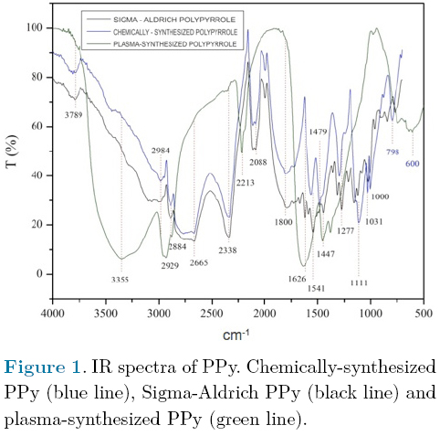
The chemical structure of polymers employed in the experiment was determined by IR spectroscopy. <a href="#f1">Figure 1</a> shows the similarity between the two chemically&#45;synthesized PPy; in particular in the region between 1750 and 4000 cm<sup>&#45;1</sup>. Small differences in the spectra can be observed between 400 and 1750 cm<sup>&#45;1</sup> which we believe are due to some substitutions in the pyrrole rings. In contrast, the same figure shows a significant difference between the chemically&#45;synthesized PPy and PPy synthesized by plasma. At 3355 cm<sup>&#45;1</sup> one can observe the different vibrations of the primary and secondary amine groups. Another difference is the vibration corresponding to nitrile groups at 2213 cm<sup>&#45;1</sup>.</font></p>  	    ]]></body>
<body><![CDATA[<p align="center"><font face="verdana" size="2"><a name="f1"></a></font></p>  	    <p align="center"><font face="verdana" size="2"><img src="/img/revistas/rmib/v36n1/a1f1.jpg"></font></p>  	    <p align="justify"><font face="verdana" size="2"><a href="#f2">Figure 2</a> shows the IR spectra of PPy/PEG copolymers synthesized electrochemically and by plasma polymerization. It can be observed that only the four absorptions highlighted in green coincide in both materials. In the copolymer synthesized by plasma, the main absorptions are in 717, 1617 2212, 2928 and 3312 cm<sup>&#45;1</sup>; it is notable that none of these absorptions were identified in the electrochemically&#45;synthesized copolymer, which shows main absorptions at 664, 2360 and 1033 cm<sup>&#45;1</sup>.</font></p>  	    <p align="center"><font face="verdana" size="2"><a name="f2"></a></font></p>  	    <p align="center"><font face="verdana" size="2"><img src="/img/revistas/rmib/v36n1/a1f2.jpg"></font></p>  	    <p align="justify"><font face="verdana" size="2">
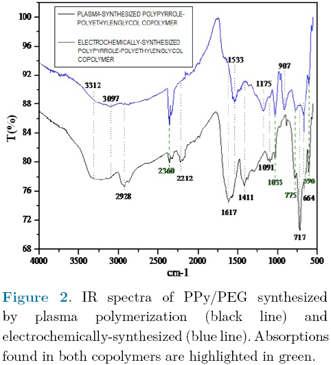
These differences are indicative of a significant difference in chemical structure between the two copolymers.</font></p>  	    <p align="justify"><font face="verdana" size="2">In the copolymer synthesized by plasma, the more intense absorption was found at 717 cm<sup>&#45;1</sup> and corresponds to &#61;C&#45;H groups, the absorption at 1617 cm<sup>&#45;1</sup> can be associated with C &#61; C groups, the absorption at 2212 cm<sup>&#45;1</sup> correspond to nitrile groups, while the absorption found at 2928 cm<sup>&#45;1</sup> indicates the presence of &#45;CH aliphatic groups which can arise from ethylene glycol segments or from fragments of pyrrole molecules which were fractionated due to the high energy of the particles in the plasma, the absorption at 3312 cm<sup>&#45;1</sup> can be associated with the presence of amine groups. These absorptions indicate that in the copolymer synthesized by plasma, the heteroaromatic character of the pyrroles predominates.</font></p>  	    <p align="justify"><font face="verdana" size="2">In the case of the electrochemically&#45;synthesized co&#45;polymer, the more intense absorption was found in 664 cm<sup>&#45;1</sup> and can be assigned to N&#45;O groups. This implies a substitution of N&#45;H groups by N&#45;O groups which could be a consequence of ethylene glycol participation during the synthesis. The absorption at 1033 cm<sup>&#45;1</sup> corresponds to C&#45;O groups that are found in ethylene glycol repeat units. These absorptions indicate that in the electrochemically&#45;synthesized copolymer, the oxygenated character of the ethylene glycol predominates.</font></p>  	    <p align="justify"><font face="verdana" size="2">&nbsp;</font></p>  	    <p align="justify"><font face="verdana" size="2"><b>Motor function recovery</b></font></p>  	    ]]></body>
<body><![CDATA[<p align="justify"><font face="verdana" size="2">The motor function was evaluated 24 h after complete transection of the spinal cord with the aim to corroborate if hind limb performance showed complete bilateral paralysis in all rats (BBB&#61;0). Afterwards, the BBB locomotor rating scale was assessed once a week during 5 weeks to evaluate the gradual improvement of the animals. Results were as follow (<a href="/img/revistas/rmib/v36n1/a1f3.jpg" target="_blank">Figure 3</a>):</font></p>  	    <blockquote> 		    <p align="justify"><font face="verdana" size="2">a) Control (3 animals alive): BBB&#61;1, meaning that animals had slight movement of 1 or 2 joints.</font></p>  		    <p align="justify"><font face="verdana" size="2">b) PPy (3 animals alive): BBB&#61;5.33, meaning that animals were able to move the 3 joints of the hind limb (hip, knee and ankle), and in two them, the movements were extensive.</font></p>  		    <p align="justify"><font face="verdana" size="2">c) ChPPy1 (3 animals alive and 2 dead animals, non&#45;evaluable): BBB&#61;1, meaning that the animals had slight movement of 1 or 2 joints. Although one of the animals had an improvement of 4 points in the BBB scale in the third week, score decreased with time.</font></p>  		    <p align="justify"><font face="verdana" size="2">d) ChPPy2 (3 animals alive and 4 dead animals, non&#45;evaluable): BBB&#61;1.33, meaning that the animal had slight movement in one or two joints. But just as in the ChPPy1 group, one animal presented improvement until the fourth week and subsequently decreased the effect of the implant.</font></p>  		    <p align="justify"><font face="verdana" size="2">e) PPy&#45;PEG (3 animals alive): BBB&#61;3.66, meaning that animals had extensive movement in 1 or 2 joints of the hind limb and slight movement of other joint (hip, knee and ankle).</font></p>  		    <p align="justify"><font face="verdana" size="2">f) EPPy&#45;PEG (3 animals alive): BBB&#61;2.6, meaning that animals had extensive movement of 1 joint and slight movement of 2 joints of the hind limbs.</font></p> 	</blockquote>  	    <p align="justify"><font face="verdana" size="2">The dead animals did not show infection of the urinary or respiratory tract, hypertrophy of urethral <i>meatus</i>, or shallow or deep wounds. Thus, the cause of death is unknown.</font></p>  	    <p align="justify"><font face="verdana" size="2">Animals implanted with materials synthesized by plasma demonstrated greater motor function recovery comparing with animals implanted with materials obtained by chemical or electrochemical synthesis and with control group animals. Animals with implants synthesized by plasma showed significant differences with animals from the control group; PPy (p &#61; 0.028) and PPy / PEG (p &#61; 0.05).</font></p>  	    ]]></body>
<body><![CDATA[<p align="justify"><font face="verdana" size="2">&nbsp;</font></p>  	    <p align="justify"><font face="verdana" size="2"><b>Histological analysis</b></font></p>  	    <p align="justify"><font face="verdana" size="2">Four weeks after SCI, animals were sacrificed to analyze the integration of implants to the spinal cord tissue and the inflammatory response. The control group (<a href="/img/revistas/rmib/v36n1/a1f4.jpg" target="_blank">Fig. 4A</a>) showed greater tissue destruction compared with implanted animals. Analysis of implants integration to the spinal cord tissue showed that PPy implants synthesized by plasma polymerization were well integrated to the tissue and that the surrounding tissue showed little destruction (<a href="/img/revistas/rmib/v36n1/a1f4.jpg" target="_blank">Fig. 4B</a>). The chemically&#45;synthesized implants ChPPy1 and ChPPy2 (<a href="/img/revistas/rmib/v36n1/a1f4.jpg" target="_blank">Fig. 4C</a> and <a href="/img/revistas/rmib/v36n1/a1f4.jpg" target="_blank">4D</a> respectively) had similar amount of tissue destruction and complete lack of implant integration, with a great cyst at the injury epicenter and a complete loss of histological architecture.</font></p>  	    <p align="justify"><font face="verdana" size="2">Regarding the inflammatory response, morphometric analysis showed the presence of 142 polymorphonuclear cells per every 4 microns in the animals with ChPPy1 implant and 128 polymorphonuclear cells in the animals with ChPPy2 implant (obtained from Sigma&#45;Aldrich). In contrast, the morphometric analysis of samples with implants synthesized by plasma for both PPy and PPy/PEG showed about 74 inflammatory cells at the tissue surrounding the implant, and if PPy was doped with iodine (PPy/I) 95 inflammatory cells were found in the tissue that surrounded the implant.</font></p>  	    <p align="justify"><font face="verdana" size="2">&nbsp;</font></p>  	    <p align="justify"><font face="verdana" size="2"><b>DISCUSSION AND CONCLUSION</b></font></p>  	    <p align="justify"><font face="verdana" size="2">The present work compared the effect of different implants derived from pyrrole (PPy and PPy/PEG) and obtained by standard chemical, electrochemical, and plasma polymerizations. The implants were analyzed according to their chemical structure, integration with the nervous tissue, and their effect on functional recovery in rats with complete section of the spinal cord.</font></p>  	    <p align="justify"><font face="verdana" size="2">The PPy implants that were chemically synthesized as ChPPy1 and ChPPy2 have a similar chemical structure. None of them were integrated to the nervous tissue, a large number of cysts and inflammatory cells were found on the tissue around the implants, and a complete loss of cytoarchitecture of the spinal cord was observed. Animals that received ChPPy1 or ChPPy2 showed some functional recovery between the third and fourth week post injury. This recovery, however, decreased afterwards. Meanwhile, animals implanted with PPy synthesized by plasma showed implant integration to the nerve tissue, little destruction of nervous tissue and significant functional recovery when comparing with animals with implants obtained by chemical methods and animals from control group (p &#61; 0.028).</font></p>  	    <p align="justify"><font face="verdana" size="2">Although both implants of PPy copolymerized with PEG and synthesized by plasma or electrochemically were well integrated to the nervous tissue, the functional recovery was different. Animals that received implants of PPy/PEG synthesized by plasma showed greater motor recovery (p &#61; 0.05 with respect to animals from control group) than those which received implants obtained electrochemically.</font></p>  	    <p align="justify"><font face="verdana" size="2">The results show significant differences in the effects of the method of synthesis on functional recovery and implant integration to the nervous tissue. Although the polymers had the same molecules of origin, IR analysis showed that method of synthesis affects the chemical structure of the material.</font></p>  	    ]]></body>
<body><![CDATA[<p align="justify"><font face="verdana" size="2">Wangh et al., showed that the chemical structure of PPy and of polythiophene synthesized by plasma are different from the ones chemically synthesized because the materials obtained by plasma are highly crosslinked and branched &#91;45&#93; and they can form three&#45;dimensional network which can support and promote neural cells grown while the presence of heteroaromatic amine groups and nitrile groups can favor neuroprotection after an injury in the central nervous system. Furthermore, the polymers synthesized by plasma are insoluble, thermally stable and chemically inert &#91;46&#93;, characteristics that make them more desirable for implants in a biological system.</font></p>  	    <p align="justify"><font face="verdana" size="2">Pyrrole belongs to a class of heterocyclic compounds that are in various natural components such as the heme group, chlorophyll and vitamin B12 &#91;47&#93; and have been used in various applications of chemical medicine as anti&#45;inflammatory, antibacterial and antihypertensive agents, as well as agents for tyrosine kinase inhibition&#91;48&#93;.</font></p>  	    <p align="justify"><font face="verdana" size="2">Currently, PPy is mainly synthesized by chemical oxidation or electrochemical polymerization &#91;16&#93;. Nevertheless, for biomedical applications, plasma synthesis might be a better option because the unique characteristics of PPy generated by plasma that allow improvement of the cellular microenvironment and favor the attachment and growth of cells. The low rate of degradation of the material maintains the cell adhesion, promotes morphological maturation and allows preservation of its properties &#91;34&#93;. It has been shown in cultures of nerve cells on surfaces treated with PPy, that the adhesion, proliferation, attachment, viability and number of synapses is increased compared with that observed in cultures performed on surfaces treated with compounds as Poly DL&#45;Ornithine / Laminin &#91;34&#93;. Furthermore, our research group has demonstrated neuroprotective activity of PPy synthesized by plasma, as well as greater functional recovery in animals that received this type of implant after a TSCI, compared with those who did not receive it &#91;36&#45;38&#93;.</font></p>  	    <p align="justify"><font face="verdana" size="2">The above may be due to the chemical structure of PPy obtained by plasma polymerization. IR spectroscopy of this material shows the presence of a variety of chemical groups including primary amines, nitriles and aliphatic sections &#91;34&#93;, and it has been shown that the structures consisting mainly of methyl&#45;, hydroxyl&#45;, amino&#45; and carboxyl&#45; functional groups, which are found in natural biological surfaces, favor the growth of cells &#91;49&#93;.</font></p>  	    <p align="justify"><font face="verdana" size="2">The adaptability of tissues to materials such as the implants studied in this work begins with the absorption of solutions at the surface of the material. The hydrophilic properties of PPy, which can be increased by increasing the ionization capability of the material, allow one to store solutions and favor interaction with cells &#91;50&#45;51&#93;, which generates optimal sites for cell attachment.</font></p>  	    <p align="justify"><font face="verdana" size="2">Nerve cells carry out their function by generating electrical activity. The nervous system thus responds to electric fields and the key component of the neural communication is the action potential generated in the synapse. This implies that the ideal biomaterial to implant in this system must introduce electrical stimulators to promote neuronal growth and nerve regeneration &#91;13&#93;. The material must promote regeneration of the nervous tissue at the interface by attraction or rejection of ions and polar groups between the cells and the material. In addition to this, it has been shown that electrical stimulation alters the absorption of proteins and the interaction with the nerve cells &#91;52&#93;, which could also favor nerve regeneration processes. Although the breaking of rings in the plasma polymerization process results in a polymer complex of low conductivity, when is introduced into a biological system its sensitivity to humidity increases its conductivity &#91;40&#93;. It is known that the electric conductivity of conductive polymers synthesized by chemical or electrochemical methods oscillates within a range of 10<sup>&#45;10</sup> to 10<sup>&#45;5</sup> S cm<sup>&#45;1</sup> &#91;39&#93;. The electric conductivity of the plasma&#45;synthesized PPy measured at 30 &#37; relative humidity is around 10<sup>&#45;12</sup> S cm<sup>&#45;1</sup>, while at 90 &#37; of relative humidity it is 10<sup>&#45;9</sup> S cm<sup>&#45;1</sup> &#91;40&#93;. The PPy/PEG copolymer has a conductivity of 10<sup>&#45;12</sup> S cm<sup>&#45;1</sup> at 30 &#37; of relative humidity &#91;36&#93; and of 10<sup>&#45;9</sup> to 10<sup>&#45;8</sup> S cm<sup>&#45;1</sup> when it is dampened with ionic solutions &#91;51&#93;.</font></p>  	    <p align="justify"><font face="verdana" size="2">Due to their physical and chemical properties, the PPy does not alter the biological functions of the cell cultures and provides better cell attachment and an increased rate of proliferation, which may be due to the accumulation of amino groups (&#45;NH<sub>2</sub>) and interaction with other groups generated during the process of plasma polymerization &#91;34&#93;, which may explain the better results obtained when using PPy synthesized by plasma vs the PPy synthesized by conventional chemical or electrochemical methods.</font></p>  	    <p align="justify"><font face="verdana" size="2">&nbsp;</font></p>  	    <p align="justify"><font face="verdana" size="2"><b>ACKNOWLEDGMENTS</b></font></p>  	    <p align="justify"><font face="verdana" size="2">The work was supported by CONACyT grant No. 155239 and by IMSS grant No. FOFOI 2005/1/I/149. Ana Laura &Aacute;lvarez received a scholarship from CONACyT, No. 172211. The authors want to thank to Mar&iacute;a del Carmen Baltazar for her technical assistance.</font></p>  	    ]]></body>
<body><![CDATA[<p align="justify"><font face="verdana" size="2">&nbsp;</font></p>  	    <p align="justify"><font face="verdana" size="2"><b>REFERENCES</b></font></p>      <!-- ref --><p align="justify"><font face="verdana" size="2">1. C.A. Oyinbo, "Secondary injury mechanisms in traumatic spinal cord injury: a nugget of this multiply cascade," <i>Acta Neurobiol Exp</i>, vol. 71, pp. 281&#45;299, 2011.    &nbsp;&nbsp;&nbsp;&nbsp;&nbsp;&nbsp;&nbsp;&nbsp;[&#160;<a href="javascript:void(0);" onclick="javascript: window.open('/scielo.php?script=sci_nlinks&ref=8517291&pid=S0188-9532201500010000100001&lng=','','width=640,height=500,resizable=yes,scrollbars=1,menubar=yes,');">Links</a>&#160;]<!-- end-ref --></font></p>  	    <!-- ref --><p align="justify"><font face="verdana" size="2">2. W. Tetzlaff, E.B. Okon, S. Karimi&#45;Abdolrezaee, C.E. Hill, J.S. Sparling, J.R. Plemel, W.T. Plunet, E.C. Tsai, D. Baptiste, L.J. Smithson, M.D. Kawaja, M.G. Fehlings, B.K. Kwon "A systematic review of cellular transplantation therapies for spinal cord injury," <i>J Neurotrauma</i>., vol. 28, no. 8, pp. 1611&#45;1682, 2011.    &nbsp;&nbsp;&nbsp;&nbsp;&nbsp;&nbsp;&nbsp;&nbsp;[&#160;<a href="javascript:void(0);" onclick="javascript: window.open('/scielo.php?script=sci_nlinks&ref=8517293&pid=S0188-9532201500010000100002&lng=','','width=640,height=500,resizable=yes,scrollbars=1,menubar=yes,');">Links</a>&#160;]<!-- end-ref --></font></p>  	    <!-- ref --><p align="justify"><font face="verdana" size="2">3. S. Venkatachalam, "Fetal neural tissue transplantation for spinal cord injury repair," Chapter 23 in N. Bhattacharya, P. Stubblefield (eds.), <i>Human Fetal Tissue Transplantation</i>. Springer London, pp. 297&#45;305, 2013.    &nbsp;&nbsp;&nbsp;&nbsp;&nbsp;&nbsp;&nbsp;&nbsp;[&#160;<a href="javascript:void(0);" onclick="javascript: window.open('/scielo.php?script=sci_nlinks&ref=8517295&pid=S0188-9532201500010000100003&lng=','','width=640,height=500,resizable=yes,scrollbars=1,menubar=yes,');">Links</a>&#160;]<!-- end-ref --></font></p>  	    <!-- ref --><p align="justify"><font face="verdana" size="2">4. H. Salgado&#45;Ceballos, I. Grijalva, G. Guizar&#45;Sahagun, A. L. Espitia, A. Mart&iacute;nez, A. Feria&#45;Velasco, "Predegenerated peripheral nerve graft with and without methylprednisolone administration after traumatic spinal cord injury in adult rats," <i>Neurosci. Res. Comm</i>., vol. 33, no. 2, pp. 77&#45;85, 2003.    &nbsp;&nbsp;&nbsp;&nbsp;&nbsp;&nbsp;&nbsp;&nbsp;[&#160;<a href="javascript:void(0);" onclick="javascript: window.open('/scielo.php?script=sci_nlinks&ref=8517297&pid=S0188-9532201500010000100004&lng=','','width=640,height=500,resizable=yes,scrollbars=1,menubar=yes,');">Links</a>&#160;]<!-- end-ref --></font></p>  	    ]]></body>
<body><![CDATA[<!-- ref --><p align="justify"><font face="verdana" size="2">5. M.P. C&ocirc;t&eacute;, A.A. Amin, V.J. Tom, J.D. Houle "Peripheral nerve grafts support regeneration after spinal cord injury," <i>Neurotherapeutics</i>, vol. 8, no. 2, pp. 294&#45;303, 2011.    &nbsp;&nbsp;&nbsp;&nbsp;&nbsp;&nbsp;&nbsp;&nbsp;[&#160;<a href="javascript:void(0);" onclick="javascript: window.open('/scielo.php?script=sci_nlinks&ref=8517299&pid=S0188-9532201500010000100005&lng=','','width=640,height=500,resizable=yes,scrollbars=1,menubar=yes,');">Links</a>&#160;]<!-- end-ref --></font></p>  	    <!-- ref --><p align="justify"><font face="verdana" size="2">6. H. Kanno, Y. Pressman, A. Moody, R. Berg, E.M. Muir, J.H. Rogers, H. Ozawa, E. Itoi, D.D. Pearse, M.B. Bunge, "Combination of engineered Schwann cell grafts to secrete neurotrophin and chondroitinase promotes axonal regeneration and locomotion after spinal cord injury," <i>J. Neurosci.</i>, vol. 34, no. 5, pp. 1838&#45;1855, 2014.    &nbsp;&nbsp;&nbsp;&nbsp;&nbsp;&nbsp;&nbsp;&nbsp;[&#160;<a href="javascript:void(0);" onclick="javascript: window.open('/scielo.php?script=sci_nlinks&ref=8517301&pid=S0188-9532201500010000100006&lng=','','width=640,height=500,resizable=yes,scrollbars=1,menubar=yes,');">Links</a>&#160;]<!-- end-ref --></font></p>  	    <!-- ref --><p align="justify"><font face="verdana" size="2">7. Y.J. Rao, W.X. Zhu, Z.Q. Du, C.X. Jia, T.X. Du, Q.A. Zhao, X.Y. Cao, Y.J. Wang "Effectiveness of olfactory ensheathing cell transplantation for treatment of spinal cord injury," <i>Genet. Mol. Res</i>., vol. 13, no. 2, pp. 4124&#45;4129, 2014.    &nbsp;&nbsp;&nbsp;&nbsp;&nbsp;&nbsp;&nbsp;&nbsp;[&#160;<a href="javascript:void(0);" onclick="javascript: window.open('/scielo.php?script=sci_nlinks&ref=8517303&pid=S0188-9532201500010000100007&lng=','','width=640,height=500,resizable=yes,scrollbars=1,menubar=yes,');">Links</a>&#160;]<!-- end-ref --></font></p>  	    <!-- ref --><p align="justify"><font face="verdana" size="2">8. Z.A, Sobani, S.A. Quadri, S.A. Enam, "Stem cells for spinal cord regeneration: Current status," <i>Surg Neurol Int</i>., vol. 1, pp. 93, 2010.    &nbsp;&nbsp;&nbsp;&nbsp;&nbsp;&nbsp;&nbsp;&nbsp;[&#160;<a href="javascript:void(0);" onclick="javascript: window.open('/scielo.php?script=sci_nlinks&ref=8517305&pid=S0188-9532201500010000100008&lng=','','width=640,height=500,resizable=yes,scrollbars=1,menubar=yes,');">Links</a>&#160;]<!-- end-ref --></font></p>  	    <!-- ref --><p align="justify"><font face="verdana" size="2">9. H. Nakajima, K. Uchida, Rodriguez A. Guerrero, S. Watanabe, D. Sugita, N. Takeura, A. Yoshida, G. Long, K. Wright, E. Johnson, H. Baba, "Transplantation of mesenchymal stem cells Promotes the alternative pathway of macrophage activation and functional recovery after spinal cord injury," <i>J. Neurotrauma</i>., vol. 29, no. 8, pp. 1614&#45;1625, 2012.    &nbsp;&nbsp;&nbsp;&nbsp;&nbsp;&nbsp;&nbsp;&nbsp;[&#160;<a href="javascript:void(0);" onclick="javascript: window.open('/scielo.php?script=sci_nlinks&ref=8517307&pid=S0188-9532201500010000100009&lng=','','width=640,height=500,resizable=yes,scrollbars=1,menubar=yes,');">Links</a>&#160;]<!-- end-ref --></font></p>  	    ]]></body>
<body><![CDATA[<!-- ref --><p align="justify"><font face="verdana" size="2">10. J. Ai, A. Kiasat&#45;Dolatabadi, S. Ebrahimi&#45;Barough, A. Ai, N. Lotfibakhshaiesh, A. Norouzi&#45;Javidan, H. Saberi, B. Arjmand, HR. Aghayan, "Polymeric scaffolds in neural tissue engineering: A review," <i>Arch. Neuro Sci</i>., vol. 1, no. 1, pp. 15&#45;20, 2013.    &nbsp;&nbsp;&nbsp;&nbsp;&nbsp;&nbsp;&nbsp;&nbsp;[&#160;<a href="javascript:void(0);" onclick="javascript: window.open('/scielo.php?script=sci_nlinks&ref=8517309&pid=S0188-9532201500010000100010&lng=','','width=640,height=500,resizable=yes,scrollbars=1,menubar=yes,');">Links</a>&#160;]<!-- end-ref --></font></p>  	    <!-- ref --><p align="justify"><font face="verdana" size="2">11. V. Krishna, S. Konakondla, J. Nicholas, A. Varma, M. Kindy, X. Wen, "Biomaterial&#45;based interventions for neuronal regeneration and functional recovery in rodent model of spinal cord injury: A systematic review," <i>J Spinal Cord Med</i>., vol. 36, no. 3, pp. 174&#45;190, 2013.    &nbsp;&nbsp;&nbsp;&nbsp;&nbsp;&nbsp;&nbsp;&nbsp;[&#160;<a href="javascript:void(0);" onclick="javascript: window.open('/scielo.php?script=sci_nlinks&ref=8517311&pid=S0188-9532201500010000100011&lng=','','width=640,height=500,resizable=yes,scrollbars=1,menubar=yes,');">Links</a>&#160;]<!-- end-ref --></font></p>  	    <!-- ref --><p align="justify"><font face="verdana" size="2">12. M. Wang, P. Zhai, X. Chen, D.J. Schreyer, X. Sun, F. Cui "Bioengineered scaffolds for spinal cord repair," <i>Tissue Eng. Part B Rev</i>., vol. 17, no. 3, pp. 177&#45;194, 2011.    &nbsp;&nbsp;&nbsp;&nbsp;&nbsp;&nbsp;&nbsp;&nbsp;[&#160;<a href="javascript:void(0);" onclick="javascript: window.open('/scielo.php?script=sci_nlinks&ref=8517313&pid=S0188-9532201500010000100012&lng=','','width=640,height=500,resizable=yes,scrollbars=1,menubar=yes,');">Links</a>&#160;]<!-- end-ref --></font></p>  	    <!-- ref --><p align="justify"><font face="verdana" size="2">13. L. Ghasemi&#45;Mobarakeh, M.P. Prabhakaran, M. Morshed, M.H. Nasr&#45;Esfahani, H. Baharvand, S. Kiani, S.S. Al&#45;Deyab, S. Ramakrishna "Application of conductive polymers, scaffolds and electrical stimulation for nerve tissue engineering," <i>J. Tissue Eng Regen Med</i>, vol. 5, no. 4, 17&#45;35, 2011.    &nbsp;&nbsp;&nbsp;&nbsp;&nbsp;&nbsp;&nbsp;&nbsp;[&#160;<a href="javascript:void(0);" onclick="javascript: window.open('/scielo.php?script=sci_nlinks&ref=8517315&pid=S0188-9532201500010000100013&lng=','','width=640,height=500,resizable=yes,scrollbars=1,menubar=yes,');">Links</a>&#160;]<!-- end-ref --></font></p>  	    <!-- ref --><p align="justify"><font face="verdana" size="2">14. I.S. Chronakis, S. Grapenson, A. Jakob, "Conductive polypyrrole nanofibers via electrospinning: electrical and morphological properties," <i>Polymer</i>, vol. 47, no. 5, pp. 1597&#45;1603, 2006.    &nbsp;&nbsp;&nbsp;&nbsp;&nbsp;&nbsp;&nbsp;&nbsp;[&#160;<a href="javascript:void(0);" onclick="javascript: window.open('/scielo.php?script=sci_nlinks&ref=8517317&pid=S0188-9532201500010000100014&lng=','','width=640,height=500,resizable=yes,scrollbars=1,menubar=yes,');">Links</a>&#160;]<!-- end-ref --></font></p>  	    ]]></body>
<body><![CDATA[<!-- ref --><p align="justify"><font face="verdana" size="2">15. Q. Zhang, Y. Yan, S. Li, <i>et al.</i>, "The synthesis and characterization of a novel biodegradable and electroactive polyphosphazene for nerve regeneration," <i>Mater. Sci. Eng. C</i>, vol. 30, no. 1, pp. 160&#45;166, 2010.    &nbsp;&nbsp;&nbsp;&nbsp;&nbsp;&nbsp;&nbsp;&nbsp;[&#160;<a href="javascript:void(0);" onclick="javascript: window.open('/scielo.php?script=sci_nlinks&ref=8517319&pid=S0188-9532201500010000100015&lng=','','width=640,height=500,resizable=yes,scrollbars=1,menubar=yes,');">Links</a>&#160;]<!-- end-ref --></font></p>  	    <!-- ref --><p align="justify"><font face="verdana" size="2">16. M.A. Chougule, S.G. Pawar, P.R. Godse, R.N. Mulik, S. Sen, V.B. Patil, "Synthesis and characterization of polypyrrole (PPy) thin films," <i>Soft Nanoscience Letters</i>, vol. 1, no. 1, pp. 6&#45;10, 2011.    &nbsp;&nbsp;&nbsp;&nbsp;&nbsp;&nbsp;&nbsp;&nbsp;[&#160;<a href="javascript:void(0);" onclick="javascript: window.open('/scielo.php?script=sci_nlinks&ref=8517321&pid=S0188-9532201500010000100016&lng=','','width=640,height=500,resizable=yes,scrollbars=1,menubar=yes,');">Links</a>&#160;]<!-- end-ref --></font></p>  	    <!-- ref --><p align="justify"><font face="verdana" size="2">17. J.C. Vidal, E. Garcia, J.R. Castillo "<i>In situ</i> preparation of a cholesterol biosensor: entrapment of cholesterol oxidase in an overoxidized polypyrrole film electrodeposited in a flow system: determination of total cholesterol in serum," <i>Analytica Chimica Acta</i>, vol. 385, no. 1&#45;3, pp. 213&#45;222, 1999.    &nbsp;&nbsp;&nbsp;&nbsp;&nbsp;&nbsp;&nbsp;&nbsp;[&#160;<a href="javascript:void(0);" onclick="javascript: window.open('/scielo.php?script=sci_nlinks&ref=8517323&pid=S0188-9532201500010000100017&lng=','','width=640,height=500,resizable=yes,scrollbars=1,menubar=yes,');">Links</a>&#160;]<!-- end-ref --></font></p>  	    <!-- ref --><p align="justify"><font face="verdana" size="2">18. E. Lopez&#45;Crapez, T. Livache, J. Marchand, J. Grenier "K&#45;ras mutation detection by hybridization to a polypyrrole DNA chip," <i>Clin Chem</i>., vol. 47, no. 2, pp. 186&#45;194, 2001.    &nbsp;&nbsp;&nbsp;&nbsp;&nbsp;&nbsp;&nbsp;&nbsp;[&#160;<a href="javascript:void(0);" onclick="javascript: window.open('/scielo.php?script=sci_nlinks&ref=8517325&pid=S0188-9532201500010000100018&lng=','','width=640,height=500,resizable=yes,scrollbars=1,menubar=yes,');">Links</a>&#160;]<!-- end-ref --></font></p>  	    <!-- ref --><p align="justify"><font face="verdana" size="2">19. T.E. Campbell, A.J. Hodgson, G.G. Wallace "Incorporation of eythrocytes into polypyrrole to form the basis of a biosensor to screen for rhesus (D) blood groups and rhesus (D) antibodies," <i>Electroanalysis</i>, vol. 11, no. 4, pp. 215&#45;222, 1999.    &nbsp;&nbsp;&nbsp;&nbsp;&nbsp;&nbsp;&nbsp;&nbsp;[&#160;<a href="javascript:void(0);" onclick="javascript: window.open('/scielo.php?script=sci_nlinks&ref=8517327&pid=S0188-9532201500010000100019&lng=','','width=640,height=500,resizable=yes,scrollbars=1,menubar=yes,');">Links</a>&#160;]<!-- end-ref --></font></p>  	    ]]></body>
<body><![CDATA[<!-- ref --><p align="justify"><font face="verdana" size="2">20. R.H.M. van de Leur, A. van der Waal, "Gas and vapour detection using polypyrrole," <i>Synthetic Metals</i>, vol. 102, no. 1&#45;3, pp. 1330&#45;1331, 1999.    &nbsp;&nbsp;&nbsp;&nbsp;&nbsp;&nbsp;&nbsp;&nbsp;[&#160;<a href="javascript:void(0);" onclick="javascript: window.open('/scielo.php?script=sci_nlinks&ref=8517329&pid=S0188-9532201500010000100020&lng=','','width=640,height=500,resizable=yes,scrollbars=1,menubar=yes,');">Links</a>&#160;]<!-- end-ref --></font></p>  	    <!-- ref --><p align="justify"><font face="verdana" size="2">21. A.B. Sanghvi, K.P. Miller, A.M. Belcher, C.E. Schmidt, "Biomaterials fictionalization using a novel peptide that selectively binds to a conducting polymer," <i>Nat Mater,</i> vol. 4, no. 6, pp. 496&#45;502, 2005.    &nbsp;&nbsp;&nbsp;&nbsp;&nbsp;&nbsp;&nbsp;&nbsp;[&#160;<a href="javascript:void(0);" onclick="javascript: window.open('/scielo.php?script=sci_nlinks&ref=8517331&pid=S0188-9532201500010000100021&lng=','','width=640,height=500,resizable=yes,scrollbars=1,menubar=yes,');">Links</a>&#160;]<!-- end-ref --></font></p>  	    <!-- ref --><p align="justify"><font face="verdana" size="2">22. J.R. Reynolds, H. Ly, F. Selampinar, P.J. Kinlen, "Controlled drug and biomolecule release from electroactive host polymer systems," <i>Polymer Preprints</i>, vol. 40, no. 1, pp. 307, 1999.    &nbsp;&nbsp;&nbsp;&nbsp;&nbsp;&nbsp;&nbsp;&nbsp;[&#160;<a href="javascript:void(0);" onclick="javascript: window.open('/scielo.php?script=sci_nlinks&ref=8517333&pid=S0188-9532201500010000100022&lng=','','width=640,height=500,resizable=yes,scrollbars=1,menubar=yes,');">Links</a>&#160;]<!-- end-ref --></font></p>  	    <!-- ref --><p align="justify"><font face="verdana" size="2">23. T.F. Otero, M.T. Cort&eacute;s, "Artificial muscles with tactile sensitivity," <i>Adv. Mater</i>., vol. 15, no. 3, 279&#45;289, 2003.    &nbsp;&nbsp;&nbsp;&nbsp;&nbsp;&nbsp;&nbsp;&nbsp;[&#160;<a href="javascript:void(0);" onclick="javascript: window.open('/scielo.php?script=sci_nlinks&ref=8517335&pid=S0188-9532201500010000100023&lng=','','width=640,height=500,resizable=yes,scrollbars=1,menubar=yes,');">Links</a>&#160;]<!-- end-ref --></font></p>  	    <!-- ref --><p align="justify"><font face="verdana" size="2">24. X. Wang, X. Gu, C. Yuan, S. Chen, P. Zhang, T. Zhang, J. Yao, F. Chen, G. Chen, "Evaluation of biocompatibility of polypyrrole <i>in vitro</i> and <i>in vivo</i>," <i>J Biomed Mater Res A</i>, vol. 68A, no. 3, pp. 411&#45;422, 2004.    &nbsp;&nbsp;&nbsp;&nbsp;&nbsp;&nbsp;&nbsp;&nbsp;[&#160;<a href="javascript:void(0);" onclick="javascript: window.open('/scielo.php?script=sci_nlinks&ref=8517337&pid=S0188-9532201500010000100024&lng=','','width=640,height=500,resizable=yes,scrollbars=1,menubar=yes,');">Links</a>&#160;]<!-- end-ref --></font></p>  	    ]]></body>
<body><![CDATA[<!-- ref --><p align="justify"><font face="verdana" size="2">25. R.L. Williams, P.J. Doherty, "A preliminary assessment of poly(pyrrole) in nerve guide studies," <i>J. Mater Sci&#45;Mater M</i>., vol. 5, no. 6&#45;7, pp. 429&#45;433, 1994.    &nbsp;&nbsp;&nbsp;&nbsp;&nbsp;&nbsp;&nbsp;&nbsp;[&#160;<a href="javascript:void(0);" onclick="javascript: window.open('/scielo.php?script=sci_nlinks&ref=8517339&pid=S0188-9532201500010000100025&lng=','','width=640,height=500,resizable=yes,scrollbars=1,menubar=yes,');">Links</a>&#160;]<!-- end-ref --></font></p>  	    <!-- ref --><p align="justify"><font face="verdana" size="2">26. H. Castano, E.A. O'Rear, P.S. McFetridge, V.I. Sikavitsas "Polypyrrole thin films formed by admicellar polymerization support the osteogenic differentiation of mesenchymal stem cells," <i>Macromol Biosci</i>, vol. 4, no. 8, pp. 785&#45;794, 2004.    &nbsp;&nbsp;&nbsp;&nbsp;&nbsp;&nbsp;&nbsp;&nbsp;[&#160;<a href="javascript:void(0);" onclick="javascript: window.open('/scielo.php?script=sci_nlinks&ref=8517341&pid=S0188-9532201500010000100026&lng=','','width=640,height=500,resizable=yes,scrollbars=1,menubar=yes,');">Links</a>&#160;]<!-- end-ref --></font></p>  	    <!-- ref --><p align="justify"><font face="verdana" size="2">27. H.K. Song, B. Toste, K. Ahmann, D. Hoffman&#45;Kim, G.T. Palmore, "Micropatterns of positive guidance cues anchored to polypyrrole dopedwith polyglutamic acid: a new platform for characterzing neurite extension in complex environments," <i>Biomaterials</i>, vol. 27, no. 3, pp. 473&#45;484, 2006.    &nbsp;&nbsp;&nbsp;&nbsp;&nbsp;&nbsp;&nbsp;&nbsp;[&#160;<a href="javascript:void(0);" onclick="javascript: window.open('/scielo.php?script=sci_nlinks&ref=8517343&pid=S0188-9532201500010000100027&lng=','','width=640,height=500,resizable=yes,scrollbars=1,menubar=yes,');">Links</a>&#160;]<!-- end-ref --></font></p>  	    <!-- ref --><p align="justify"><font face="verdana" size="2">28. D.D. Ateh, P. Vadgama, H.A. Navasaria, "Polypyrrole&#45;based conducting polymers and interactions with biological tissues," <i>J. R. Soc. Interface</i>, vol. 3, no. 11, pp. 741&#45;752, 2006.    &nbsp;&nbsp;&nbsp;&nbsp;&nbsp;&nbsp;&nbsp;&nbsp;[&#160;<a href="javascript:void(0);" onclick="javascript: window.open('/scielo.php?script=sci_nlinks&ref=8517345&pid=S0188-9532201500010000100028&lng=','','width=640,height=500,resizable=yes,scrollbars=1,menubar=yes,');">Links</a>&#160;]<!-- end-ref --></font></p>  	    <!-- ref --><p align="justify"><font face="verdana" size="2">29. N. Gomez, C.E. Schmidt, "Nerve growth factor&#45;immobilized polypyrrole: bioactive electrically conducting polymer for enhanced neurite extension," <i>J. Biomed. Mater. Res</i>, vol. 81, no. 1, pp. 135&#45;149. 2007.    &nbsp;&nbsp;&nbsp;&nbsp;&nbsp;&nbsp;&nbsp;&nbsp;[&#160;<a href="javascript:void(0);" onclick="javascript: window.open('/scielo.php?script=sci_nlinks&ref=8517347&pid=S0188-9532201500010000100029&lng=','','width=640,height=500,resizable=yes,scrollbars=1,menubar=yes,');">Links</a>&#160;]<!-- end-ref --></font></p>  	    ]]></body>
<body><![CDATA[<!-- ref --><p align="justify"><font face="verdana" size="2">30. J.Y. Lee, C.A. Bashur, A.S. Goldstein, C.E. Schmidt, "Polypyrrole&#45;coated electrospun PLGA nanofibers for neural tissue applications," <i>Biomaterials</i>, vol. 30, no. 26, pp. 4325&#45;4334, 2009.    &nbsp;&nbsp;&nbsp;&nbsp;&nbsp;&nbsp;&nbsp;&nbsp;[&#160;<a href="javascript:void(0);" onclick="javascript: window.open('/scielo.php?script=sci_nlinks&ref=8517349&pid=S0188-9532201500010000100030&lng=','','width=640,height=500,resizable=yes,scrollbars=1,menubar=yes,');">Links</a>&#160;]<!-- end-ref --></font></p>  	    <!-- ref --><p align="justify"><font face="verdana" size="2">31. D. Kai, M.P. Prabhakaran, G. Jin, S. Ramakrishna, "Polypyrrole&#45;contained electrospun conductive nanofibrous membranes for cardiac tissue engineering," <i>J. Biomed. Mater. Res. A</i>, vol. 99, no. 3, pp. 376&#45;385, 2011.    &nbsp;&nbsp;&nbsp;&nbsp;&nbsp;&nbsp;&nbsp;&nbsp;[&#160;<a href="javascript:void(0);" onclick="javascript: window.open('/scielo.php?script=sci_nlinks&ref=8517351&pid=S0188-9532201500010000100031&lng=','','width=640,height=500,resizable=yes,scrollbars=1,menubar=yes,');">Links</a>&#160;]<!-- end-ref --></font></p>  	    <!-- ref --><p align="justify"><font face="verdana" size="2">32. A.D. Bendrea, L. Cianga, I. Cianga, "Review paper: progress in the field of conducting polymers for tissue engineering applications," <i>J. Biomater. Appl.,</i> vol. 26, no. 1, pp. 3&#45;84, 2011.    &nbsp;&nbsp;&nbsp;&nbsp;&nbsp;&nbsp;&nbsp;&nbsp;[&#160;<a href="javascript:void(0);" onclick="javascript: window.open('/scielo.php?script=sci_nlinks&ref=8517353&pid=S0188-9532201500010000100032&lng=','','width=640,height=500,resizable=yes,scrollbars=1,menubar=yes,');">Links</a>&#160;]<!-- end-ref --></font></p>  	    <!-- ref --><p align="justify"><font face="verdana" size="2">33. Z. Zhang, M. Rouabhia, Z. Wang, C. Roberge, G. Shi, P. Roche, J. Li, L.H. Dao, "Electrically conductive biodegradable polymer composite for nerve regeneration: electricity&#45;stimulated neurite outgrowth and axon regeneration," <i>Artif Organs</i>, vol. 31, no. 1, pp. 13&#45;22, 2007.    &nbsp;&nbsp;&nbsp;&nbsp;&nbsp;&nbsp;&nbsp;&nbsp;[&#160;<a href="javascript:void(0);" onclick="javascript: window.open('/scielo.php?script=sci_nlinks&ref=8517355&pid=S0188-9532201500010000100033&lng=','','width=640,height=500,resizable=yes,scrollbars=1,menubar=yes,');">Links</a>&#160;]<!-- end-ref --></font></p>  	    <!-- ref --><p align="justify"><font face="verdana" size="2">34. E. Zu&ntilde;iga&#45;Aguilar, R. Olayo, O. Ram&iacute;rez&#45;Fern&aacute;ndez, J. Morales, R. God&iacute;nez, "Nerve cells culture from lumbar spinal cord on surfaces modified by plasma pyrrole polymerization," <i>J Biomater Sci Polym Ed.</i>, vol. 25, no. 7, pp. 729&#45;747, 2014.    &nbsp;&nbsp;&nbsp;&nbsp;&nbsp;&nbsp;&nbsp;&nbsp;[&#160;<a href="javascript:void(0);" onclick="javascript: window.open('/scielo.php?script=sci_nlinks&ref=8517357&pid=S0188-9532201500010000100034&lng=','','width=640,height=500,resizable=yes,scrollbars=1,menubar=yes,');">Links</a>&#160;]<!-- end-ref --></font></p>  	    ]]></body>
<body><![CDATA[<!-- ref --><p align="justify"><font face="verdana" size="2">35. X. Wang, X. Gu, C. Yuan, S. Chen, P. Zhang, T. Zhang, J. Yao, F. Chen, G. Chen "Evaluation of biocompatibility of polypyrrole in vitro and <i>in vivo</i>," <i>J Biomed Mater Res A</i>, vol. 68, no. 3, pp. 411&#45;422, 2004.    &nbsp;&nbsp;&nbsp;&nbsp;&nbsp;&nbsp;&nbsp;&nbsp;[&#160;<a href="javascript:void(0);" onclick="javascript: window.open('/scielo.php?script=sci_nlinks&ref=8517359&pid=S0188-9532201500010000100035&lng=','','width=640,height=500,resizable=yes,scrollbars=1,menubar=yes,');">Links</a>&#160;]<!-- end-ref --></font></p>  	    <!-- ref --><p align="justify"><font face="verdana" size="2">36. R. Olayo, C. R&iacute;os, H. Salgado&#45;Ceballos, G. Cruz, J. Morales, G. Olayo, L. Alvarez, R. Mondrag&oacute;n, A. Morales&#45;Guadarrama, G. Guizar&#45;Sahagun, A. Diaz&#45;Ruiz, "Tissue spinal cord response in rats after implants of polypyrrole and polyethylene glicol obtained by plasma," <i>J Mater Sci:Mater Med</i>, vol. 19, no. 2, pp. 817&#45;826, 2008.    &nbsp;&nbsp;&nbsp;&nbsp;&nbsp;&nbsp;&nbsp;&nbsp;[&#160;<a href="javascript:void(0);" onclick="javascript: window.open('/scielo.php?script=sci_nlinks&ref=8517361&pid=S0188-9532201500010000100036&lng=','','width=640,height=500,resizable=yes,scrollbars=1,menubar=yes,');">Links</a>&#160;]<!-- end-ref --></font></p>  	    <!-- ref --><p align="justify"><font face="verdana" size="2">37. G.J. Cruz, R. Mondrag&oacute;n&#45;Lozano, A. Diaz&#45;Ruiz, J. Manjarrez, R. Olayo, H. Salgado&#45;Ceballos, M.G. Olayo, J. Morales, L. Alvarez&#45;Mej&iacute;a, A. Morales, M. M&eacute;ndez&#45;Armenta, N. Plascencia, M. Fernandez, C. R&iacute;os "Plasma polypyrrole implants recover motor function in rats after spinal cord transection," <i>J Mater Sci:Mater Med</i>, vol. 23, no. 10, pp. 2583&#45;2592, 2012.    &nbsp;&nbsp;&nbsp;&nbsp;&nbsp;&nbsp;&nbsp;&nbsp;[&#160;<a href="javascript:void(0);" onclick="javascript: window.open('/scielo.php?script=sci_nlinks&ref=8517363&pid=S0188-9532201500010000100037&lng=','','width=640,height=500,resizable=yes,scrollbars=1,menubar=yes,');">Links</a>&#160;]<!-- end-ref --></font></p>  	    <!-- ref --><p align="justify"><font face="verdana" size="2">38. A. Morales&#45;Guadarrama, H. Salgado&#45;Ceballos, J. Morales, C. R&iacute;os, G.J. Cruz, A. Diaz&#45;Ruiz, M.G. Olayo, L. Alvarez&#45;Mejia, R. Mondrag&oacute;n&#45;Lozano, R. Olayo, "CAT and MRI studies of spinal cord injured rats implanted with PPy/I," <i>Revista Mexicana en Ingenier&iacute;a Biom&eacute;dica</i>, vol. 34, no. 2, pp. 145&#45;155, 2013.    &nbsp;&nbsp;&nbsp;&nbsp;&nbsp;&nbsp;&nbsp;&nbsp;[&#160;<a href="javascript:void(0);" onclick="javascript: window.open('/scielo.php?script=sci_nlinks&ref=8517365&pid=S0188-9532201500010000100038&lng=','','width=640,height=500,resizable=yes,scrollbars=1,menubar=yes,');">Links</a>&#160;]<!-- end-ref --></font></p>  	    <!-- ref --><p align="justify"><font face="verdana" size="2">39. T.V. Vernitskaya, O.N. Efimov, "Polypyrrole: a conducting polymer; its synthesis, properties and applications," <i>Russian Chemical Reviews</i>, vol. 66, no. 5, pp. 443&#45;457, 1997.    &nbsp;&nbsp;&nbsp;&nbsp;&nbsp;&nbsp;&nbsp;&nbsp;[&#160;<a href="javascript:void(0);" onclick="javascript: window.open('/scielo.php?script=sci_nlinks&ref=8517367&pid=S0188-9532201500010000100039&lng=','','width=640,height=500,resizable=yes,scrollbars=1,menubar=yes,');">Links</a>&#160;]<!-- end-ref --></font></p>  	    ]]></body>
<body><![CDATA[<!-- ref --><p align="justify"><font face="verdana" size="2">40. G.J. Cruz, J. Morales, R. Olayo, "Films obtained by plasma polymerization of pyrrole," <i>Thin solid films</i>, vol. 342, no. 1&#45;2, pp. 119&#45;126, 1999.    &nbsp;&nbsp;&nbsp;&nbsp;&nbsp;&nbsp;&nbsp;&nbsp;[&#160;<a href="javascript:void(0);" onclick="javascript: window.open('/scielo.php?script=sci_nlinks&ref=8517369&pid=S0188-9532201500010000100040&lng=','','width=640,height=500,resizable=yes,scrollbars=1,menubar=yes,');">Links</a>&#160;]<!-- end-ref --></font></p>  	    <!-- ref --><p align="justify"><font face="verdana" size="2">41. J. Morales, E. P&eacute;rez&#45;Tejada, C.R. Montiel, V.H. Torres, R. Olayo, "Modificaci&oacute;n superficial por plasma aplicada a biomateriales Surface modification by plasma applied to biomaterials," In: <i>La F&iacute;sica Biol&oacute;gica en M&eacute;xico</i>: Temas Selectos 2 &#91;Biological Physics in Mexico:Selected Items 2&#93;. M&eacute;xico D. F.: Colegio Nacional, pp. 241&#45;257, 2008.    &nbsp;&nbsp;&nbsp;&nbsp;&nbsp;&nbsp;&nbsp;&nbsp;[&#160;<a href="javascript:void(0);" onclick="javascript: window.open('/scielo.php?script=sci_nlinks&ref=8517371&pid=S0188-9532201500010000100041&lng=','','width=640,height=500,resizable=yes,scrollbars=1,menubar=yes,');">Links</a>&#160;]<!-- end-ref --></font></p>  	    <!-- ref --><p align="justify"><font face="verdana" size="2">42. J. Luo, R. Borgens, R. Shi, "Polyethylene glycol immediately repairs neuronal membranes and inhibits free radical production after acute spinal cord injury," <i>J Neurochem</i>, vol. 83, no. 2, pp. 471&#45;480, 2002.    &nbsp;&nbsp;&nbsp;&nbsp;&nbsp;&nbsp;&nbsp;&nbsp;[&#160;<a href="javascript:void(0);" onclick="javascript: window.open('/scielo.php?script=sci_nlinks&ref=8517373&pid=S0188-9532201500010000100042&lng=','','width=640,height=500,resizable=yes,scrollbars=1,menubar=yes,');">Links</a>&#160;]<!-- end-ref --></font></p>  	    <!-- ref --><p align="justify"><font face="verdana" size="2">43. R. Shi "Polyethylene glycol repairs membrane damage and enhances functional recovery: a tissue engineering approach to spinal cord injury," <i>Neurosci Bull</i>, vol. 29, no. 4, 460&#45;466, 2013.    &nbsp;&nbsp;&nbsp;&nbsp;&nbsp;&nbsp;&nbsp;&nbsp;[&#160;<a href="javascript:void(0);" onclick="javascript: window.open('/scielo.php?script=sci_nlinks&ref=8517375&pid=S0188-9532201500010000100043&lng=','','width=640,height=500,resizable=yes,scrollbars=1,menubar=yes,');">Links</a>&#160;]<!-- end-ref --></font></p>  	    <!-- ref --><p align="justify"><font face="verdana" size="2">44. D.M. Basso, M.S. Beattie, J.C. Bresnahan "A sensitive and reliable locomotor rating scale for open field testing in rats," <i>J. Neurotrauma</i>, vol. 12, pp. 1&#45;21, 1995.    &nbsp;&nbsp;&nbsp;&nbsp;&nbsp;&nbsp;&nbsp;&nbsp;[&#160;<a href="javascript:void(0);" onclick="javascript: window.open('/scielo.php?script=sci_nlinks&ref=8517377&pid=S0188-9532201500010000100044&lng=','','width=640,height=500,resizable=yes,scrollbars=1,menubar=yes,');">Links</a>&#160;]<!-- end-ref --></font></p>  	    ]]></body>
<body><![CDATA[<!-- ref --><p align="justify"><font face="verdana" size="2">45. J. Wang, K.G. Neoh, E.T. Kang, "Comparative study of chemically synthesized and plasma polymerized pyrrole and thiophene thin films," <i>Thin Solid Films</i>, vol. 446, no. 2, pp. 205&#45;217, 2004.    &nbsp;&nbsp;&nbsp;&nbsp;&nbsp;&nbsp;&nbsp;&nbsp;[&#160;<a href="javascript:void(0);" onclick="javascript: window.open('/scielo.php?script=sci_nlinks&ref=8517379&pid=S0188-9532201500010000100045&lng=','','width=640,height=500,resizable=yes,scrollbars=1,menubar=yes,');">Links</a>&#160;]<!-- end-ref --></font></p>  	    <!-- ref --><p align="justify"><font face="verdana" size="2">46. M.M. Kamal, A.H. Bhuiyan, "Structural and optical characterization of plasma polymerized pyrrole monolayer thin films," <i>Advances in Optoelectronic Materials (AOM)</i>, vol. 1, no. 2, pp. 11&#45;17, 2013.    &nbsp;&nbsp;&nbsp;&nbsp;&nbsp;&nbsp;&nbsp;&nbsp;[&#160;<a href="javascript:void(0);" onclick="javascript: window.open('/scielo.php?script=sci_nlinks&ref=8517381&pid=S0188-9532201500010000100046&lng=','','width=640,height=500,resizable=yes,scrollbars=1,menubar=yes,');">Links</a>&#160;]<!-- end-ref --></font></p>  	    <!-- ref --><p align="justify"><font face="verdana" size="2">47. C.Y. De Leon, B. Garem, "A New Approach to porphobilinogen and its analogs," <i>Tetrahedron</i>, vol. 23, no. 9, pp. 7731&#45;7752, 1997.    &nbsp;&nbsp;&nbsp;&nbsp;&nbsp;&nbsp;&nbsp;&nbsp;[&#160;<a href="javascript:void(0);" onclick="javascript: window.open('/scielo.php?script=sci_nlinks&ref=8517383&pid=S0188-9532201500010000100047&lng=','','width=640,height=500,resizable=yes,scrollbars=1,menubar=yes,');">Links</a>&#160;]<!-- end-ref --></font></p>  	    <!-- ref --><p align="justify"><font face="verdana" size="2">48. A.M. Manning, D.J. Davis, "Targeting JNK for therapeutic benefit: from junk to gold&#63;," <i>Nat Rev Drug Discov</i>., vol. 2, no. 7, pp. 554&#45;565, 2003.    &nbsp;&nbsp;&nbsp;&nbsp;&nbsp;&nbsp;&nbsp;&nbsp;[&#160;<a href="javascript:void(0);" onclick="javascript: window.open('/scielo.php?script=sci_nlinks&ref=8517385&pid=S0188-9532201500010000100048&lng=','','width=640,height=500,resizable=yes,scrollbars=1,menubar=yes,');">Links</a>&#160;]<!-- end-ref --></font></p>  	    <!-- ref --><p align="justify"><font face="verdana" size="2">49. P. Roach, D. Eglin, K. Rohde, CC. Perry, "Modern biomaterials: a review &#45; bulk properties and implications of surface modifications," <i>J Mater Sci Mater Med.</i>, vol. 18, no. 7, pp. 1263&#45;1277, 2007.    &nbsp;&nbsp;&nbsp;&nbsp;&nbsp;&nbsp;&nbsp;&nbsp;[&#160;<a href="javascript:void(0);" onclick="javascript: window.open('/scielo.php?script=sci_nlinks&ref=8517387&pid=S0188-9532201500010000100049&lng=','','width=640,height=500,resizable=yes,scrollbars=1,menubar=yes,');">Links</a>&#160;]<!-- end-ref --></font></p>  	    ]]></body>
<body><![CDATA[<!-- ref --><p align="justify"><font face="verdana" size="2">50. L.M. G&oacute;mez, M.G. Olayo, G.J. Cruz, M. Gonz&aacute;lez&#45;Torres, O.G. L&oacute;pez, C. De Jes&uacute;s, "Interaction of plasma polypyrrole particles with ionic solutions," <i>Macromolecular Symposia</i>, vol. 325&#45;326, no. 1, pp. 112&#45;119, 2013.    &nbsp;&nbsp;&nbsp;&nbsp;&nbsp;&nbsp;&nbsp;&nbsp;[&#160;<a href="javascript:void(0);" onclick="javascript: window.open('/scielo.php?script=sci_nlinks&ref=8517389&pid=S0188-9532201500010000100050&lng=','','width=640,height=500,resizable=yes,scrollbars=1,menubar=yes,');">Links</a>&#160;]<!-- end-ref --></font></p>  	    <!-- ref --><p align="justify"><font face="verdana" size="2">51. E. Col&iacute;n, M.G. Olayo, G.J. Cruz, L. Carapia, J. Morales, R. Olayo, "Affinity of amine&#45;functionalized plasma polymers with ionic solutions similar to those in the human body," <i>Progress in Organic Coatings</i>, pp. 64322&#45;326, 2009.    &nbsp;&nbsp;&nbsp;&nbsp;&nbsp;&nbsp;&nbsp;&nbsp;[&#160;<a href="javascript:void(0);" onclick="javascript: window.open('/scielo.php?script=sci_nlinks&ref=8517391&pid=S0188-9532201500010000100051&lng=','','width=640,height=500,resizable=yes,scrollbars=1,menubar=yes,');">Links</a>&#160;]<!-- end-ref --></font></p>  	    <!-- ref --><p align="justify"><font face="verdana" size="2">52. A. Kotwal, C.E. Schmidt, "Electrical stimulation alters protein adsorption and nerve cell interactions with electrically conducting biomaterials," <i>Biomaterials</i> vol. 22, no. 10, pp. 1055&#45;1064, 2001.    &nbsp;&nbsp;&nbsp;&nbsp;&nbsp;&nbsp;&nbsp;&nbsp;[&#160;<a href="javascript:void(0);" onclick="javascript: window.open('/scielo.php?script=sci_nlinks&ref=8517393&pid=S0188-9532201500010000100052&lng=','','width=640,height=500,resizable=yes,scrollbars=1,menubar=yes,');">Links</a>&#160;]<!-- end-ref --></font></p>      ]]></body><back>
<ref-list>
<ref id="B1">
<label>1</label><nlm-citation citation-type="journal">
<person-group person-group-type="author">
<name>
<surname><![CDATA[Oyinbo]]></surname>
<given-names><![CDATA[C.A.]]></given-names>
</name>
</person-group>
<article-title xml:lang="en"><![CDATA[Secondary injury mechanisms in traumatic spinal cord injury: a nugget of this multiply cascade]]></article-title>
<source><![CDATA[Acta Neurobiol Exp]]></source>
<year>2011</year>
<volume>71</volume>
<page-range>281-299</page-range></nlm-citation>
</ref>
<ref id="B2">
<label>2</label><nlm-citation citation-type="journal">
<person-group person-group-type="author">
<name>
<surname><![CDATA[Tetzlaff]]></surname>
<given-names><![CDATA[W.]]></given-names>
</name>
<name>
<surname><![CDATA[Okon]]></surname>
<given-names><![CDATA[E.B.]]></given-names>
</name>
<name>
<surname><![CDATA[Karimi-Abdolrezaee]]></surname>
<given-names><![CDATA[S.]]></given-names>
</name>
<name>
<surname><![CDATA[Hill]]></surname>
<given-names><![CDATA[C.E.]]></given-names>
</name>
<name>
<surname><![CDATA[Sparling]]></surname>
<given-names><![CDATA[J.S.]]></given-names>
</name>
<name>
<surname><![CDATA[Plemel]]></surname>
<given-names><![CDATA[J.R.]]></given-names>
</name>
<name>
<surname><![CDATA[Plunet]]></surname>
<given-names><![CDATA[W.T.]]></given-names>
</name>
<name>
<surname><![CDATA[Tsai]]></surname>
<given-names><![CDATA[E.C.]]></given-names>
</name>
<name>
<surname><![CDATA[Baptiste]]></surname>
<given-names><![CDATA[D.]]></given-names>
</name>
<name>
<surname><![CDATA[Smithson]]></surname>
<given-names><![CDATA[L.J.]]></given-names>
</name>
<name>
<surname><![CDATA[Kawaja]]></surname>
<given-names><![CDATA[M.D.]]></given-names>
</name>
<name>
<surname><![CDATA[Fehlings]]></surname>
<given-names><![CDATA[M.G.]]></given-names>
</name>
<name>
<surname><![CDATA[Kwon]]></surname>
<given-names><![CDATA[B.K.]]></given-names>
</name>
</person-group>
<article-title xml:lang="en"><![CDATA[A systematic review of cellular transplantation therapies for spinal cord injury]]></article-title>
<source><![CDATA[J Neurotrauma]]></source>
<year>2011</year>
<volume>28</volume>
<numero>8</numero>
<issue>8</issue>
<page-range>1611-1682</page-range></nlm-citation>
</ref>
<ref id="B3">
<label>3</label><nlm-citation citation-type="book">
<person-group person-group-type="author">
<name>
<surname><![CDATA[Venkatachalam]]></surname>
<given-names><![CDATA[S.]]></given-names>
</name>
</person-group>
<article-title xml:lang="en"><![CDATA[Fetal neural tissue transplantation for spinal cord injury repair]]></article-title>
<person-group person-group-type="editor">
<name>
<surname><![CDATA[Bhattacharya]]></surname>
<given-names><![CDATA[N.]]></given-names>
</name>
<name>
<surname><![CDATA[Stubblefield]]></surname>
<given-names><![CDATA[P.]]></given-names>
</name>
</person-group>
<source><![CDATA[Human Fetal Tissue Transplantation]]></source>
<year>2013</year>
<page-range>297-305</page-range><publisher-loc><![CDATA[London ]]></publisher-loc>
<publisher-name><![CDATA[Springer]]></publisher-name>
</nlm-citation>
</ref>
<ref id="B4">
<label>4</label><nlm-citation citation-type="journal">
<person-group person-group-type="author">
<name>
<surname><![CDATA[Salgado-Ceballos]]></surname>
<given-names><![CDATA[H.]]></given-names>
</name>
<name>
<surname><![CDATA[Grijalva]]></surname>
<given-names><![CDATA[I.]]></given-names>
</name>
<name>
<surname><![CDATA[Guizar-Sahagun]]></surname>
<given-names><![CDATA[G.]]></given-names>
</name>
<name>
<surname><![CDATA[Espitia]]></surname>
<given-names><![CDATA[A. L.]]></given-names>
</name>
<name>
<surname><![CDATA[Martínez]]></surname>
<given-names><![CDATA[A.]]></given-names>
</name>
<name>
<surname><![CDATA[Feria-Velasco]]></surname>
<given-names><![CDATA[A.]]></given-names>
</name>
</person-group>
<article-title xml:lang="en"><![CDATA[Predegenerated peripheral nerve graft with and without methylprednisolone administration after traumatic spinal cord injury in adult rats]]></article-title>
<source><![CDATA[Neurosci. Res. Comm.]]></source>
<year>2003</year>
<volume>33</volume>
<numero>2</numero>
<issue>2</issue>
<page-range>77-85</page-range></nlm-citation>
</ref>
<ref id="B5">
<label>5</label><nlm-citation citation-type="journal">
<person-group person-group-type="author">
<name>
<surname><![CDATA[Côté]]></surname>
<given-names><![CDATA[M.P.]]></given-names>
</name>
<name>
<surname><![CDATA[Amin]]></surname>
<given-names><![CDATA[A.A.]]></given-names>
</name>
<name>
<surname><![CDATA[Tom]]></surname>
<given-names><![CDATA[V.J.]]></given-names>
</name>
<name>
<surname><![CDATA[Houle]]></surname>
<given-names><![CDATA[J.D.]]></given-names>
</name>
</person-group>
<article-title xml:lang="en"><![CDATA[Peripheral nerve grafts support regeneration after spinal cord injury]]></article-title>
<source><![CDATA[Neurotherapeutics]]></source>
<year>2011</year>
<volume>8</volume>
<numero>2</numero>
<issue>2</issue>
<page-range>294-303</page-range></nlm-citation>
</ref>
<ref id="B6">
<label>6</label><nlm-citation citation-type="journal">
<person-group person-group-type="author">
<name>
<surname><![CDATA[Kanno]]></surname>
<given-names><![CDATA[H.]]></given-names>
</name>
<name>
<surname><![CDATA[Pressman]]></surname>
<given-names><![CDATA[Y.]]></given-names>
</name>
<name>
<surname><![CDATA[Moody]]></surname>
<given-names><![CDATA[A.]]></given-names>
</name>
<name>
<surname><![CDATA[Berg]]></surname>
<given-names><![CDATA[R.]]></given-names>
</name>
<name>
<surname><![CDATA[Muir]]></surname>
<given-names><![CDATA[E.M.]]></given-names>
</name>
<name>
<surname><![CDATA[Rogers]]></surname>
<given-names><![CDATA[J.H.]]></given-names>
</name>
<name>
<surname><![CDATA[Ozawa]]></surname>
<given-names><![CDATA[H.]]></given-names>
</name>
<name>
<surname><![CDATA[Itoi]]></surname>
<given-names><![CDATA[E.]]></given-names>
</name>
<name>
<surname><![CDATA[Pearse]]></surname>
<given-names><![CDATA[D.D.]]></given-names>
</name>
<name>
<surname><![CDATA[Bunge]]></surname>
<given-names><![CDATA[M.B.]]></given-names>
</name>
</person-group>
<article-title xml:lang="en"><![CDATA[Combination of engineered Schwann cell grafts to secrete neurotrophin and chondroitinase promotes axonal regeneration and locomotion after spinal cord injury]]></article-title>
<source><![CDATA[J. Neurosci.]]></source>
<year>2014</year>
<volume>34</volume>
<numero>5</numero>
<issue>5</issue>
<page-range>1838-1855</page-range></nlm-citation>
</ref>
<ref id="B7">
<label>7</label><nlm-citation citation-type="journal">
<person-group person-group-type="author">
<name>
<surname><![CDATA[Rao]]></surname>
<given-names><![CDATA[Y.J.]]></given-names>
</name>
<name>
<surname><![CDATA[Zhu]]></surname>
<given-names><![CDATA[W.X.]]></given-names>
</name>
<name>
<surname><![CDATA[Du]]></surname>
<given-names><![CDATA[Z.Q.]]></given-names>
</name>
<name>
<surname><![CDATA[Jia]]></surname>
<given-names><![CDATA[C.X.]]></given-names>
</name>
<name>
<surname><![CDATA[Du]]></surname>
<given-names><![CDATA[T.X.]]></given-names>
</name>
<name>
<surname><![CDATA[Zhao]]></surname>
<given-names><![CDATA[Q.A.]]></given-names>
</name>
<name>
<surname><![CDATA[Cao]]></surname>
<given-names><![CDATA[X.Y.]]></given-names>
</name>
<name>
<surname><![CDATA[Wang]]></surname>
<given-names><![CDATA[Y.J.]]></given-names>
</name>
</person-group>
<article-title xml:lang="en"><![CDATA[Effectiveness of olfactory ensheathing cell transplantation for treatment of spinal cord injury]]></article-title>
<source><![CDATA[Genet. Mol. Res.]]></source>
<year>2014</year>
<volume>13</volume>
<numero>2</numero>
<issue>2</issue>
<page-range>4124-4129</page-range></nlm-citation>
</ref>
<ref id="B8">
<label>8</label><nlm-citation citation-type="journal">
<person-group person-group-type="author">
<name>
<surname><![CDATA[Sobani]]></surname>
<given-names><![CDATA[Z.A]]></given-names>
</name>
<name>
<surname><![CDATA[Quadri]]></surname>
<given-names><![CDATA[S.A.]]></given-names>
</name>
<name>
<surname><![CDATA[Enam]]></surname>
<given-names><![CDATA[S.A.]]></given-names>
</name>
</person-group>
<article-title xml:lang="en"><![CDATA[Stem cells for spinal cord regeneration: Current status]]></article-title>
<source><![CDATA[Surg Neurol Int.]]></source>
<year>2010</year>
<volume>1</volume>
<page-range>93</page-range></nlm-citation>
</ref>
<ref id="B9">
<label>9</label><nlm-citation citation-type="journal">
<person-group person-group-type="author">
<name>
<surname><![CDATA[Nakajima]]></surname>
<given-names><![CDATA[H.]]></given-names>
</name>
<name>
<surname><![CDATA[Uchida]]></surname>
<given-names><![CDATA[K.]]></given-names>
</name>
<name>
<surname><![CDATA[Guerrero]]></surname>
<given-names><![CDATA[Rodriguez A.]]></given-names>
</name>
<name>
<surname><![CDATA[Watanabe]]></surname>
<given-names><![CDATA[S.]]></given-names>
</name>
<name>
<surname><![CDATA[Sugita]]></surname>
<given-names><![CDATA[D.]]></given-names>
</name>
<name>
<surname><![CDATA[Takeura]]></surname>
<given-names><![CDATA[N.]]></given-names>
</name>
<name>
<surname><![CDATA[Yoshida]]></surname>
<given-names><![CDATA[A.]]></given-names>
</name>
<name>
<surname><![CDATA[Long]]></surname>
<given-names><![CDATA[G.]]></given-names>
</name>
<name>
<surname><![CDATA[Wright]]></surname>
<given-names><![CDATA[K.]]></given-names>
</name>
<name>
<surname><![CDATA[Johnson]]></surname>
<given-names><![CDATA[E.]]></given-names>
</name>
<name>
<surname><![CDATA[Baba]]></surname>
<given-names><![CDATA[H.]]></given-names>
</name>
</person-group>
<article-title xml:lang="en"><![CDATA[Transplantation of mesenchymal stem cells Promotes the alternative pathway of macrophage activation and functional recovery after spinal cord injury]]></article-title>
<source><![CDATA[J. Neurotrauma]]></source>
<year></year>
<volume>29</volume>
<numero>8</numero>
<issue>8</issue>
<page-range>1614-1625</page-range></nlm-citation>
</ref>
<ref id="B10">
<label>10</label><nlm-citation citation-type="journal">
<person-group person-group-type="author">
<name>
<surname><![CDATA[Ai]]></surname>
<given-names><![CDATA[J.]]></given-names>
</name>
<name>
<surname><![CDATA[Kiasat-Dolatabadi]]></surname>
<given-names><![CDATA[A.]]></given-names>
</name>
<name>
<surname><![CDATA[Ebrahimi-Barough]]></surname>
<given-names><![CDATA[S.]]></given-names>
</name>
<name>
<surname><![CDATA[Ai]]></surname>
<given-names><![CDATA[A.]]></given-names>
</name>
<name>
<surname><![CDATA[Lotfibakhshaiesh]]></surname>
<given-names><![CDATA[N.]]></given-names>
</name>
<name>
<surname><![CDATA[Norouzi-Javidan]]></surname>
<given-names><![CDATA[A.]]></given-names>
</name>
<name>
<surname><![CDATA[Saberi]]></surname>
<given-names><![CDATA[H.]]></given-names>
</name>
<name>
<surname><![CDATA[Arjmand]]></surname>
<given-names><![CDATA[B.]]></given-names>
</name>
<name>
<surname><![CDATA[Aghayan]]></surname>
<given-names><![CDATA[HR]]></given-names>
</name>
</person-group>
<article-title xml:lang="en"><![CDATA[Polymeric scaffolds in neural tissue engineering: A review]]></article-title>
<source><![CDATA[Arch. Neuro Sci.]]></source>
<year>2013</year>
<volume>1</volume>
<numero>1</numero>
<issue>1</issue>
<page-range>15-20</page-range></nlm-citation>
</ref>
<ref id="B11">
<label>11</label><nlm-citation citation-type="journal">
<person-group person-group-type="author">
<name>
<surname><![CDATA[Krishna]]></surname>
<given-names><![CDATA[V.]]></given-names>
</name>
<name>
<surname><![CDATA[Konakondla]]></surname>
<given-names><![CDATA[S.]]></given-names>
</name>
<name>
<surname><![CDATA[Nicholas]]></surname>
<given-names><![CDATA[J.]]></given-names>
</name>
<name>
<surname><![CDATA[Varma]]></surname>
<given-names><![CDATA[A.]]></given-names>
</name>
<name>
<surname><![CDATA[Kindy]]></surname>
<given-names><![CDATA[M.]]></given-names>
</name>
<name>
<surname><![CDATA[Wen]]></surname>
<given-names><![CDATA[X.]]></given-names>
</name>
</person-group>
<article-title xml:lang="en"><![CDATA[Biomaterial-based interventions for neuronal regeneration and functional recovery in rodent model of spinal cord injury: A systematic review]]></article-title>
<source><![CDATA[J Spinal Cord Med.]]></source>
<year>2013</year>
<volume>36</volume>
<numero>3</numero>
<issue>3</issue>
<page-range>174-190</page-range></nlm-citation>
</ref>
<ref id="B12">
<label>12</label><nlm-citation citation-type="journal">
<person-group person-group-type="author">
<name>
<surname><![CDATA[Wang]]></surname>
<given-names><![CDATA[M.]]></given-names>
</name>
<name>
<surname><![CDATA[Zhai]]></surname>
<given-names><![CDATA[P.]]></given-names>
</name>
<name>
<surname><![CDATA[Chen]]></surname>
<given-names><![CDATA[X.]]></given-names>
</name>
<name>
<surname><![CDATA[Schreyer]]></surname>
<given-names><![CDATA[D.J.]]></given-names>
</name>
<name>
<surname><![CDATA[Sun]]></surname>
<given-names><![CDATA[X.]]></given-names>
</name>
<name>
<surname><![CDATA[Cui]]></surname>
<given-names><![CDATA[F.]]></given-names>
</name>
</person-group>
<article-title xml:lang="en"><![CDATA[Bioengineered scaffolds for spinal cord repair]]></article-title>
<source><![CDATA[Tissue Eng. Part B Rev.]]></source>
<year>2011</year>
<volume>17</volume>
<numero>3</numero>
<issue>3</issue>
<page-range>177-194</page-range></nlm-citation>
</ref>
<ref id="B13">
<label>13</label><nlm-citation citation-type="journal">
<person-group person-group-type="author">
<name>
<surname><![CDATA[Ghasemi-Mobarakeh]]></surname>
<given-names><![CDATA[L.]]></given-names>
</name>
<name>
<surname><![CDATA[Prabhakaran]]></surname>
<given-names><![CDATA[M.P.]]></given-names>
</name>
<name>
<surname><![CDATA[Morshed]]></surname>
<given-names><![CDATA[M.]]></given-names>
</name>
<name>
<surname><![CDATA[Nasr-Esfahani]]></surname>
<given-names><![CDATA[M.H.]]></given-names>
</name>
<name>
<surname><![CDATA[Baharvand]]></surname>
<given-names><![CDATA[H.]]></given-names>
</name>
<name>
<surname><![CDATA[Kiani]]></surname>
<given-names><![CDATA[S.]]></given-names>
</name>
<name>
<surname><![CDATA[Al-Deyab]]></surname>
<given-names><![CDATA[S.S.]]></given-names>
</name>
<name>
<surname><![CDATA[Ramakrishna]]></surname>
<given-names><![CDATA[S.]]></given-names>
</name>
</person-group>
<article-title xml:lang="en"><![CDATA[Application of conductive polymers, scaffolds and electrical stimulation for nerve tissue engineering]]></article-title>
<source><![CDATA[J. Tissue Eng Regen Med]]></source>
<year>2011</year>
<volume>5</volume>
<numero>4</numero>
<issue>4</issue>
<page-range>17-35</page-range></nlm-citation>
</ref>
<ref id="B14">
<label>14</label><nlm-citation citation-type="journal">
<person-group person-group-type="author">
<name>
<surname><![CDATA[Chronakis]]></surname>
<given-names><![CDATA[I.S.]]></given-names>
</name>
<name>
<surname><![CDATA[Grapenson]]></surname>
<given-names><![CDATA[S.]]></given-names>
</name>
<name>
<surname><![CDATA[Jakob]]></surname>
<given-names><![CDATA[A.]]></given-names>
</name>
</person-group>
<article-title xml:lang="en"><![CDATA[Conductive polypyrrole nanofibers via electrospinning: electrical and morphological properties]]></article-title>
<source><![CDATA[Polymer]]></source>
<year>2006</year>
<volume>47</volume>
<numero>5</numero>
<issue>5</issue>
<page-range>1597-1603</page-range></nlm-citation>
</ref>
<ref id="B15">
<label>15</label><nlm-citation citation-type="journal">
<person-group person-group-type="author">
<name>
<surname><![CDATA[Zhang]]></surname>
<given-names><![CDATA[Q.]]></given-names>
</name>
<name>
<surname><![CDATA[Yan]]></surname>
<given-names><![CDATA[Y.]]></given-names>
</name>
<name>
<surname><![CDATA[Li]]></surname>
<given-names><![CDATA[S.]]></given-names>
</name>
</person-group>
<article-title xml:lang="en"><![CDATA[The synthesis and characterization of a novel biodegradable and electroactive polyphosphazene for nerve regeneration]]></article-title>
<source><![CDATA[Mater. Sci. Eng. C]]></source>
<year>2010</year>
<volume>30</volume>
<numero>1</numero>
<issue>1</issue>
<page-range>160-166</page-range></nlm-citation>
</ref>
<ref id="B16">
<label>16</label><nlm-citation citation-type="journal">
<person-group person-group-type="author">
<name>
<surname><![CDATA[Chougule]]></surname>
<given-names><![CDATA[M.A.]]></given-names>
</name>
<name>
<surname><![CDATA[Pawar]]></surname>
<given-names><![CDATA[S.G.]]></given-names>
</name>
<name>
<surname><![CDATA[Godse]]></surname>
<given-names><![CDATA[P.R.]]></given-names>
</name>
<name>
<surname><![CDATA[Mulik]]></surname>
<given-names><![CDATA[R.N.]]></given-names>
</name>
<name>
<surname><![CDATA[Sen]]></surname>
<given-names><![CDATA[S.]]></given-names>
</name>
<name>
<surname><![CDATA[Patil]]></surname>
<given-names><![CDATA[V.B.]]></given-names>
</name>
</person-group>
<article-title xml:lang="en"><![CDATA[Synthesis and characterization of polypyrrole (PPy) thin films]]></article-title>
<source><![CDATA[Soft Nanoscience Letters]]></source>
<year>2011</year>
<volume>1</volume>
<numero>1</numero>
<issue>1</issue>
<page-range>6-10</page-range></nlm-citation>
</ref>
<ref id="B17">
<label>17</label><nlm-citation citation-type="journal">
<person-group person-group-type="author">
<name>
<surname><![CDATA[Vidal]]></surname>
<given-names><![CDATA[J.C.]]></given-names>
</name>
<name>
<surname><![CDATA[Garcia]]></surname>
<given-names><![CDATA[E.]]></given-names>
</name>
<name>
<surname><![CDATA[Castillo]]></surname>
<given-names><![CDATA[J.R.]]></given-names>
</name>
</person-group>
<article-title xml:lang="en"><![CDATA[In situ preparation of a cholesterol biosensor: entrapment of cholesterol oxidase in an overoxidized polypyrrole film electrodeposited in a flow system: determination of total cholesterol in serum]]></article-title>
<source><![CDATA[Analytica Chimica Acta]]></source>
<year>1999</year>
<volume>385</volume>
<numero>1-3</numero>
<issue>1-3</issue>
<page-range>213-222</page-range></nlm-citation>
</ref>
<ref id="B18">
<label>18</label><nlm-citation citation-type="journal">
<person-group person-group-type="author">
<name>
<surname><![CDATA[Lopez-Crapez]]></surname>
<given-names><![CDATA[E.]]></given-names>
</name>
<name>
<surname><![CDATA[Livache]]></surname>
<given-names><![CDATA[T.]]></given-names>
</name>
<name>
<surname><![CDATA[Marchand]]></surname>
<given-names><![CDATA[J.]]></given-names>
</name>
<name>
<surname><![CDATA[Grenier]]></surname>
<given-names><![CDATA[J.]]></given-names>
</name>
</person-group>
<article-title xml:lang="en"><![CDATA[K-ras mutation detection by hybridization to a polypyrrole DNA chip]]></article-title>
<source><![CDATA[Clin Chem.]]></source>
<year>2001</year>
<volume>47</volume>
<numero>2</numero>
<issue>2</issue>
<page-range>186-194</page-range></nlm-citation>
</ref>
<ref id="B19">
<label>19</label><nlm-citation citation-type="journal">
<person-group person-group-type="author">
<name>
<surname><![CDATA[Campbell]]></surname>
<given-names><![CDATA[T.E.]]></given-names>
</name>
<name>
<surname><![CDATA[Hodgson]]></surname>
<given-names><![CDATA[A.J.]]></given-names>
</name>
<name>
<surname><![CDATA[Wallace]]></surname>
<given-names><![CDATA[G.G.]]></given-names>
</name>
</person-group>
<article-title xml:lang="en"><![CDATA[Incorporation of eythrocytes into polypyrrole to form the basis of a biosensor to screen for rhesus (D) blood groups and rhesus (D) antibodies]]></article-title>
<source><![CDATA[Electroanalysis]]></source>
<year>1999</year>
<volume>11</volume>
<numero>4</numero>
<issue>4</issue>
<page-range>215-222</page-range></nlm-citation>
</ref>
<ref id="B20">
<label>20</label><nlm-citation citation-type="journal">
<person-group person-group-type="author">
<name>
<surname><![CDATA[van de Leur]]></surname>
<given-names><![CDATA[R.H.M.]]></given-names>
</name>
<name>
<surname><![CDATA[van der Waal]]></surname>
<given-names><![CDATA[A.]]></given-names>
</name>
</person-group>
<article-title xml:lang="en"><![CDATA[Gas and vapour detection using polypyrrole]]></article-title>
<source><![CDATA[Synthetic Metals]]></source>
<year>1999</year>
<volume>102</volume>
<numero>1-3</numero>
<issue>1-3</issue>
<page-range>1330-1331</page-range></nlm-citation>
</ref>
<ref id="B21">
<label>21</label><nlm-citation citation-type="journal">
<person-group person-group-type="author">
<name>
<surname><![CDATA[Sanghvi]]></surname>
<given-names><![CDATA[A.B.]]></given-names>
</name>
<name>
<surname><![CDATA[Miller]]></surname>
<given-names><![CDATA[K.P.]]></given-names>
</name>
<name>
<surname><![CDATA[Belcher]]></surname>
<given-names><![CDATA[A.M.]]></given-names>
</name>
<name>
<surname><![CDATA[Schmidt]]></surname>
<given-names><![CDATA[C.E.]]></given-names>
</name>
</person-group>
<article-title xml:lang="en"><![CDATA[Biomaterials fictionalization using a novel peptide that selectively binds to a conducting polymer]]></article-title>
<source><![CDATA[Nat Mater]]></source>
<year>2005</year>
<volume>4</volume>
<numero>6</numero>
<issue>6</issue>
<page-range>496-502</page-range></nlm-citation>
</ref>
<ref id="B22">
<label>22</label><nlm-citation citation-type="journal">
<person-group person-group-type="author">
<name>
<surname><![CDATA[Reynolds]]></surname>
<given-names><![CDATA[J.R.]]></given-names>
</name>
<name>
<surname><![CDATA[Ly]]></surname>
<given-names><![CDATA[H.]]></given-names>
</name>
<name>
<surname><![CDATA[Selampinar]]></surname>
<given-names><![CDATA[F.]]></given-names>
</name>
<name>
<surname><![CDATA[Kinlen]]></surname>
<given-names><![CDATA[P.J.]]></given-names>
</name>
</person-group>
<article-title xml:lang="en"><![CDATA[Controlled drug and biomolecule release from electroactive host polymer systems]]></article-title>
<source><![CDATA[Polymer Preprints]]></source>
<year>1999</year>
<volume>40</volume>
<numero>1</numero>
<issue>1</issue>
<page-range>307</page-range></nlm-citation>
</ref>
<ref id="B23">
<label>23</label><nlm-citation citation-type="journal">
<person-group person-group-type="author">
<name>
<surname><![CDATA[Otero]]></surname>
<given-names><![CDATA[T.F.]]></given-names>
</name>
<name>
<surname><![CDATA[Cortés]]></surname>
<given-names><![CDATA[M.T.]]></given-names>
</name>
</person-group>
<article-title xml:lang="en"><![CDATA[Artificial muscles with tactile sensitivity]]></article-title>
<source><![CDATA[Adv. Mater.]]></source>
<year>2003</year>
<volume>15</volume>
<numero>3</numero>
<issue>3</issue>
<page-range>279-289</page-range></nlm-citation>
</ref>
<ref id="B24">
<label>24</label><nlm-citation citation-type="journal">
<person-group person-group-type="author">
<name>
<surname><![CDATA[Wang]]></surname>
<given-names><![CDATA[X.]]></given-names>
</name>
<name>
<surname><![CDATA[Gu]]></surname>
<given-names><![CDATA[X.]]></given-names>
</name>
<name>
<surname><![CDATA[Yuan]]></surname>
<given-names><![CDATA[C.]]></given-names>
</name>
<name>
<surname><![CDATA[Chen]]></surname>
<given-names><![CDATA[S.]]></given-names>
</name>
<name>
<surname><![CDATA[Zhang]]></surname>
<given-names><![CDATA[P.]]></given-names>
</name>
<name>
<surname><![CDATA[Zhang]]></surname>
<given-names><![CDATA[T.]]></given-names>
</name>
<name>
<surname><![CDATA[Yao]]></surname>
<given-names><![CDATA[J.]]></given-names>
</name>
<name>
<surname><![CDATA[Chen]]></surname>
<given-names><![CDATA[F.]]></given-names>
</name>
<name>
<surname><![CDATA[Chen]]></surname>
<given-names><![CDATA[G.]]></given-names>
</name>
</person-group>
<article-title xml:lang="en"><![CDATA[Evaluation of biocompatibility of polypyrrole in vitro and in vivo]]></article-title>
<source><![CDATA[J Biomed Mater Res A]]></source>
<year>2004</year>
<volume>68A</volume>
<numero>3</numero>
<issue>3</issue>
<page-range>411-422</page-range></nlm-citation>
</ref>
<ref id="B25">
<label>25</label><nlm-citation citation-type="journal">
<person-group person-group-type="author">
<name>
<surname><![CDATA[Williams]]></surname>
<given-names><![CDATA[R.L.]]></given-names>
</name>
<name>
<surname><![CDATA[Doherty]]></surname>
<given-names><![CDATA[P.J.]]></given-names>
</name>
</person-group>
<article-title xml:lang="en"><![CDATA[A preliminary assessment of poly(pyrrole) in nerve guide studies]]></article-title>
<source><![CDATA[J. Mater Sci-Mater M.]]></source>
<year>1994</year>
<volume>5</volume>
<page-range>6-7</page-range><page-range>429-433</page-range></nlm-citation>
</ref>
<ref id="B26">
<label>26</label><nlm-citation citation-type="journal">
<person-group person-group-type="author">
<name>
<surname><![CDATA[Castano]]></surname>
<given-names><![CDATA[H.]]></given-names>
</name>
<name>
<surname><![CDATA[O'Rear]]></surname>
<given-names><![CDATA[E.A.]]></given-names>
</name>
<name>
<surname><![CDATA[McFetridge]]></surname>
<given-names><![CDATA[P.S.]]></given-names>
</name>
<name>
<surname><![CDATA[Sikavitsas]]></surname>
<given-names><![CDATA[V.I.]]></given-names>
</name>
</person-group>
<article-title xml:lang="en"><![CDATA[Polypyrrole thin films formed by admicellar polymerization support the osteogenic differentiation of mesenchymal stem cells]]></article-title>
<source><![CDATA[Macromol Biosci]]></source>
<year>2004</year>
<volume>4</volume>
<numero>8</numero>
<issue>8</issue>
<page-range>785-794</page-range></nlm-citation>
</ref>
<ref id="B27">
<label>27</label><nlm-citation citation-type="journal">
<person-group person-group-type="author">
<name>
<surname><![CDATA[Song]]></surname>
<given-names><![CDATA[H.K.]]></given-names>
</name>
<name>
<surname><![CDATA[Toste]]></surname>
<given-names><![CDATA[B.]]></given-names>
</name>
<name>
<surname><![CDATA[Ahmann]]></surname>
<given-names><![CDATA[K.]]></given-names>
</name>
<name>
<surname><![CDATA[Hoffman-Kim]]></surname>
<given-names><![CDATA[D.]]></given-names>
</name>
<name>
<surname><![CDATA[Palmore]]></surname>
<given-names><![CDATA[G.T.]]></given-names>
</name>
</person-group>
<article-title xml:lang="en"><![CDATA[Micropatterns of positive guidance cues anchored to polypyrrole dopedwith polyglutamic acid: a new platform for characterzing neurite extension in complex environments]]></article-title>
<source><![CDATA[Biomaterials]]></source>
<year>2006</year>
<volume>27</volume>
<numero>3</numero>
<issue>3</issue>
<page-range>473-484</page-range></nlm-citation>
</ref>
<ref id="B28">
<label>28</label><nlm-citation citation-type="journal">
<person-group person-group-type="author">
<name>
<surname><![CDATA[Ateh]]></surname>
<given-names><![CDATA[D.D.]]></given-names>
</name>
<name>
<surname><![CDATA[Vadgama]]></surname>
<given-names><![CDATA[P.]]></given-names>
</name>
<name>
<surname><![CDATA[Navasaria]]></surname>
<given-names><![CDATA[H.A.]]></given-names>
</name>
</person-group>
<article-title xml:lang="en"><![CDATA[Polypyrrole-based conducting polymers and interactions with biological tissues]]></article-title>
<source><![CDATA[J. R. Soc. Interface]]></source>
<year>2006</year>
<volume>3</volume>
<numero>11</numero>
<issue>11</issue>
<page-range>741-752</page-range></nlm-citation>
</ref>
<ref id="B29">
<label>29</label><nlm-citation citation-type="journal">
<person-group person-group-type="author">
<name>
<surname><![CDATA[Gomez]]></surname>
<given-names><![CDATA[N.]]></given-names>
</name>
<name>
<surname><![CDATA[Schmidt]]></surname>
<given-names><![CDATA[C.E.]]></given-names>
</name>
</person-group>
<article-title xml:lang="en"><![CDATA[Nerve growth factor-immobilized polypyrrole: bioactive electrically conducting polymer for enhanced neurite extension]]></article-title>
<source><![CDATA[J. Biomed. Mater. Res]]></source>
<year>2007</year>
<volume>81</volume>
<numero>1</numero>
<issue>1</issue>
<page-range>135-149</page-range></nlm-citation>
</ref>
<ref id="B30">
<label>30</label><nlm-citation citation-type="journal">
<person-group person-group-type="author">
<name>
<surname><![CDATA[Lee]]></surname>
<given-names><![CDATA[J.Y.]]></given-names>
</name>
<name>
<surname><![CDATA[Bashur]]></surname>
<given-names><![CDATA[C.A.]]></given-names>
</name>
<name>
<surname><![CDATA[Goldstein]]></surname>
<given-names><![CDATA[A.S.]]></given-names>
</name>
<name>
<surname><![CDATA[Schmidt]]></surname>
<given-names><![CDATA[C.E.]]></given-names>
</name>
</person-group>
<article-title xml:lang="en"><![CDATA[Polypyrrole-coated electrospun PLGA nanofibers for neural tissue applications]]></article-title>
<source><![CDATA[Biomaterials]]></source>
<year>2009</year>
<volume>30</volume>
<numero>26</numero>
<issue>26</issue>
<page-range>4325-4334</page-range></nlm-citation>
</ref>
<ref id="B31">
<label>31</label><nlm-citation citation-type="journal">
<person-group person-group-type="author">
<name>
<surname><![CDATA[Kai]]></surname>
<given-names><![CDATA[D.]]></given-names>
</name>
<name>
<surname><![CDATA[Prabhakaran]]></surname>
<given-names><![CDATA[M.P.]]></given-names>
</name>
<name>
<surname><![CDATA[Jin]]></surname>
<given-names><![CDATA[G.]]></given-names>
</name>
<name>
<surname><![CDATA[Ramakrishna]]></surname>
<given-names><![CDATA[S.]]></given-names>
</name>
</person-group>
<article-title xml:lang="en"><![CDATA[Polypyrrole-contained electrospun conductive nanofibrous membranes for cardiac tissue engineering]]></article-title>
<source><![CDATA[J. Biomed. Mater. Res. A]]></source>
<year>2011</year>
<volume>99</volume>
<numero>3</numero>
<issue>3</issue>
<page-range>376-385</page-range></nlm-citation>
</ref>
<ref id="B32">
<label>32</label><nlm-citation citation-type="journal">
<person-group person-group-type="author">
<name>
<surname><![CDATA[Bendrea]]></surname>
<given-names><![CDATA[A.D.]]></given-names>
</name>
<name>
<surname><![CDATA[Cianga]]></surname>
<given-names><![CDATA[L.]]></given-names>
</name>
<name>
<surname><![CDATA[Cianga]]></surname>
<given-names><![CDATA[I.]]></given-names>
</name>
</person-group>
<article-title xml:lang="en"><![CDATA[Review paper: progress in the field of conducting polymers for tissue engineering applications]]></article-title>
<source><![CDATA[J. Biomater. Appl.]]></source>
<year>2011</year>
<volume>26</volume>
<numero>1</numero>
<issue>1</issue>
<page-range>3-84</page-range></nlm-citation>
</ref>
<ref id="B33">
<label>33</label><nlm-citation citation-type="journal">
<person-group person-group-type="author">
<name>
<surname><![CDATA[Zhang]]></surname>
<given-names><![CDATA[Z.]]></given-names>
</name>
<name>
<surname><![CDATA[Rouabhia]]></surname>
<given-names><![CDATA[M.]]></given-names>
</name>
<name>
<surname><![CDATA[Wang]]></surname>
<given-names><![CDATA[Z.]]></given-names>
</name>
<name>
<surname><![CDATA[Roberge]]></surname>
<given-names><![CDATA[C.]]></given-names>
</name>
<name>
<surname><![CDATA[Shi]]></surname>
<given-names><![CDATA[G.]]></given-names>
</name>
<name>
<surname><![CDATA[Roche]]></surname>
<given-names><![CDATA[P.]]></given-names>
</name>
<name>
<surname><![CDATA[Li]]></surname>
<given-names><![CDATA[J.]]></given-names>
</name>
<name>
<surname><![CDATA[Dao]]></surname>
<given-names><![CDATA[L.H.]]></given-names>
</name>
</person-group>
<article-title xml:lang="en"><![CDATA[Electrically conductive biodegradable polymer composite for nerve regeneration: electricity-stimulated neurite outgrowth and axon regeneration]]></article-title>
<source><![CDATA[Artif Organs]]></source>
<year>2007</year>
<volume>31</volume>
<numero>1</numero>
<issue>1</issue>
<page-range>13-22</page-range></nlm-citation>
</ref>
<ref id="B34">
<label>34</label><nlm-citation citation-type="journal">
<person-group person-group-type="author">
<name>
<surname><![CDATA[Zuñiga-Aguilar]]></surname>
<given-names><![CDATA[E.]]></given-names>
</name>
<name>
<surname><![CDATA[Olayo]]></surname>
<given-names><![CDATA[R.]]></given-names>
</name>
<name>
<surname><![CDATA[Ramírez-Fernández]]></surname>
<given-names><![CDATA[O.]]></given-names>
</name>
<name>
<surname><![CDATA[Morales]]></surname>
<given-names><![CDATA[J.]]></given-names>
</name>
<name>
<surname><![CDATA[Godínez]]></surname>
<given-names><![CDATA[R.]]></given-names>
</name>
</person-group>
<article-title xml:lang="en"><![CDATA[Nerve cells culture from lumbar spinal cord on surfaces modified by plasma pyrrole polymerization]]></article-title>
<source><![CDATA[J Biomater Sci Polym]]></source>
<year>2014</year>
<volume>25</volume>
<numero>7</numero>
<issue>7</issue>
<page-range>729-747</page-range></nlm-citation>
</ref>
<ref id="B35">
<label>35</label><nlm-citation citation-type="journal">
<person-group person-group-type="author">
<name>
<surname><![CDATA[Wang]]></surname>
<given-names><![CDATA[X.]]></given-names>
</name>
<name>
<surname><![CDATA[Gu]]></surname>
<given-names><![CDATA[X.]]></given-names>
</name>
<name>
<surname><![CDATA[Yuan]]></surname>
<given-names><![CDATA[C.]]></given-names>
</name>
<name>
<surname><![CDATA[Chen]]></surname>
<given-names><![CDATA[S.]]></given-names>
</name>
<name>
<surname><![CDATA[Zhang]]></surname>
<given-names><![CDATA[P.]]></given-names>
</name>
<name>
<surname><![CDATA[Zhang]]></surname>
<given-names><![CDATA[T.]]></given-names>
</name>
<name>
<surname><![CDATA[Yao]]></surname>
<given-names><![CDATA[J.]]></given-names>
</name>
<name>
<surname><![CDATA[Chen]]></surname>
<given-names><![CDATA[F.]]></given-names>
</name>
<name>
<surname><![CDATA[Chen]]></surname>
<given-names><![CDATA[G.]]></given-names>
</name>
</person-group>
<article-title xml:lang="en"><![CDATA[Evaluation of biocompatibility of polypyrrole in vitro and in vivo]]></article-title>
<source><![CDATA[J Biomed Mater Res A]]></source>
<year>2004</year>
<volume>68</volume>
<numero>3</numero>
<issue>3</issue>
<page-range>411-422</page-range></nlm-citation>
</ref>
<ref id="B36">
<label>36</label><nlm-citation citation-type="journal">
<person-group person-group-type="author">
<name>
<surname><![CDATA[Olayo]]></surname>
<given-names><![CDATA[R.]]></given-names>
</name>
<name>
<surname><![CDATA[Ríos]]></surname>
<given-names><![CDATA[C.]]></given-names>
</name>
<name>
<surname><![CDATA[Salgado-Ceballos]]></surname>
<given-names><![CDATA[H.]]></given-names>
</name>
<name>
<surname><![CDATA[Cruz]]></surname>
<given-names><![CDATA[G.]]></given-names>
</name>
<name>
<surname><![CDATA[Morales]]></surname>
<given-names><![CDATA[J.]]></given-names>
</name>
<name>
<surname><![CDATA[Olayo]]></surname>
<given-names><![CDATA[G.]]></given-names>
</name>
<name>
<surname><![CDATA[Alvarez]]></surname>
<given-names><![CDATA[L.]]></given-names>
</name>
<name>
<surname><![CDATA[Mondragón]]></surname>
<given-names><![CDATA[R.]]></given-names>
</name>
<name>
<surname><![CDATA[Morales-Guadarrama]]></surname>
<given-names><![CDATA[A.]]></given-names>
</name>
<name>
<surname><![CDATA[Guizar-Sahagun]]></surname>
<given-names><![CDATA[G.]]></given-names>
</name>
<name>
<surname><![CDATA[Diaz-Ruiz]]></surname>
<given-names><![CDATA[A.]]></given-names>
</name>
</person-group>
<article-title xml:lang="en"><![CDATA[Tissue spinal cord response in rats after implants of polypyrrole and polyethylene glicol obtained by plasma]]></article-title>
<source><![CDATA[J Mater Sci:Mater Med]]></source>
<year>2008</year>
<volume>19</volume>
<numero>2</numero>
<issue>2</issue>
<page-range>817-826</page-range></nlm-citation>
</ref>
<ref id="B37">
<label>37</label><nlm-citation citation-type="journal">
<person-group person-group-type="author">
<name>
<surname><![CDATA[Cruz]]></surname>
<given-names><![CDATA[G.J.]]></given-names>
</name>
<name>
<surname><![CDATA[Mondragón-Lozano]]></surname>
<given-names><![CDATA[R.]]></given-names>
</name>
<name>
<surname><![CDATA[Diaz-Ruiz]]></surname>
<given-names><![CDATA[A.]]></given-names>
</name>
<name>
<surname><![CDATA[Manjarrez]]></surname>
<given-names><![CDATA[J.]]></given-names>
</name>
<name>
<surname><![CDATA[Olayo]]></surname>
<given-names><![CDATA[R.]]></given-names>
</name>
<name>
<surname><![CDATA[Salgado-Ceballos]]></surname>
<given-names><![CDATA[H.]]></given-names>
</name>
<name>
<surname><![CDATA[Olayo]]></surname>
<given-names><![CDATA[M.G.]]></given-names>
</name>
<name>
<surname><![CDATA[Morales]]></surname>
<given-names><![CDATA[J.]]></given-names>
</name>
<name>
<surname><![CDATA[Alvarez-Mejía]]></surname>
<given-names><![CDATA[L.]]></given-names>
</name>
<name>
<surname><![CDATA[Morales]]></surname>
<given-names><![CDATA[A.]]></given-names>
</name>
<name>
<surname><![CDATA[Méndez-Armenta]]></surname>
<given-names><![CDATA[M.]]></given-names>
</name>
<name>
<surname><![CDATA[Plascencia]]></surname>
<given-names><![CDATA[N.]]></given-names>
</name>
<name>
<surname><![CDATA[Fernandez]]></surname>
<given-names><![CDATA[M.]]></given-names>
</name>
<name>
<surname><![CDATA[Ríos]]></surname>
<given-names><![CDATA[C.]]></given-names>
</name>
</person-group>
<article-title xml:lang="en"><![CDATA[Plasma polypyrrole implants recover motor function in rats after spinal cord transection]]></article-title>
<source><![CDATA[J Mater Sci:Mater Med]]></source>
<year>2012</year>
<volume>23</volume>
<numero>10</numero>
<issue>10</issue>
<page-range>2583-2592</page-range></nlm-citation>
</ref>
<ref id="B38">
<label>38</label><nlm-citation citation-type="journal">
<person-group person-group-type="author">
<name>
<surname><![CDATA[Morales-Guadarrama]]></surname>
<given-names><![CDATA[A.]]></given-names>
</name>
<name>
<surname><![CDATA[Salgado-Ceballos]]></surname>
<given-names><![CDATA[H.]]></given-names>
</name>
<name>
<surname><![CDATA[Morales]]></surname>
<given-names><![CDATA[J.]]></given-names>
</name>
<name>
<surname><![CDATA[Ríos]]></surname>
<given-names><![CDATA[C.]]></given-names>
</name>
<name>
<surname><![CDATA[Cruz]]></surname>
<given-names><![CDATA[G.J.]]></given-names>
</name>
<name>
<surname><![CDATA[Diaz-Ruiz]]></surname>
<given-names><![CDATA[A.]]></given-names>
</name>
<name>
<surname><![CDATA[Olayo]]></surname>
<given-names><![CDATA[M.G.]]></given-names>
</name>
<name>
<surname><![CDATA[Alvarez-Mejia]]></surname>
<given-names><![CDATA[L.]]></given-names>
</name>
<name>
<surname><![CDATA[Mondragón-Lozano]]></surname>
<given-names><![CDATA[R.]]></given-names>
</name>
<name>
<surname><![CDATA[Olayo]]></surname>
<given-names><![CDATA[R.]]></given-names>
</name>
</person-group>
<article-title xml:lang="en"><![CDATA[CAT and MRI studies of spinal cord injured rats implanted with PPy/I]]></article-title>
<source><![CDATA[Revista Mexicana en Ingeniería Biomédica]]></source>
<year>2013</year>
<volume>34</volume>
<numero>2</numero>
<issue>2</issue>
<page-range>145-155</page-range></nlm-citation>
</ref>
<ref id="B39">
<label>39</label><nlm-citation citation-type="journal">
<person-group person-group-type="author">
<name>
<surname><![CDATA[Vernitskaya]]></surname>
<given-names><![CDATA[T.V.]]></given-names>
</name>
<name>
<surname><![CDATA[Efimov]]></surname>
<given-names><![CDATA[O.N.]]></given-names>
</name>
</person-group>
<article-title xml:lang="en"><![CDATA[Polypyrrole: a conducting polymer; its synthesis, properties and applications]]></article-title>
<source><![CDATA[Russian Chemical Reviews]]></source>
<year></year>
<volume>66</volume>
<numero>5</numero>
<issue>5</issue>
<page-range>443-457</page-range></nlm-citation>
</ref>
<ref id="B40">
<label>40</label><nlm-citation citation-type="journal">
<person-group person-group-type="author">
<name>
<surname><![CDATA[Cruz]]></surname>
<given-names><![CDATA[G.J.]]></given-names>
</name>
<name>
<surname><![CDATA[Morales]]></surname>
<given-names><![CDATA[J.]]></given-names>
</name>
<name>
<surname><![CDATA[Olayo]]></surname>
<given-names><![CDATA[R.]]></given-names>
</name>
</person-group>
<article-title xml:lang="en"><![CDATA[Films obtained by plasma polymerization of pyrrole]]></article-title>
<source><![CDATA[Thin solid films]]></source>
<year>1999</year>
<volume>342</volume>
<numero>1-2</numero>
<issue>1-2</issue>
<page-range>119-126</page-range></nlm-citation>
</ref>
<ref id="B41">
<label>41</label><nlm-citation citation-type="book">
<person-group person-group-type="author">
<name>
<surname><![CDATA[Morales]]></surname>
<given-names><![CDATA[J.]]></given-names>
</name>
<name>
<surname><![CDATA[Pérez-Tejada]]></surname>
<given-names><![CDATA[E.]]></given-names>
</name>
<name>
<surname><![CDATA[Montiel]]></surname>
<given-names><![CDATA[C.R.]]></given-names>
</name>
<name>
<surname><![CDATA[Torres]]></surname>
<given-names><![CDATA[V.H.]]></given-names>
</name>
<name>
<surname><![CDATA[Olayo]]></surname>
<given-names><![CDATA[R.]]></given-names>
</name>
</person-group>
<article-title xml:lang="en"><![CDATA[Modificación superficial por plasma aplicada a biomateriales Surface modification by plasma applied to biomaterials]]></article-title>
<source><![CDATA[La Física Biológica en México: Temas Selectos 2]]></source>
<year>2008</year>
<page-range>241-257</page-range><publisher-loc><![CDATA[México^eD. F. D. F.]]></publisher-loc>
<publisher-name><![CDATA[Colegio Nacional]]></publisher-name>
</nlm-citation>
</ref>
<ref id="B42">
<label>42</label><nlm-citation citation-type="journal">
<person-group person-group-type="author">
<name>
<surname><![CDATA[Luo]]></surname>
<given-names><![CDATA[J.]]></given-names>
</name>
<name>
<surname><![CDATA[Borgens]]></surname>
<given-names><![CDATA[R.]]></given-names>
</name>
<name>
<surname><![CDATA[Shi]]></surname>
<given-names><![CDATA[R.]]></given-names>
</name>
</person-group>
<article-title xml:lang="en"><![CDATA[Polyethylene glycol immediately repairs neuronal membranes and inhibits free radical production after acute spinal cord injury]]></article-title>
<source><![CDATA[J Neurochem]]></source>
<year>2002</year>
<volume>83</volume>
<numero>2</numero>
<issue>2</issue>
<page-range>471-480</page-range></nlm-citation>
</ref>
<ref id="B43">
<label>43</label><nlm-citation citation-type="journal">
<person-group person-group-type="author">
<name>
<surname><![CDATA[Shi]]></surname>
<given-names><![CDATA[R.]]></given-names>
</name>
</person-group>
<article-title xml:lang="en"><![CDATA[Polyethylene glycol repairs membrane damage and enhances functional recovery: a tissue engineering approach to spinal cord injury]]></article-title>
<source><![CDATA[Neurosci Bull]]></source>
<year>2013</year>
<volume>29</volume>
<numero>4</numero>
<issue>4</issue>
<page-range>460-466</page-range></nlm-citation>
</ref>
<ref id="B44">
<label>44</label><nlm-citation citation-type="journal">
<person-group person-group-type="author">
<name>
<surname><![CDATA[Basso]]></surname>
<given-names><![CDATA[D.M.]]></given-names>
</name>
<name>
<surname><![CDATA[Beattie]]></surname>
<given-names><![CDATA[M.S.]]></given-names>
</name>
<name>
<surname><![CDATA[Bresnahan]]></surname>
<given-names><![CDATA[J.C.]]></given-names>
</name>
</person-group>
<article-title xml:lang="en"><![CDATA[A sensitive and reliable locomotor rating scale for open field testing in rats]]></article-title>
<source><![CDATA[J. Neurotrauma]]></source>
<year>1995</year>
<volume>12</volume>
<page-range>1-21</page-range></nlm-citation>
</ref>
<ref id="B45">
<label>45</label><nlm-citation citation-type="journal">
<person-group person-group-type="author">
<name>
<surname><![CDATA[Wang]]></surname>
<given-names><![CDATA[J.]]></given-names>
</name>
<name>
<surname><![CDATA[Neoh]]></surname>
<given-names><![CDATA[K.G.]]></given-names>
</name>
<name>
<surname><![CDATA[Kang]]></surname>
<given-names><![CDATA[E.T.]]></given-names>
</name>
</person-group>
<article-title xml:lang="en"><![CDATA[Comparative study of chemically synthesized and plasma polymerized pyrrole and thiophene thin films]]></article-title>
<source><![CDATA[Thin Solid Films]]></source>
<year>2004</year>
<volume>446</volume>
<numero>2</numero>
<issue>2</issue>
<page-range>205-217</page-range></nlm-citation>
</ref>
<ref id="B46">
<label>46</label><nlm-citation citation-type="journal">
<person-group person-group-type="author">
<name>
<surname><![CDATA[Kamal]]></surname>
<given-names><![CDATA[M.M.]]></given-names>
</name>
<name>
<surname><![CDATA[Bhuiyan]]></surname>
<given-names><![CDATA[A.H.]]></given-names>
</name>
</person-group>
<article-title xml:lang="en"><![CDATA[Structural and optical characterization of plasma polymerized pyrrole monolayer thin films]]></article-title>
<source><![CDATA[Advances in Optoelectronic Materials (AOM)]]></source>
<year>2013</year>
<volume>1</volume>
<numero>2</numero>
<issue>2</issue>
<page-range>11-17</page-range></nlm-citation>
</ref>
<ref id="B47">
<label>47</label><nlm-citation citation-type="journal">
<person-group person-group-type="author">
<name>
<surname><![CDATA[De Leon]]></surname>
<given-names><![CDATA[C.Y.]]></given-names>
</name>
<name>
<surname><![CDATA[Garem]]></surname>
<given-names><![CDATA[B.]]></given-names>
</name>
</person-group>
<article-title xml:lang="en"><![CDATA[A New Approach to porphobilinogen and its analogs]]></article-title>
<source><![CDATA[Tetrahedron]]></source>
<year>1997</year>
<volume>23</volume>
<numero>9</numero>
<issue>9</issue>
<page-range>7731-7752</page-range></nlm-citation>
</ref>
<ref id="B48">
<label>48</label><nlm-citation citation-type="journal">
<person-group person-group-type="author">
<name>
<surname><![CDATA[Manning]]></surname>
<given-names><![CDATA[A.M.]]></given-names>
</name>
<name>
<surname><![CDATA[Davis]]></surname>
<given-names><![CDATA[D.J.]]></given-names>
</name>
</person-group>
<article-title xml:lang="en"><![CDATA[Targeting JNK for therapeutic benefit: from junk to gold?]]></article-title>
<source><![CDATA[Nat Rev Drug Discov.]]></source>
<year>2003</year>
<volume>2</volume>
<numero>7</numero>
<issue>7</issue>
<page-range>554-565</page-range></nlm-citation>
</ref>
<ref id="B49">
<label>49</label><nlm-citation citation-type="journal">
<person-group person-group-type="author">
<name>
<surname><![CDATA[Roach]]></surname>
<given-names><![CDATA[P.]]></given-names>
</name>
<name>
<surname><![CDATA[Eglin]]></surname>
<given-names><![CDATA[D.]]></given-names>
</name>
<name>
<surname><![CDATA[Rohde]]></surname>
<given-names><![CDATA[K.]]></given-names>
</name>
<name>
<surname><![CDATA[Perry]]></surname>
<given-names><![CDATA[CC.]]></given-names>
</name>
</person-group>
<article-title xml:lang="en"><![CDATA[Modern biomaterials: a review - bulk properties and implications of surface modifications]]></article-title>
<source><![CDATA[J Mater Sci Mater Med.]]></source>
<year>2007</year>
<volume>18</volume>
<numero>7</numero>
<issue>7</issue>
<page-range>1263-1277</page-range></nlm-citation>
</ref>
<ref id="B50">
<label>50</label><nlm-citation citation-type="journal">
<person-group person-group-type="author">
<name>
<surname><![CDATA[Gómez]]></surname>
<given-names><![CDATA[L.M.]]></given-names>
</name>
<name>
<surname><![CDATA[Olayo]]></surname>
<given-names><![CDATA[M.G.]]></given-names>
</name>
<name>
<surname><![CDATA[Cruz]]></surname>
<given-names><![CDATA[G.J.]]></given-names>
</name>
<name>
<surname><![CDATA[González-Torres]]></surname>
<given-names><![CDATA[M.]]></given-names>
</name>
<name>
<surname><![CDATA[López]]></surname>
<given-names><![CDATA[O.G.]]></given-names>
</name>
<name>
<surname><![CDATA[Jesús]]></surname>
<given-names><![CDATA[C. De]]></given-names>
</name>
</person-group>
<article-title xml:lang="en"><![CDATA[Interaction of plasma polypyrrole particles with ionic solutions]]></article-title>
<source><![CDATA[Macromolecular Symposia]]></source>
<year>2013</year>
<volume>325-326</volume>
<numero>1</numero>
<issue>1</issue>
<page-range>112-119</page-range></nlm-citation>
</ref>
<ref id="B51">
<label>51</label><nlm-citation citation-type="journal">
<person-group person-group-type="author">
<name>
<surname><![CDATA[Colín]]></surname>
<given-names><![CDATA[E.]]></given-names>
</name>
<name>
<surname><![CDATA[Olayo]]></surname>
<given-names><![CDATA[M.G.]]></given-names>
</name>
<name>
<surname><![CDATA[Cruz]]></surname>
<given-names><![CDATA[G.J.]]></given-names>
</name>
<name>
<surname><![CDATA[Carapia]]></surname>
<given-names><![CDATA[L.]]></given-names>
</name>
<name>
<surname><![CDATA[Morales]]></surname>
<given-names><![CDATA[J.]]></given-names>
</name>
<name>
<surname><![CDATA[Olayo]]></surname>
<given-names><![CDATA[R.]]></given-names>
</name>
</person-group>
<article-title xml:lang="en"><![CDATA[Affinity of amine-functionalized plasma polymers with ionic solutions similar to those in the human body]]></article-title>
<source><![CDATA[Progress in Organic Coatings]]></source>
<year>2009</year>
<page-range>64322-326</page-range></nlm-citation>
</ref>
<ref id="B52">
<label>52</label><nlm-citation citation-type="journal">
<person-group person-group-type="author">
<name>
<surname><![CDATA[Kotwal]]></surname>
<given-names><![CDATA[A.]]></given-names>
</name>
<name>
<surname><![CDATA[Schmidt]]></surname>
<given-names><![CDATA[C.E.]]></given-names>
</name>
</person-group>
<article-title xml:lang="en"><![CDATA[Electrical stimulation alters protein adsorption and nerve cell interactions with electrically conducting biomaterials]]></article-title>
<source><![CDATA[Biomaterials]]></source>
<year>2001</year>
<volume>22</volume>
<numero>10</numero>
<issue>10</issue>
<page-range>1055-1064</page-range></nlm-citation>
</ref>
</ref-list>
</back>
</article>
