<?xml version="1.0" encoding="ISO-8859-1"?><article xmlns:mml="http://www.w3.org/1998/Math/MathML" xmlns:xlink="http://www.w3.org/1999/xlink" xmlns:xsi="http://www.w3.org/2001/XMLSchema-instance">
<front>
<journal-meta>
<journal-id>0036-3634</journal-id>
<journal-title><![CDATA[Salud Pública de México]]></journal-title>
<abbrev-journal-title><![CDATA[Salud pública Méx]]></abbrev-journal-title>
<issn>0036-3634</issn>
<publisher>
<publisher-name><![CDATA[Instituto Nacional de Salud Pública]]></publisher-name>
</publisher>
</journal-meta>
<article-meta>
<article-id>S0036-36342009000700005</article-id>
<title-group>
<article-title xml:lang="en"><![CDATA[Quantitative ultrasound and bone health]]></article-title>
<article-title xml:lang="es"><![CDATA[Ultrasonido cuantitativo y salud ósea]]></article-title>
</title-group>
<contrib-group>
<contrib contrib-type="author">
<name>
<surname><![CDATA[Knapp]]></surname>
<given-names><![CDATA[Karen M]]></given-names>
</name>
<xref ref-type="aff" rid="A01"/>
</contrib>
</contrib-group>
<aff id="A01">
<institution><![CDATA[,University of Exeter  ]]></institution>
<addr-line><![CDATA[ ]]></addr-line>
<country>United Kingdom</country>
</aff>
<pub-date pub-type="pub">
<day>00</day>
<month>00</month>
<year>2009</year>
</pub-date>
<pub-date pub-type="epub">
<day>00</day>
<month>00</month>
<year>2009</year>
</pub-date>
<volume>51</volume>
<fpage>s18</fpage>
<lpage>s24</lpage>
<copyright-statement/>
<copyright-year/>
<self-uri xlink:href="http://www.scielo.org.mx/scielo.php?script=sci_arttext&amp;pid=S0036-36342009000700005&amp;lng=en&amp;nrm=iso"></self-uri><self-uri xlink:href="http://www.scielo.org.mx/scielo.php?script=sci_abstract&amp;pid=S0036-36342009000700005&amp;lng=en&amp;nrm=iso"></self-uri><self-uri xlink:href="http://www.scielo.org.mx/scielo.php?script=sci_pdf&amp;pid=S0036-36342009000700005&amp;lng=en&amp;nrm=iso"></self-uri><abstract abstract-type="short" xml:lang="en"><p><![CDATA[This review of quantiative ultrasound (QUS) and bone health uses the current literature to summarise the clinical and research effectiveness of QUS. QUS has been demonstrated to have the ability to predict fracture, particularly at the hip. However, the magnitude of prediction is fracture-site, measurement-site and device dependent. The correlations between dual X-ray absorptiometry (DXA) and bone mineral density (BMD) are weak to moderate, resulting in different subjects being identified as being at risk of fracture by the two different methods. QUS is sensitive to age and menopause-related changes and to clinical risk factors and lifestyle factors associated with osteoporosis. Whilst a limited ability of QUS to monitor therapeutic intervention has been demonstrated, this is still an area where it's poorer precision, in comparison to DXA, results in limited applicability. Whilst DXA remains the gold standard for the diagnosis of osteoporosis, QUS may be of use for the prediction of those at risk of future fracture in areas where there is limited availability of DXA.]]></p></abstract>
<abstract abstract-type="short" xml:lang="es"><p><![CDATA[En esta revisión sobre el Ultrasonido Cuantitativo (QUS) y su aplicación en la evaluación de la salud de los huesos, se analiza detalladamente la literatura disponible para conocer su papel y efectividad en la clínica cotidiana y en los programas de investigación. El QUS ha probado ser útil para predecir fracturas, especialmente de la cadera. Sin embargo, la exactitud de la predicción depende del sitio de fractura que se desea evaluar, del sitio anatómico donde se realiza la medición y de los diferentes instrumentos. La correlación que existe entre densitometría de rayos X (DXA) y QUS puede ser débil a moderada, porque ambos métodos determinan diferentes componentes de la masa ósea relacionados con la presentación de las fracturas. El resultado del QUS como el del DXA también es sensible a la edad, cambios relacionados con la menopausia, a factores de riesgo clínicos y de estilo de vida relacionados con la osteoporosis. Se ha demostrado que el QUS puede servir para monitoreo de las intervenciones terapéuticas, de manera menos sensible que el DXA, lo que limita su aplicación con este propósito. El DXA sigue siendo el estándar diagnóstico de oro para la osteoporosis; la capacidad del QUS para predecir fracturas, lo vuelve una buena alternativa en lugares y/o países donde el acceso a DXA tiene limitaciones.]]></p></abstract>
<kwd-group>
<kwd lng="en"><![CDATA[osteoporosis]]></kwd>
<kwd lng="en"><![CDATA[ultrasound]]></kwd>
<kwd lng="en"><![CDATA[bone density]]></kwd>
<kwd lng="en"><![CDATA[fracture]]></kwd>
<kwd lng="es"><![CDATA[osteoporosis]]></kwd>
<kwd lng="es"><![CDATA[ultrasonido]]></kwd>
<kwd lng="es"><![CDATA[densidad ósea]]></kwd>
<kwd lng="es"><![CDATA[fractura]]></kwd>
</kwd-group>
</article-meta>
</front><body><![CDATA[ <p align="right"><font size="2" face="Verdana"><b>ART&Iacute;CULO DE REVISI&Oacute;N</b></font></p>     <p>&nbsp;</p>     <p><font size="4" face="verdana"><b>Quantitative ultrasound and bone health</b></font></p>     <p>&nbsp;</p>     <p><b><font size="3" face="verdana">Ultrasonido cuantitativo y salud &oacute;sea</font></b></p>     <p>&nbsp;</p>     <p>&nbsp;</p>     <p><font size="2" face="Verdana"><b>Karen M Knapp, PhD</b></font></p>     <p><font size="2" face="Verdana">University of Exeter, United Kingdom</font></p>     <p>&nbsp;</p>     ]]></body>
<body><![CDATA[<p>&nbsp;</p> <hr size="1" noshade>     <p><font size="2" face="VERDANA"><b>ABSTRACT</b></font></p>     <p><font size="2" face="Verdana">This review of quantiative ultrasound (QUS) and    bone health uses the current literature to summarise the clinical and research    effectiveness of QUS. QUS has been demonstrated to have the ability to predict    fracture, particularly at the hip. However, the magnitude of prediction is fracture&#45;site,    measurement&#45;site and device dependent. The correlations between dual X&#45;ray absorptiometry    (DXA) and bone mineral density (BMD) are weak to moderate, resulting in different    subjects being identified as being at risk of fracture by the two different    methods. QUS is sensitive to age and menopause&#45;related changes and to clinical    risk factors and lifestyle factors associated with osteoporosis. Whilst a limited    ability of QUS to monitor therapeutic intervention has been demonstrated, this    is still an area where it's poorer precision, in comparison to DXA, results    in limited applicability. Whilst DXA remains the gold standard for the diagnosis    of osteoporosis, QUS may be of use for the prediction of those at risk of future    fracture in areas where there is limited availability of DXA.</font></p>     <p><font size="2" face="Verdana"><b>Key words:</b> osteoporosis; ultrasound; bone    density; fracture</font></p> <hr size="1" noshade>     <p><font size="2" face="Verdana"><b>RESUMEN</b></font></p>     <p><font size="2" face="Verdana">En esta revisi&oacute;n sobre el Ultrasonido    Cuantitativo (QUS) y su aplicaci&oacute;n en la evaluaci&oacute;n de la salud    de los huesos, se analiza detalladamente la literatura disponible para conocer    su papel y efectividad en la cl&iacute;nica cotidiana y en los programas de    investigaci&oacute;n. El QUS ha probado ser &uacute;til para predecir fracturas,    especialmente de la cadera. Sin embargo, la exactitud de la predicci&oacute;n    depende del sitio de fractura que se desea evaluar, del sitio anat&oacute;mico    donde se realiza la medici&oacute;n y de los diferentes instrumentos. La correlaci&oacute;n    que existe entre densitometr&iacute;a de rayos X (DXA) y QUS puede ser d&eacute;bil    a moderada, porque ambos m&eacute;todos determinan diferentes componentes de    la masa &oacute;sea relacionados con la presentaci&oacute;n de las fracturas.    El resultado del QUS como el del DXA tambi&eacute;n es sensible a la edad, cambios    relacionados con la menopausia, a factores de riesgo cl&iacute;nicos y de estilo    de vida relacionados con la osteoporosis. Se ha demostrado que el QUS puede    servir para monitoreo de las intervenciones terap&eacute;uticas, de manera menos    sensible que el DXA, lo que limita su aplicaci&oacute;n con este prop&oacute;sito.    El DXA sigue siendo el est&aacute;ndar diagn&oacute;stico de oro para la osteoporosis;    la capacidad del QUS para predecir fracturas, lo vuelve una buena alternativa    en lugares y/o pa&iacute;ses donde el acceso a DXA tiene limitaciones.</font></p>     <p><font size="2" face="Verdana"><b>Palabras clave:</b> osteoporosis; ultrasonido;    densidad &oacute;sea; fractura</font></p> <hr size="1" noshade>     <p>&nbsp;</p>     <p>&nbsp;</p>     <p><font size="2" face="Verdana">The use of quantitative ultrasound (QUS) for    the measurement of bone was first reported by Langton <I>et al.</I> in 1984.    Since 1984, the use of QUS has expanded vastly, and it has been widely used    for research and clinical purposes.<SUP>1</SUP> The first ultrasound system    reported by Langton used transmission mode ultrasound with a transmitting and    a receiving transducer placed either side of the calcaneus, which was placed    within a water bath.<SUP>1</SUP> The calcaneus was chosen as a site for measurement    since it is easily accessible, with the medial and lateral aspects being relatively    flat and parallel. It contains approximately 90% trabecular bone, which has    a high metabolic turnover rate and a pattern of bone loss similar to the spine.<SUP>2,3</SUP>    The majority of subsequent ultrasound systems have been based upon this prototype,    although dry systems which use ultrasound gel as a coupling medium instead of    water have also been introduced. Much of the research done into QUS has therefore    been performed using ultrasound measurements of the calcaneus. However, there    have also been devices introduced that measure the patella, tibia, phalanges,    radius and metatarsal, using a range of techniques including transmission and    semi&#45;reflection or axial transmission mode ultrasound.<SUP>4&#45;10</sup></font></p>     ]]></body>
<body><![CDATA[<p><font size="2" face="Verdana"> Dual X&#45;ray absorptiometry (DXA) is currently    the most widely used tool in the United Kingdom for the measurement of bone    mineral density (BMD) and is widely regarded as the gold standard for the diagnosis    of osteoporosis and fracture prediction. However, in many areas there are inadequate    resources to meet the demand.<SUP>11</SUP> Quantitative ultrasound offers a    radiation&#45;free, inexpensive, non&#45;invasive alternative to DXA.<SUP>11,12</SUP>    </font></p>     <p>&nbsp;</p>     <p><font size="3" face="verdana"><b>What QUS measures</b></font></p>     <p><font size="2" face="Verdana">A number of authors have investigated the ability    of QUS to measure the density and micro&#45;architectural properties of bone.<SUP>3,13&#45;18</SUP>    Results from in&#45;vitro studies in bovine bone have found the ultrasound derived    modulus of elasticity correlates strongly with values of bone breaking strength    derived from static loading.<SUP>19&#45;21</SUP> A number of studies have reported    QUS parameters to be significantly associated with bone structure independently    of BMD.<SUP>3,16&#45;18,20</SUP> Whilst Mehta <I>et al.</I> reported ultrasound    velocity and elasticity to be strongly connected with material elasticity as    measured by mechanical testing.<SUP>14</SUP> Broadband ultrasound attenuation    (BUA) values have been reported to be dependent upon trabecular orientation    <I>in vitro</I>.<SUP>22,23</SUP> However, high correlation's <I>in&#45;vivo </I>of    r=0.75 to r=0.90 between BUA and BMD at the calcaneus using QUS and DXA with    matched regions of interest have been reported,<SUP>24,25</SUP> suggesting that    QUS may reflect micro&#45;architecture, but only to a small extent. Njeh<I> et al.</I>    in a comprehensive review of whether QUS is dependent on structure concluded    that ultrasound attenuation is due to structural parameters as well as dependent    on density.<SUP>15</sup></font></p>     <p>&nbsp;</p>     <p><font size="3" face="verdana"><b>Precision</b></font></p>     <p><font size="2" face="Verdana">QUS has demonstrated limited use in the monitoring    of patients undergoing treatment, primarily due to its poor precision in comparison    with DXA, leading to long time intervals being required to detect changes in    bone.<SUP>26&#45;28</SUP> The precision of QUS is generally reported to be poorer    than that of DXA. There are currently QUS devices available for measuring a    range of anatomical sites including both predominantly trabecular and predominantly    cortical bone sites. The calcaneus is a site with a high trabecular bone content,    whilst some other sites of measurement are primarily cortical. When measuring    the speed of sound (SOS), the coefficient of variation can appear particularly    favourable for cortical sites due to the higher SOS in cortical bone compared    to trabecular bone. When the coefficient of variation is calculated for cortical    bone, division by a larger denominator is applied than for measurements in trabecular    bone, giving the appearance of better precision at the cortical sites. Broadband    ultrasound attenuation precision also appears to be poorer than its corresponding    SOS precision in the same devices for the identical reason. As such, it is difficult    to compare precision results between ultrasound devices and different anatomical    regions. In order to make a useful comparison the precision results need to    be standardised (standardised precision &#150; SP) to the population standard deviation,    signifying the error within the useful clinical range, of the individual device.    Frost <I>et al. </I>found short term precision to range from 0.3&#45;0.4% (SP 0.16&#45;0.23)    in the Hologic Sahara<SUP>29</SUP> and 1.21&#45;1.62% (SP 0.14&#45;0.19) for the Osteometer    DTU&#45;1.<SUP>30</SUP> Precision results for the Sunlight Omnisense have been reported    to range from 0.2 to 1.48% depending on the anatomical site measured.<SUP>31&#45;33</SUP>    </font></p>     <p><font size="2" face="Verdana"> As a result of the poor precision of QUS in    comparison to DXA, optimisation of measurements to reduce precision errors is    of utmost importance. Parametric imaging has been introduced on some calcaneal    scanners in an attempt to improve precision.<SUP>30,34</SUP> In contrast to    the fixed transducers of many calcaneal systems, the Sunlight Omnisense uses    hand&#45;held probes to enable measurement of multiple peripheral sites (<a href="#fig01">figure    1</a>).<SUP>33</SUP> This has the potential to influence the precision results,    especially inter&#45;operator precision. Operator training is therefore particularly    important on this device to minimise precision errors. </font></p>     <p><a name="fig01"></a></p>     <p>&nbsp;</p>     ]]></body>
<body><![CDATA[<p align="center"><img src="/img/revistas/spm/v51s1/a05fig01.gif"></p>     <p>&nbsp;</p>     <p><font size="3" face="verdana"><b>Age&#45;related changes</b></font></p>     <p><font size="2" face="Verdana">QUS has been demonstrated to be sensitive to    age&#45;related, pubertal stage and menopause&#45;related changes in bone.<SUP>5,27,32,33,35&#45;37</SUP>    Multi&#45;site quantitative ultrasound has demonstrated differing peak bone mass    for different sites, with the weight bearing sites tending to peak earlier and    have a lower rate of bone loss postmenopausally.<SUP>32,33</SUP> Studies evaluating    premenopausal age related changes at the phalanges using the DBM&#45;sonic have    also found age&#45;related bone loss premenopausally.<SUP>4,5</SUP> </font></p>     <p><font size="2" face="Verdana"><i>Correlation with DXA </i></font></p>     <p><font size="2" face="Verdana">The correlation of QUS measurements with BMD    has generally yielded weak to moderate correlations, particularly when peripheral    QUS measurement sites have been correlated with axial BMD.<SUP>7,33</SUP> Faulkner    <I>et al. </I>found correlations ranging from 0.26&#45;0.63 for calcaneal QUS and    DXA of the spine, hip, forearm and whole body.<SUP>38</SUP> He et al found correlations    ranging from 0.30&#45;0.41 for calcaneal QUS parameters and whole body DXA and correlations    ranging from 0.28&#45;0.41 between calcaneal QUS and proximal femur BMD regions    of interest (ROI's).<SUP>39</SUP> Site matched correlations between BUA, velocity    of sound (VOS) and BMD at the calcaneus have given better correlations ranging    from 0.66&#45;0.73.<SUP>20</SUP> Some of the highest site matched correlations have    been reported by Chappard <I>et al.</I> and Laugier<I> et al. </I>Chappard <I>et    al.</I> reported high correlations in vivo of 0.78&#45;0.90 between BUA and BMD    at the calcaneus from their study using imaging QUS and DXA where site matched    regions of interest were able to be correlated.<SUP>24</SUP> In a similar study    Laugier <I>et al</I> used imaging QUS and QCT in cadavers to investigate the    relationship between site matched measurements of BUA and BMD and reported high    correlations of r=0.75&#45;0.88.<SUP>25</SUP> </font></p>     <p>&nbsp;</p>     <p><font size="3" face="verdana"><b>Fracture discrimination</b></font></p>     <p><font size="2" face="Verdana">Quantitative ultrasound measurements at the calcaneus    have been demonstrated to be able to discriminate between cases and controls    with vertebral crush fractures and non&#45;spine fractures.<SUP>1,10,27,39&#45;45</sup></font></p>     <p><font size="2" face="Verdana"> 
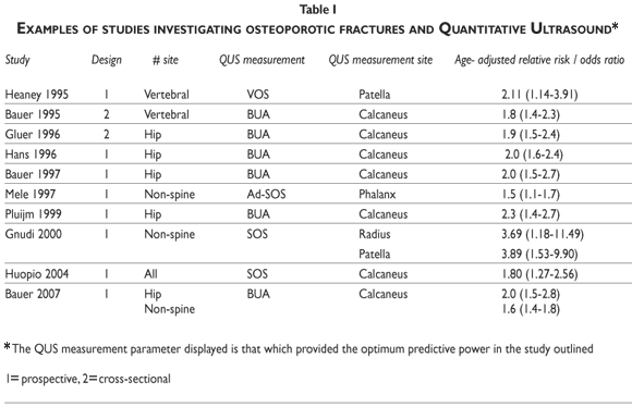
Some of the earliest strongest evidence to support    the use of QUS for discriminating between osteoporotic fractures and controls    has come from large prospective studies. The first of these was published in    1990, when Porter <I>et al.</I> measured 1414 women over the age of 69 who were    in residential accommodation.<SUP>46</SUP> Measurements were performed at the    calcaneus using the original Langton device. The women were followed up over    a period of two years during which time a total of 73 women suffered a hip fracture.    The fracture group was found to have a lower BUA than the non&#45;fracture group    and were also found to be more active and have a lower cognisance score. <a href="#tab01">Table    I</a> outlines a number of cross&#45;sectional and prospective studies demonstrating    the ability of QUS measurements at various sites to predict osteoporotic fracture.    In all these studies, QUS is a good predictor or discriminator of hip or non&#45;spine    fractures.<SUP>9,45,47&#45;53</SUP> In a meta&#45;analysis of QUS and fractures, Mar&iacute;n    <I>et al. </I>reported the strength of association between QUS with non&#45;spinal    fractures to be similar to axial or peripheral BMD measurements. However, QUS    was shown to be inferior to the association between BMD measured at the hip    and hip fracture.<SUP>54</SUP> Despite its ability to predict non&#45;spine and    hip fractures, the data to support the ability of QUS to predict vertebral fractures    is variable. However, calcaneal QUS generally has better prediction and discrimination    of vertebral fracture than other sites.<SUP>27,33,55&#45;64</sup></font></p>     ]]></body>
<body><![CDATA[<p><a name="tab01"></a></p>     <p>&nbsp;</p>     <p align="center"><img src="/img/revistas/spm/v51s1/a05tab01.gif"></p>     <p>&nbsp;</p>     <p><font size="3" face="verdana"><b>Risk factors and secondary osteoporosis</b></font></p>     <p><font size="2" face="Verdana">Studies have found QUS to be sensitive to clinical    risk factors for osteoporosis and secondary causes of osteoporosis. Frost <I>et    al.</I> reported calcaneal QUS to be as sensitive as BMD measurements to clinical    risk factors for osteoporosis.<SUP>65</SUP> Stewart<I> et al. </I>also reported    QUS measurements to be sensitive to clinical risk factors for osteoporosis,    but reported the strength of the association to be dependent upon the type of    QUS device and the variable measured.<SUP>66</sup></font></p>     <p><font size="2" face="Verdana"> Other studies have reported QUS parameters to    be decreased in subjects with renal disease,<SUP>67,68</SUP> Crohn's disease,<SUP>69</SUP>    primary hyperparathyroism,<SUP>70&#45;73</SUP> Rheumatoid arthritis<SUP>74</SUP>    and glucocorticoid use.<SUP>6,75</SUP> Children and young adults with severe    cerebral palsy and taking anticonvulsant therapy (with and without fractures)    have been demonstrated to have reduced QUS measurements at the calcaneus.<SUP>76</SUP>    Damilakis <I>et al.</I> reported a negative correlation of SOS measurements    at the radius and tibia with duration of type 1 diabetes in adolescents.<SUP>77</SUP>    However, other authors have reported limited usefulness of QUS in patients with    rheumatoid arthritis and with inflammatory bowel disease.<SUP>78&#45;80</sup></font></p>     <p>&nbsp;</p>     <p><font size="3" face="verdana"><b>Lifestyle factors</b></font></p>     <p><font size="2" face="Verdana">Physical activity has been demonstrated to have    a positive effect on calcaneal QUS parameters.<SUP>81&#45;86</SUP> QUS measurements    have also been demonstrated to be reduced in smokers compared to non&#45;smokers.<SUP>87,88</SUP>    </font></p>     ]]></body>
<body><![CDATA[<p>&nbsp;</p>     <p><font size="3" face="verdana"><b>T&#45;score equivalence with DXA</b></font></p>     <p><font size="2" face="Verdana">The World Health Organisation (WHO) criteria<SUP>89</SUP>    cannot be used with QUS to diagnose osteoporosis,<SUP>90</SUP> although QUS    can be useful as an indication of osteoporosis. A number of authors have reported    site and device specific T&#45;score equivalence to DXA.<SUP>91&#45;99</SUP> These studies    demonstrate that a single diagnostic threshold for QUS is not appropriate and    T&#45;score equivalence to DXA is site&#45; and device&#45;specific. </font></p>     <p>&nbsp;</p>     <p><font size="3" face="verdana"><b>Monitoring therapy</b></font></p>     <p><font size="2" face="Verdana">The poorer precision of QUS in comparison to    DXA has resulted in limited applicability for longitudinal use to monitor disease    progression or therapeutic intervention. The international QUS consensus group    report in 1997 recommends further study of the use of QUS to monitor disease    progression or response to therapy,<SUP>100</SUP> whilst the National Osteoporosis    Society in the UK at present does not recommend the use of QUS for monitoring    treatment and states that DXA should be used instead.<SUP>101</SUP> </font></p>     <p><font size="2" face="Verdana"> Cross&#45;sectional studies of QUS have demonstrated    its ability to differentiate between patients treated with HRT compared to age&#45;matched    controls.<SUP>102&#45;104</SUP> These data are at variance with some of the research    into the effect of HRT on calcaneal QUS parameters, which have found limited    effects.<SUP>10,105,106</SUP> However, Sahotal <I>et al.</I> found a positive    effect of HRT at the calcaneus in a longitudinal study over a four year period,    although the individual increases for BUA and SOS were not as great as found    for lumbar spine and total hip BMD.<SUP>26</SUP> </font></p>     <p><font size="2" face="Verdana"> A number of longitudinal studies have demonstrated    a response of QUS to patients treated with bisphosphonates at the calcaneus    and tibia.<SUP>107&#45;110</SUP> However, as a result of the poorer precision, the    follow&#45;up period is generally required to be longer than that of DXA.<SUP>109</sup></font></p>     <p><font size="2" face="Verdana"> In conclusion, QUS has been extensively researched    and has been demonstrated to have the ability to predict fracture, particularly    at the hip. However, the predictive ability of QUS appears to be fracture&#45;site,    measurement&#45;site and device specific. The correlations between DXA and BMD are    weak to moderate, resulting in different subjects being identified as being    at risk of fracture by the two different methods. QUS is sensitive to age and    menopause&#45;related changes and to clinical risk factors lifestyle factors associated    with osteoporosis. Whilst a limited ability of QUS to monitor therapeutic intervention    has been demonstrated, this is still an area where its poorer precision, in    comparison to DXA, results in limited applicability. Whilst DXA remains the    gold standard for the diagnosis of osteoporosis, QUS may be of use for the prediction    of those at risk of future fracture in areas where there is limited availability    of DXA. </font></p>     <p>&nbsp;</p>     ]]></body>
<body><![CDATA[<p><font size="3" face="verdana"><b>References</b></font></p>     <!-- ref --><p><font size="2" face="Verdana">1. Langton C M, Palmer S B, Porter R W. The measurement    of broadband ultrasonic attenuation in cancellous bone. London: MEP Ltd 1984;13:89&#45;91.</font>&nbsp;&nbsp;&nbsp;&nbsp;&nbsp;&nbsp;&nbsp;&nbsp;[&#160;<a href="javascript:void(0);" onclick="javascript: window.open('/scielo.php?script=sci_nlinks&ref=9281617&pid=S0036-3634200900070000500001&lng=','','width=640,height=500,resizable=yes,scrollbars=1,menubar=yes,');">Links</a>&#160;]<!-- end-ref --><!-- ref --><p><font size="2" face="Verdana">2. Blake G M, Wahner H W, Fogelman I. The evaluation    of osteoporosis: dual x&#45;ray absorptiometry and ultrasound in clinical practice.    London: Martin Dunitz Ltd; 1999 .</font>&nbsp;&nbsp;&nbsp;&nbsp;&nbsp;&nbsp;&nbsp;&nbsp;[&#160;<a href="javascript:void(0);" onclick="javascript: window.open('/scielo.php?script=sci_nlinks&ref=9281618&pid=S0036-3634200900070000500002&lng=','','width=640,height=500,resizable=yes,scrollbars=1,menubar=yes,');">Links</a>&#160;]<!-- end-ref --><!-- ref --><p><font size="2" face="Verdana">3. Gl&uuml;er C&#45;C, Wu C Y, Jergas M, Goldstein    S A, Genant H K. The quantitative ultrasound parameters reflect bone structure.    Calcif Tissue Int 1994;55:46&#45;5.</font>&nbsp;&nbsp;&nbsp;&nbsp;&nbsp;&nbsp;&nbsp;&nbsp;[&#160;<a href="javascript:void(0);" onclick="javascript: window.open('/scielo.php?script=sci_nlinks&ref=9281619&pid=S0036-3634200900070000500003&lng=','','width=640,height=500,resizable=yes,scrollbars=1,menubar=yes,');">Links</a>&#160;]<!-- end-ref --><!-- ref --><p><font size="2" face="Verdana">4. Joly J, Westhovens R, Borghs H, Peeters H,    Tirry J, Nijs J, <I>et al.</I> Reference curve and diagnostic sensitivity for    a new ultrasound device for the phalanges, the DBMsonic 1200, in Belgian women.    Osteoporos Int 1999;9:284&#45;9.</font>&nbsp;&nbsp;&nbsp;&nbsp;&nbsp;&nbsp;&nbsp;&nbsp;[&#160;<a href="javascript:void(0);" onclick="javascript: window.open('/scielo.php?script=sci_nlinks&ref=9281620&pid=S0036-3634200900070000500004&lng=','','width=640,height=500,resizable=yes,scrollbars=1,menubar=yes,');">Links</a>&#160;]<!-- end-ref --><!-- ref --><p><font size="2" face="Verdana">5. Pluskiewicz W, Drozdzowska B. Ultrasound measurements    of proximal phalanges in Polish early postmenopausal women. Osteoporos Int 1998;8:578&#45;83.</font>&nbsp;&nbsp;&nbsp;&nbsp;&nbsp;&nbsp;&nbsp;&nbsp;[&#160;<a href="javascript:void(0);" onclick="javascript: window.open('/scielo.php?script=sci_nlinks&ref=9281621&pid=S0036-3634200900070000500005&lng=','','width=640,height=500,resizable=yes,scrollbars=1,menubar=yes,');">Links</a>&#160;]<!-- end-ref --><!-- ref --><p><font size="2" face="Verdana">6. Orgee JM, Foster H, McCloskey EV, Khan S,    Coombes G, Kanis JA. A precise method for the assessment of tibial ultrasound    velocity. Osteoporos Int 1996;6:1&#45;7.</font>&nbsp;&nbsp;&nbsp;&nbsp;&nbsp;&nbsp;&nbsp;&nbsp;[&#160;<a href="javascript:void(0);" onclick="javascript: window.open('/scielo.php?script=sci_nlinks&ref=9281622&pid=S0036-3634200900070000500006&lng=','','width=640,height=500,resizable=yes,scrollbars=1,menubar=yes,');">Links</a>&#160;]<!-- end-ref --><!-- ref --><p><font size="2" face="Verdana">7. Tromp AM, Smit JH, Deeg DJ, Lips P. Quantitative    ultrasound measurements of the tibia and calcaneus in comparison with DXA measurements    at various skeletal sites. Osteoporos Int 1999;9:230&#45;</font>&nbsp;&nbsp;&nbsp;&nbsp;&nbsp;&nbsp;&nbsp;&nbsp;[&#160;<a href="javascript:void(0);" onclick="javascript: window.open('/scielo.php?script=sci_nlinks&ref=9281623&pid=S0036-3634200900070000500007&lng=','','width=640,height=500,resizable=yes,scrollbars=1,menubar=yes,');">Links</a>&#160;]<!-- end-ref --><!-- ref --><p><font size="2" face="Verdana">8. Lehmann R, Wapniarz M, Kvasnicka HM, Klein    K, Allolio B. Velocity of ultrasound at the patella: influence of age, menopause    and estrogen replacement therapy. Osteoporos Int 1993;3:308&#45;13.</font>&nbsp;&nbsp;&nbsp;&nbsp;&nbsp;&nbsp;&nbsp;&nbsp;[&#160;<a href="javascript:void(0);" onclick="javascript: window.open('/scielo.php?script=sci_nlinks&ref=9281624&pid=S0036-3634200900070000500008&lng=','','width=640,height=500,resizable=yes,scrollbars=1,menubar=yes,');">Links</a>&#160;]<!-- end-ref --><p><font size="2" face="Verdana">9. Heaney RP, Avioli LV, Chesnut CH 3rd, Lappe    J, Recker RR, Brandenburger GH. Ultrasound velocity, through bone predicts incident    vertebral deformity. J Bone Miner Res. 1995;10:341&#45;5.</font></p>     ]]></body>
<body><![CDATA[<!-- ref --><p><font size="2" face="Verdana">10. Stegman MR, Heaney RP, Recker RR. Comparison    of speed of sound ultrasound with single photon absorptiometry for determining    fracture odds ratios. J Bone Miner Res 1995;10:346&#45;52.</font>&nbsp;&nbsp;&nbsp;&nbsp;&nbsp;&nbsp;&nbsp;&nbsp;[&#160;<a href="javascript:void(0);" onclick="javascript: window.open('/scielo.php?script=sci_nlinks&ref=9281626&pid=S0036-3634200900070000500010&lng=','','width=640,height=500,resizable=yes,scrollbars=1,menubar=yes,');">Links</a>&#160;]<!-- end-ref --><!-- ref --><p><font size="2" face="Verdana">11. Blake G M, Patel R, Fogelman I. Peripheral    or axial bone density measurements. J Clin Densitom 1998;1:55&#45;63.</font>&nbsp;&nbsp;&nbsp;&nbsp;&nbsp;&nbsp;&nbsp;&nbsp;[&#160;<a href="javascript:void(0);" onclick="javascript: window.open('/scielo.php?script=sci_nlinks&ref=9281627&pid=S0036-3634200900070000500011&lng=','','width=640,height=500,resizable=yes,scrollbars=1,menubar=yes,');">Links</a>&#160;]<!-- end-ref --><!-- ref --><p><font size="2" face="Verdana">12. Thompson P, Taylor J, Fisher A, Oliver R.    Quantitative heel ultrasound in 3180 women between 45 and 75 years of age: compliance,    normal ranges and relationship to fracture history. Osteoporos Int 1998;8:211&#45;4.</font>&nbsp;&nbsp;&nbsp;&nbsp;&nbsp;&nbsp;&nbsp;&nbsp;[&#160;<a href="javascript:void(0);" onclick="javascript: window.open('/scielo.php?script=sci_nlinks&ref=9281628&pid=S0036-3634200900070000500012&lng=','','width=640,height=500,resizable=yes,scrollbars=1,menubar=yes,');">Links</a>&#160;]<!-- end-ref --><!-- ref --><p><font size="2" face="Verdana">13. Gl&uuml;er C C, Vahlensieck M, Faulkner K    G, Engelke K, Black D, Genant H K. Site&#45;matched calcaneal measurements of broad&#45;band    ultrasound attenuation and single x&#45;ray absorptiometry: do they measure different    skeletal properties? J Bone Miner Res 1992 7;9:1071&#45;1079.</font>&nbsp;&nbsp;&nbsp;&nbsp;&nbsp;&nbsp;&nbsp;&nbsp;[&#160;<a href="javascript:void(0);" onclick="javascript: window.open('/scielo.php?script=sci_nlinks&ref=9281629&pid=S0036-3634200900070000500013&lng=','','width=640,height=500,resizable=yes,scrollbars=1,menubar=yes,');">Links</a>&#160;]<!-- end-ref --><!-- ref --><p><font size="2" face="Verdana">14. Mehta S S, Orham K O, Antich P P. Bone elasticity    and ultrasound velocity are affected by subtle changes in the organic matrix.    J Bone Miner Res 1998;13:114&#45;121.</font>&nbsp;&nbsp;&nbsp;&nbsp;&nbsp;&nbsp;&nbsp;&nbsp;[&#160;<a href="javascript:void(0);" onclick="javascript: window.open('/scielo.php?script=sci_nlinks&ref=9281630&pid=S0036-3634200900070000500014&lng=','','width=640,height=500,resizable=yes,scrollbars=1,menubar=yes,');">Links</a>&#160;]<!-- end-ref --><!-- ref --><p><font size="2" face="Verdana">15. Njeh C F, Fuerst T, Diessel E, Genant H K.    Is quantitative ultrasound dependent on bone structure? A reflection. Osteoporos    Int 2001;12:1&#45;15.</font>&nbsp;&nbsp;&nbsp;&nbsp;&nbsp;&nbsp;&nbsp;&nbsp;[&#160;<a href="javascript:void(0);" onclick="javascript: window.open('/scielo.php?script=sci_nlinks&ref=9281631&pid=S0036-3634200900070000500015&lng=','','width=640,height=500,resizable=yes,scrollbars=1,menubar=yes,');">Links</a>&#160;]<!-- end-ref --><!-- ref --><p><font size="2" face="Verdana">16. Wuster C, de Terlizzi F, Becker S, Cadossi    M, Cadossi R, Muller R. Usefulness of quantitative ultrasound in evaluating    structural and mechanical properties of bone: comparison of ultrasound, dual&#45;energy    X&#45;ray absorptiometry, micro&#45;computed tomography, and mechanical testing of human    phalanges in vitro. Technol Health Care 2005;13:497&#45;510.</font>&nbsp;&nbsp;&nbsp;&nbsp;&nbsp;&nbsp;&nbsp;&nbsp;[&#160;<a href="javascript:void(0);" onclick="javascript: window.open('/scielo.php?script=sci_nlinks&ref=9281632&pid=S0036-3634200900070000500016&lng=','','width=640,height=500,resizable=yes,scrollbars=1,menubar=yes,');">Links</a>&#160;]<!-- end-ref --><!-- ref --><p><font size="2" face="Verdana">17. Portero NR, Arlot ME, Roux JP, Duboeuf F,    Chavassieux PM, Meunier PJ. Evaluation and development of automatic two&#45;dimensional    measurements of histomorphometric parameters reflecting trabecular bone connectivity:    correlations with dual&#45;energy x&#45;ray absorptiometry and quantitative ultrasound    in human calcaneum. Calcif Tissue Int 2005;77:195&#45;204.</font>&nbsp;&nbsp;&nbsp;&nbsp;&nbsp;&nbsp;&nbsp;&nbsp;[&#160;<a href="javascript:void(0);" onclick="javascript: window.open('/scielo.php?script=sci_nlinks&ref=9281633&pid=S0036-3634200900070000500017&lng=','','width=640,height=500,resizable=yes,scrollbars=1,menubar=yes,');">Links</a>&#160;]<!-- end-ref --><!-- ref --><p><font size="2" face="Verdana">18. Cortet B, Boutry N, Dubois P, Legroux&#45;Gerot    I, Cotten A, Marchandise X. Does quantitative ultrasound of bone reflect more    bone mineral density than bone microarchitecture? Calcif Tissue Int 2004;74:60&#45;7.</font>&nbsp;&nbsp;&nbsp;&nbsp;&nbsp;&nbsp;&nbsp;&nbsp;[&#160;<a href="javascript:void(0);" onclick="javascript: window.open('/scielo.php?script=sci_nlinks&ref=9281634&pid=S0036-3634200900070000500018&lng=','','width=640,height=500,resizable=yes,scrollbars=1,menubar=yes,');">Links</a>&#160;]<!-- end-ref --><!-- ref --><p><font size="2" face="Verdana">19. Turner C H, Eich M. Ultrasonic velocity as    a predictor of strength in bovine cancellous bone. Calcif Tissue Int 1991;49:116&#45;119.</font>&nbsp;&nbsp;&nbsp;&nbsp;&nbsp;&nbsp;&nbsp;&nbsp;[&#160;<a href="javascript:void(0);" onclick="javascript: window.open('/scielo.php?script=sci_nlinks&ref=9281635&pid=S0036-3634200900070000500019&lng=','','width=640,height=500,resizable=yes,scrollbars=1,menubar=yes,');">Links</a>&#160;]<!-- end-ref --><!-- ref --><p><font size="2" face="Verdana">20. Waud C E, Lew R and Baran D T. The relationship    between ultrasound and densitometric measurements of bone mass at the calcaneus    in women. Calcif Tissue Int 1992;51:415&#45;418.</font>&nbsp;&nbsp;&nbsp;&nbsp;&nbsp;&nbsp;&nbsp;&nbsp;[&#160;<a href="javascript:void(0);" onclick="javascript: window.open('/scielo.php?script=sci_nlinks&ref=9281636&pid=S0036-3634200900070000500020&lng=','','width=640,height=500,resizable=yes,scrollbars=1,menubar=yes,');">Links</a>&#160;]<!-- end-ref --><!-- ref --><p><font size="2" face="Verdana">21. Antich P P. Ultrasound study of bone in&#45;vitro.    Calcif Tissue Int 1993; 53:S157&#45;S161.</font>&nbsp;&nbsp;&nbsp;&nbsp;&nbsp;&nbsp;&nbsp;&nbsp;[&#160;<a href="javascript:void(0);" onclick="javascript: window.open('/scielo.php?script=sci_nlinks&ref=9281637&pid=S0036-3634200900070000500021&lng=','','width=640,height=500,resizable=yes,scrollbars=1,menubar=yes,');">Links</a>&#160;]<!-- end-ref --><!-- ref --><p><font size="2" face="Verdana">22. Gl&uuml;er C&#45;C, Wu C Y, Genant H K. Broadband    ultrasound attenuation signals depend on trabecular orientation: An in&#45;vitro    study. Osteoporos Int 1993;3:185&#45;191.</font>&nbsp;&nbsp;&nbsp;&nbsp;&nbsp;&nbsp;&nbsp;&nbsp;[&#160;<a href="javascript:void(0);" onclick="javascript: window.open('/scielo.php?script=sci_nlinks&ref=9281638&pid=S0036-3634200900070000500022&lng=','','width=640,height=500,resizable=yes,scrollbars=1,menubar=yes,');">Links</a>&#160;]<!-- end-ref --><!-- ref --><p><font size="2" face="Verdana">23. Wu C, Gl&uuml;er C, Lu Y, Fuerst T, Hans    D, Genant H K. Ultrasound characterisation of bone demineralisation. Calcif    Tissue Int 1998;62:133&#45;139.</font>&nbsp;&nbsp;&nbsp;&nbsp;&nbsp;&nbsp;&nbsp;&nbsp;[&#160;<a href="javascript:void(0);" onclick="javascript: window.open('/scielo.php?script=sci_nlinks&ref=9281639&pid=S0036-3634200900070000500023&lng=','','width=640,height=500,resizable=yes,scrollbars=1,menubar=yes,');">Links</a>&#160;]<!-- end-ref --><!-- ref --><p><font size="2" face="Verdana">24. Chappard C, Laugier P, Fournier B, Roux C,    Berger G. Assessment of the relationship between broadband ultrasound attenuation    and bone mineral density at the calcaneus using BUA Imaging and DXA. Osteoporos    Int 1997;7:316&#45;322.</font>&nbsp;&nbsp;&nbsp;&nbsp;&nbsp;&nbsp;&nbsp;&nbsp;[&#160;<a href="javascript:void(0);" onclick="javascript: window.open('/scielo.php?script=sci_nlinks&ref=9281640&pid=S0036-3634200900070000500024&lng=','','width=640,height=500,resizable=yes,scrollbars=1,menubar=yes,');">Links</a>&#160;]<!-- end-ref --><!-- ref --><p><font size="2" face="Verdana">25. Laugier P, Droin P, Laval&#45;Jeantet A M, Berger    G. In&#45;vitro assessment of the relationship between acoustic properties and bone    mass density of the calcaneus by comparison of ultrasound parametric imaging    and quantitative computed tomography. Bone 1997;20:157&#45;165.</font>&nbsp;&nbsp;&nbsp;&nbsp;&nbsp;&nbsp;&nbsp;&nbsp;[&#160;<a href="javascript:void(0);" onclick="javascript: window.open('/scielo.php?script=sci_nlinks&ref=9281641&pid=S0036-3634200900070000500025&lng=','','width=640,height=500,resizable=yes,scrollbars=1,menubar=yes,');">Links</a>&#160;]<!-- end-ref --><!-- ref --><p><font size="2" face="Verdana">26. Sahota O, San P, Cawte S A, Pearson D, Hosking    D J. A comparison of the longitudinal changes in quantitative ultrasound with    dual&#45;energy x&#45;ray absorptiometry: The four&#45;ar effects of hormone replacement    therapy. Osteoporos Int 2000;11:52&#45;58.</font>&nbsp;&nbsp;&nbsp;&nbsp;&nbsp;&nbsp;&nbsp;&nbsp;[&#160;<a href="javascript:void(0);" onclick="javascript: window.open('/scielo.php?script=sci_nlinks&ref=9281642&pid=S0036-3634200900070000500026&lng=','','width=640,height=500,resizable=yes,scrollbars=1,menubar=yes,');">Links</a>&#160;]<!-- end-ref --><!-- ref --><p><font size="2" face="Verdana">27. Ravaud P, Reny JL, Giraudeau B, Porcher R,    Dougados M, Roux C. Individual smallest detectable difference in bone mineral    density measurements. J Bone Miner Res 1999;14:1449&#45;56.</font>&nbsp;&nbsp;&nbsp;&nbsp;&nbsp;&nbsp;&nbsp;&nbsp;[&#160;<a href="javascript:void(0);" onclick="javascript: window.open('/scielo.php?script=sci_nlinks&ref=9281643&pid=S0036-3634200900070000500027&lng=','','width=640,height=500,resizable=yes,scrollbars=1,menubar=yes,');">Links</a>&#160;]<!-- end-ref --><!-- ref --><p><font size="2" face="Verdana">28. Stewart A, Reid D M. Precision of quantitative    ultrasound: comparison of three commercial scanners. Bone 2000;27:139&#45;143.</font>&nbsp;&nbsp;&nbsp;&nbsp;&nbsp;&nbsp;&nbsp;&nbsp;[&#160;<a href="javascript:void(0);" onclick="javascript: window.open('/scielo.php?script=sci_nlinks&ref=9281644&pid=S0036-3634200900070000500028&lng=','','width=640,height=500,resizable=yes,scrollbars=1,menubar=yes,');">Links</a>&#160;]<!-- end-ref --><!-- ref --><p><font size="2" face="Verdana">29. Frost M L, Blake G M, Fogelman I. Contact    quantitative ultrasound: An evaluation of precision, fracture discrimination,    age&#45;related bone loss and applicability of the WHO Criteria. Osteoporos Int    1999;10:441&#45;449.</font>&nbsp;&nbsp;&nbsp;&nbsp;&nbsp;&nbsp;&nbsp;&nbsp;[&#160;<a href="javascript:void(0);" onclick="javascript: window.open('/scielo.php?script=sci_nlinks&ref=9281645&pid=S0036-3634200900070000500029&lng=','','width=640,height=500,resizable=yes,scrollbars=1,menubar=yes,');">Links</a>&#160;]<!-- end-ref --><!-- ref --><p><font size="2" face="Verdana">30. Frost M L, Blake G M, Fogelman I. Does quantitative    ultrasound imaging enhance precision and discrimination? Osteoporos Int 2000;    11:425&#45;433.</font>&nbsp;&nbsp;&nbsp;&nbsp;&nbsp;&nbsp;&nbsp;&nbsp;[&#160;<a href="javascript:void(0);" onclick="javascript: window.open('/scielo.php?script=sci_nlinks&ref=9281646&pid=S0036-3634200900070000500030&lng=','','width=640,height=500,resizable=yes,scrollbars=1,menubar=yes,');">Links</a>&#160;]<!-- end-ref --><!-- ref --><p><font size="2" face="Verdana">31. Barkmann R, Kantorovich E, Singal C, Hans    D, Genant H K, Heller M <I>et al.</i> A new method for quantitative ultrasound    measurements at multiple skeletal sites. J Clin Densitom 2000;3:1:1&#45;7.</font>&nbsp;&nbsp;&nbsp;&nbsp;&nbsp;&nbsp;&nbsp;&nbsp;[&#160;<a href="javascript:void(0);" onclick="javascript: window.open('/scielo.php?script=sci_nlinks&ref=9281647&pid=S0036-3634200900070000500031&lng=','','width=640,height=500,resizable=yes,scrollbars=1,menubar=yes,');">Links</a>&#160;]<!-- end-ref --><!-- ref --><p><font size="2" face="Verdana">32.Weiss M, Ben&#45;Shlomo A, Hagag P, Rapoport M.    Reference database for bone speed of sound measurements by a novel quantitative    multi&#45;site ultrasound device. Osteoporos Int 2000;11:688.</font>&nbsp;&nbsp;&nbsp;&nbsp;&nbsp;&nbsp;&nbsp;&nbsp;[&#160;<a href="javascript:void(0);" onclick="javascript: window.open('/scielo.php?script=sci_nlinks&ref=9281648&pid=S0036-3634200900070000500032&lng=','','width=640,height=500,resizable=yes,scrollbars=1,menubar=yes,');">Links</a>&#160;]<!-- end-ref --><!-- ref --><p><font size="2" face="Verdana">33. Knapp KM, Blake GM, Spector TD, Fogelman    I. Multisite quantitative ultrasound: precision, age&#45; and menopause&#45;related    changes, fracture discrimination, and T&#45;score equivalence with dual&#45;energy X&#45;ray    absorptiometry. Osteoporos Int 2001;12:456&#45;64.</font>&nbsp;&nbsp;&nbsp;&nbsp;&nbsp;&nbsp;&nbsp;&nbsp;[&#160;<a href="javascript:void(0);" onclick="javascript: window.open('/scielo.php?script=sci_nlinks&ref=9281649&pid=S0036-3634200900070000500033&lng=','','width=640,height=500,resizable=yes,scrollbars=1,menubar=yes,');">Links</a>&#160;]<!-- end-ref --><!-- ref --><p><font size="2" face="Verdana">34. Roux C, Fournier B, Laugier P, Chappard C,    Kolta S, Dougados M <I>et al. </i>Broadband ultrasound attenuation imaging:    a new imaging method in osteoporosis. J Bone Miner Res 1996;11:1112&#45;8.</font>&nbsp;&nbsp;&nbsp;&nbsp;&nbsp;&nbsp;&nbsp;&nbsp;[&#160;<a href="javascript:void(0);" onclick="javascript: window.open('/scielo.php?script=sci_nlinks&ref=9281650&pid=S0036-3634200900070000500034&lng=','','width=640,height=500,resizable=yes,scrollbars=1,menubar=yes,');">Links</a>&#160;]<!-- end-ref --><!-- ref --><p><font size="2" face="Verdana">35. Pettinato AA, Loud KJ, Bristol SK, Feldman    HA, Gordon CM. Effects of nutrition, puberty, and gender on bone ultrasound    measurements in adolescents and young adults. J Adolesc Health 2006;39:828&#45;34.</font>&nbsp;&nbsp;&nbsp;&nbsp;&nbsp;&nbsp;&nbsp;&nbsp;[&#160;<a href="javascript:void(0);" onclick="javascript: window.open('/scielo.php?script=sci_nlinks&ref=9281651&pid=S0036-3634200900070000500035&lng=','','width=640,height=500,resizable=yes,scrollbars=1,menubar=yes,');">Links</a>&#160;]<!-- end-ref --><!-- ref --><p><font size="2" face="Verdana">36. Vignolo M, Parodi A, Mascagni A, Torrisi    C, De Terlizzi F, Aicardi G. Longitudinal assessment of bone quality by quantitative    ultrasonography in children and adolescents. Ultrasound Med Biol 2006;32:1003&#45;10.</font>&nbsp;&nbsp;&nbsp;&nbsp;&nbsp;&nbsp;&nbsp;&nbsp;[&#160;<a href="javascript:void(0);" onclick="javascript: window.open('/scielo.php?script=sci_nlinks&ref=9281652&pid=S0036-3634200900070000500036&lng=','','width=640,height=500,resizable=yes,scrollbars=1,menubar=yes,');">Links</a>&#160;]<!-- end-ref --><!-- ref --><p><font size="2" face="Verdana">37. Yang NP, Jen I, Chuang SY, Chen SH, Chou    P. Screening for low bone mass with quantitative ultrasonography in a community    without dual&#45;energy X&#45;ray absorptiometry: population&#45;based survey. BMC Musculoskelet    Disord 2006;7:24.</font>&nbsp;&nbsp;&nbsp;&nbsp;&nbsp;&nbsp;&nbsp;&nbsp;[&#160;<a href="javascript:void(0);" onclick="javascript: window.open('/scielo.php?script=sci_nlinks&ref=9281653&pid=S0036-3634200900070000500037&lng=','','width=640,height=500,resizable=yes,scrollbars=1,menubar=yes,');">Links</a>&#160;]<!-- end-ref --><!-- ref --><p><font size="2" face="Verdana">38. Faulkner K G, McClung M R, Coleman L J, Kingston&#45;Sandahl    E. Quantitative ultrasound of the heel: correlation with densitometric measurements    at different skeletal sites. Osteoporos Int 1994;4:42&#45;47.</font>&nbsp;&nbsp;&nbsp;&nbsp;&nbsp;&nbsp;&nbsp;&nbsp;[&#160;<a href="javascript:void(0);" onclick="javascript: window.open('/scielo.php?script=sci_nlinks&ref=9281654&pid=S0036-3634200900070000500038&lng=','','width=640,height=500,resizable=yes,scrollbars=1,menubar=yes,');">Links</a>&#160;]<!-- end-ref --><!-- ref --><p><font size="2" face="Verdana">39. He Y Q, Fan B, Hans D, Li J, Wu C Y, Njeh    C F <I>et al.</I> Assessment of a new quantitative ultrasound calcaneus measurement:    precision and discrimination of hip fractures in elderly women compared with    dual X&#45;ray absorptiometry. Osteoporos Int 2000;11:354&#45;360.</font>&nbsp;&nbsp;&nbsp;&nbsp;&nbsp;&nbsp;&nbsp;&nbsp;[&#160;<a href="javascript:void(0);" onclick="javascript: window.open('/scielo.php?script=sci_nlinks&ref=9281655&pid=S0036-3634200900070000500039&lng=','','width=640,height=500,resizable=yes,scrollbars=1,menubar=yes,');">Links</a>&#160;]<!-- end-ref --><!-- ref --><p><font size="2" face="Verdana">40. Genant HK, Engelke K, Fuerst T, Gl&uuml;er    C&#45;C, Grampp S, Harris ST<I> et al.</I> Noninvasive assessment of bone mineral    and structure: state of the art. J Bone Miner Res 1996;11:707&#45;730.</font>&nbsp;&nbsp;&nbsp;&nbsp;&nbsp;&nbsp;&nbsp;&nbsp;[&#160;<a href="javascript:void(0);" onclick="javascript: window.open('/scielo.php?script=sci_nlinks&ref=9281656&pid=S0036-3634200900070000500040&lng=','','width=640,height=500,resizable=yes,scrollbars=1,menubar=yes,');">Links</a>&#160;]<!-- end-ref --><!-- ref --><p><font size="2" face="Verdana">41. Porter R W, Johnson K, McCutchan J D S. Wrist    fracture, heel bone density and thoracic kyphosis: A case control study. Bone    1990;11:211&#45;214.</font>&nbsp;&nbsp;&nbsp;&nbsp;&nbsp;&nbsp;&nbsp;&nbsp;[&#160;<a href="javascript:void(0);" onclick="javascript: window.open('/scielo.php?script=sci_nlinks&ref=9281657&pid=S0036-3634200900070000500041&lng=','','width=640,height=500,resizable=yes,scrollbars=1,menubar=yes,');">Links</a>&#160;]<!-- end-ref --><!-- ref --><p><font size="2" face="Verdana">42. Grampp S, Genant HK, Mathur A, Lang P, Jergas    Ml, Takada M <I>et al.</I> Comparisons of noninvasive bone mineral measurements    in assessing age&#45;related loss, fracture discrimination and diagnostic classification.    J Bone Miner Res 1997 12;5:697&#45;711.</font>&nbsp;&nbsp;&nbsp;&nbsp;&nbsp;&nbsp;&nbsp;&nbsp;[&#160;<a href="javascript:void(0);" onclick="javascript: window.open('/scielo.php?script=sci_nlinks&ref=9281658&pid=S0036-3634200900070000500042&lng=','','width=640,height=500,resizable=yes,scrollbars=1,menubar=yes,');">Links</a>&#160;]<!-- end-ref --><!-- ref --><p><font size="2" face="Verdana">43. Ross P, Huang C, Davis J, Imose K, Yates    J, Vogel J <I>et al.</I> Predicting vertebral deformity using bone densitometry    at various skeletal sites and calcaneus ultrasound. Bone 1995;16:325&#45;332.</font>&nbsp;&nbsp;&nbsp;&nbsp;&nbsp;&nbsp;&nbsp;&nbsp;[&#160;<a href="javascript:void(0);" onclick="javascript: window.open('/scielo.php?script=sci_nlinks&ref=9281659&pid=S0036-3634200900070000500043&lng=','','width=640,height=500,resizable=yes,scrollbars=1,menubar=yes,');">Links</a>&#160;]<!-- end-ref --><!-- ref --><p><font size="2" face="Verdana">44. Gnudi S, Ripamonti C, Malavolta N. Quantitative    ultrasound and bone densitometry to evaluate the risk of nonspine fractures:    a prospective study. Osteoporos Int 2000;11:518&#45;23.</font>&nbsp;&nbsp;&nbsp;&nbsp;&nbsp;&nbsp;&nbsp;&nbsp;[&#160;<a href="javascript:void(0);" onclick="javascript: window.open('/scielo.php?script=sci_nlinks&ref=9281660&pid=S0036-3634200900070000500044&lng=','','width=640,height=500,resizable=yes,scrollbars=1,menubar=yes,');">Links</a>&#160;]<!-- end-ref --><!-- ref --><p><font size="2" face="Verdana">45. Gluer CC, Cummings SR, Bauer DC, Stone K,    Pressman A, Mathur A <I>et al.</I> Osteoporosis: association of recent fractures    with quantitative US findings. Radiology 1996;199:725&#45;32.</font>&nbsp;&nbsp;&nbsp;&nbsp;&nbsp;&nbsp;&nbsp;&nbsp;[&#160;<a href="javascript:void(0);" onclick="javascript: window.open('/scielo.php?script=sci_nlinks&ref=9281661&pid=S0036-3634200900070000500045&lng=','','width=640,height=500,resizable=yes,scrollbars=1,menubar=yes,');">Links</a>&#160;]<!-- end-ref --><!-- ref --><p><font size="2" face="Verdana">46. Porter RW, Miller CG, Grainger D, Palmer    SB. Prediction of hip fracture in elderly women: a prospective study. BMJ 1990;301:638&#45;41.</font>&nbsp;&nbsp;&nbsp;&nbsp;&nbsp;&nbsp;&nbsp;&nbsp;[&#160;<a href="javascript:void(0);" onclick="javascript: window.open('/scielo.php?script=sci_nlinks&ref=9281662&pid=S0036-3634200900070000500046&lng=','','width=640,height=500,resizable=yes,scrollbars=1,menubar=yes,');">Links</a>&#160;]<!-- end-ref --><!-- ref --><p><font size="2" face="Verdana">47. Bauer DC, Gluer CC, Genant HK, Stone K. Quantitative    ultrasound and vertebral fracture in postmenopausal women. J Bone Miner Res    1995;10:353&#45;8.</font>&nbsp;&nbsp;&nbsp;&nbsp;&nbsp;&nbsp;&nbsp;&nbsp;[&#160;<a href="javascript:void(0);" onclick="javascript: window.open('/scielo.php?script=sci_nlinks&ref=9281663&pid=S0036-3634200900070000500047&lng=','','width=640,height=500,resizable=yes,scrollbars=1,menubar=yes,');">Links</a>&#160;]<!-- end-ref --><!-- ref --><p><font size="2" face="Verdana">48. Hans D, Dargent&#45;Molina P, Schott AM, Sebert    JL, Cormier C, Kotzki PO et al. Ultrasonographic heel measurements to predict    hip fracture in elderly women: the EPIDOS prospective study. Lancet. 1996;24:511&#45;4.</font>&nbsp;&nbsp;&nbsp;&nbsp;&nbsp;&nbsp;&nbsp;&nbsp;[&#160;<a href="javascript:void(0);" onclick="javascript: window.open('/scielo.php?script=sci_nlinks&ref=9281664&pid=S0036-3634200900070000500048&lng=','','width=640,height=500,resizable=yes,scrollbars=1,menubar=yes,');">Links</a>&#160;]<!-- end-ref --><!-- ref --><p><font size="2" face="Verdana">49. Bauer DC, Gluer CC, Cauley JA, Vogt TM, Ensrud    KE, Genant HK <I>et al</I>. Broadband ultrasound attenuation predicts fractures    strongly and independently of densitometry in older women. A prospective study.    Arch Intern Med. 1997;157:629&#45;34.</font>&nbsp;&nbsp;&nbsp;&nbsp;&nbsp;&nbsp;&nbsp;&nbsp;[&#160;<a href="javascript:void(0);" onclick="javascript: window.open('/scielo.php?script=sci_nlinks&ref=9281665&pid=S0036-3634200900070000500049&lng=','','width=640,height=500,resizable=yes,scrollbars=1,menubar=yes,');">Links</a>&#160;]<!-- end-ref --><!-- ref --><p><font size="2" face="Verdana">50. Mele R, Masci G, Ventura V, de Aloysio D,    Bicocchi M, Cadossi R. Three&#45;year longitudinal study with quantitative ultrasound    at the hand phalanx in a female population. Osteoporos Int 1997;7:550&#45;7.</font>&nbsp;&nbsp;&nbsp;&nbsp;&nbsp;&nbsp;&nbsp;&nbsp;[&#160;<a href="javascript:void(0);" onclick="javascript: window.open('/scielo.php?script=sci_nlinks&ref=9281666&pid=S0036-3634200900070000500050&lng=','','width=640,height=500,resizable=yes,scrollbars=1,menubar=yes,');">Links</a>&#160;]<!-- end-ref --><!-- ref --><p><font size="2" face="Verdana">51. Pluijm SM, Graafmans WC, Bouter LM, Lips    P. Ultrasound measurements for the prediction of osteoporotic fractures in elderly    people. Osteoporos Int 1999;9:550&#45;6.</font>&nbsp;&nbsp;&nbsp;&nbsp;&nbsp;&nbsp;&nbsp;&nbsp;[&#160;<a href="javascript:void(0);" onclick="javascript: window.open('/scielo.php?script=sci_nlinks&ref=9281667&pid=S0036-3634200900070000500051&lng=','','width=640,height=500,resizable=yes,scrollbars=1,menubar=yes,');">Links</a>&#160;]<!-- end-ref --><!-- ref --><p><font size="2" face="Verdana">52. Huopio J, Kroger H, Honkanen R, Jurvelin    J, Saarikoski S, Alhava E. Calcaneal ultrasound predicts early postmenopausal    fractures as well as axial BMD. A prospective study of 422 women. Osteoporos    Int 2004;15:190&#45;5.</font>&nbsp;&nbsp;&nbsp;&nbsp;&nbsp;&nbsp;&nbsp;&nbsp;[&#160;<a href="javascript:void(0);" onclick="javascript: window.open('/scielo.php?script=sci_nlinks&ref=9281668&pid=S0036-3634200900070000500052&lng=','','width=640,height=500,resizable=yes,scrollbars=1,menubar=yes,');">Links</a>&#160;]<!-- end-ref --><!-- ref --><p><font size="2" face="Verdana">53. Bauer DC, Ewing SK, Cauley JA, Ensrud KE,    Cummings SR, Orwoll ES. Quantitative ultrasound predicts hip and non&#45;spine fracture    in men: the MrOS study. Osteoporos Int 2007;18:771&#45;7.</font>&nbsp;&nbsp;&nbsp;&nbsp;&nbsp;&nbsp;&nbsp;&nbsp;[&#160;<a href="javascript:void(0);" onclick="javascript: window.open('/scielo.php?script=sci_nlinks&ref=9281669&pid=S0036-3634200900070000500053&lng=','','width=640,height=500,resizable=yes,scrollbars=1,menubar=yes,');">Links</a>&#160;]<!-- end-ref --><!-- ref --><p><font size="2" face="Verdana">54. Marin F, Gonzalez&#45;Macias J, Diez&#45;Perez A,    Palma S, Delgado&#45;Rodriguez M. Relationship between bone quantitative ultrasound    and fractures: a meta&#45;analysis. Bone Miner Res 2006;21:1126&#45;35.</font>&nbsp;&nbsp;&nbsp;&nbsp;&nbsp;&nbsp;&nbsp;&nbsp;[&#160;<a href="javascript:void(0);" onclick="javascript: window.open('/scielo.php?script=sci_nlinks&ref=9281670&pid=S0036-3634200900070000500054&lng=','','width=640,height=500,resizable=yes,scrollbars=1,menubar=yes,');">Links</a>&#160;]<!-- end-ref --><!-- ref --><p><font size="2" face="Verdana">55. Meszaros S, Toth E, Ferencz V, Csupor E,    Hosszu E, Horvath C. Calcaneous quantitative ultrasound measurements predicts    vertebral fractures in idiopathic male osteoporosis.Joint Bone Spine 2007;74:79&#45;84.</font>&nbsp;&nbsp;&nbsp;&nbsp;&nbsp;&nbsp;&nbsp;&nbsp;[&#160;<a href="javascript:void(0);" onclick="javascript: window.open('/scielo.php?script=sci_nlinks&ref=9281671&pid=S0036-3634200900070000500055&lng=','','width=640,height=500,resizable=yes,scrollbars=1,menubar=yes,');">Links</a>&#160;]<!-- end-ref --><!-- ref --><p><font size="2" face="Verdana">56. Frediani B, Acciai C, Falsetti P, Baldi F,    Filippou G, Siagkri C <I>et al. </I>Calcaneus ultrasonometry and dual&#45;energy    X&#45;ray absorptiometry for the evaluation of vertebral fracture risk. Calcif Tissue    Int 2006;79:223&#45;9.</font>&nbsp;&nbsp;&nbsp;&nbsp;&nbsp;&nbsp;&nbsp;&nbsp;[&#160;<a href="javascript:void(0);" onclick="javascript: window.open('/scielo.php?script=sci_nlinks&ref=9281672&pid=S0036-3634200900070000500056&lng=','','width=640,height=500,resizable=yes,scrollbars=1,menubar=yes,');">Links</a>&#160;]<!-- end-ref --><!-- ref --><p><font size="2" face="Verdana">57. Gluer MG, Minne HW, Gluer CC, Lazarescu AD,    Pfeifer M, Perschel FH<I> et al</I>. Prospective identification of postmenopausal    osteoporotic women at high vertebral fracture risk by radiography, bone densitometry,    quantitative ultrasound, and laboratory findings: results from the PIOS study.    J Clin Densitom 2005;8:386&#45;95.</font>&nbsp;&nbsp;&nbsp;&nbsp;&nbsp;&nbsp;&nbsp;&nbsp;[&#160;<a href="javascript:void(0);" onclick="javascript: window.open('/scielo.php?script=sci_nlinks&ref=9281673&pid=S0036-3634200900070000500057&lng=','','width=640,height=500,resizable=yes,scrollbars=1,menubar=yes,');">Links</a>&#160;]<!-- end-ref --><!-- ref --><p><font size="2" face="Verdana">58. Maggi S, Noale M, Giannini S, Adami S, Defeo    D, Isaia G<I> et al. </I>Quantitative heel ultrasound in a population&#45;based    study in Italy and its relationship with fracture history: the ESOPO study.    Osteoporos Int 2006;17:237&#45;44.</font>&nbsp;&nbsp;&nbsp;&nbsp;&nbsp;&nbsp;&nbsp;&nbsp;[&#160;<a href="javascript:void(0);" onclick="javascript: window.open('/scielo.php?script=sci_nlinks&ref=9281674&pid=S0036-3634200900070000500058&lng=','','width=640,height=500,resizable=yes,scrollbars=1,menubar=yes,');">Links</a>&#160;]<!-- end-ref --><!-- ref --><p><font size="2" face="Verdana">59. Clowes JA, Eastell R, Peel NF. The discriminative    ability of peripheral and axial bone measurements to identify proximal femoral,    vertebral, distal forearm and proximal humeral fractures: a case control study.    Osteoporos Int 2005;16:1794&#45;802.</font>&nbsp;&nbsp;&nbsp;&nbsp;&nbsp;&nbsp;&nbsp;&nbsp;[&#160;<a href="javascript:void(0);" onclick="javascript: window.open('/scielo.php?script=sci_nlinks&ref=9281675&pid=S0036-3634200900070000500059&lng=','','width=640,height=500,resizable=yes,scrollbars=1,menubar=yes,');">Links</a>&#160;]<!-- end-ref --><!-- ref --><p><font size="2" face="Verdana">60. Kanis JA, Johnell O, Oden A, De Laet C, de    Terlizzi F. Ten&#45;year probabilities of clinical vertebral fractures according    to phalangeal quantitative ultrasonography.Osteoporos Int 2005;16:1065&#45;70.</font>&nbsp;&nbsp;&nbsp;&nbsp;&nbsp;&nbsp;&nbsp;&nbsp;[&#160;<a href="javascript:void(0);" onclick="javascript: window.open('/scielo.php?script=sci_nlinks&ref=9281676&pid=S0036-3634200900070000500060&lng=','','width=640,height=500,resizable=yes,scrollbars=1,menubar=yes,');">Links</a>&#160;]<!-- end-ref --><!-- ref --><p><font size="2" face="Verdana">61. Gnudi S, Ripamonti C. Quantitative ultrasound    at the phalanxes discriminates osteoporotic women with vertebral but not with    hip fracture.Ultrasound Med Biol 2004;30:357&#45;61.</font>&nbsp;&nbsp;&nbsp;&nbsp;&nbsp;&nbsp;&nbsp;&nbsp;[&#160;<a href="javascript:void(0);" onclick="javascript: window.open('/scielo.php?script=sci_nlinks&ref=9281677&pid=S0036-3634200900070000500061&lng=','','width=640,height=500,resizable=yes,scrollbars=1,menubar=yes,');">Links</a>&#160;]<!-- end-ref --><!-- ref --><p><font size="2" face="Verdana">62. Drozdzowska B, Pluskiewicz W, de Terlizzi    F. The usefulness of quantitative ultrasound at the hand phalanges in the detection    of the different types of nontraumatic fractures. Ultrasound Med Biol 2003;29:1545&#45;50.</font>&nbsp;&nbsp;&nbsp;&nbsp;&nbsp;&nbsp;&nbsp;&nbsp;[&#160;<a href="javascript:void(0);" onclick="javascript: window.open('/scielo.php?script=sci_nlinks&ref=9281678&pid=S0036-3634200900070000500062&lng=','','width=640,height=500,resizable=yes,scrollbars=1,menubar=yes,');">Links</a>&#160;]<!-- end-ref --><!-- ref --><p><font size="2" face="Verdana">63. Guglielmi G, Njeh CF, de Terlizzi F, De Serio    DA, Scillitani A, Cammisa M <I>et al.</I> Palangeal quantitative ultrasound,    phalangeal morphometric variables, and vertebral fracture discrimination. Calcif    Tissue Int 2003;72:469&#45;77.</font>&nbsp;&nbsp;&nbsp;&nbsp;&nbsp;&nbsp;&nbsp;&nbsp;[&#160;<a href="javascript:void(0);" onclick="javascript: window.open('/scielo.php?script=sci_nlinks&ref=9281679&pid=S0036-3634200900070000500063&lng=','','width=640,height=500,resizable=yes,scrollbars=1,menubar=yes,');">Links</a>&#160;]<!-- end-ref --><!-- ref --><p><font size="2" face="Verdana">64. Hartl F, Tyndall A, Kraenzlin M, Bachmeier    C, Guckel C, Senn U <I>et al.</I> Discriminatory ability of quantitative ultrasound    parameters and bone mineral density in a population&#45;based sample of postmenopausal    women with vertebral fractures: results of the Basel Osteoporosis Study. J Bone    Miner Res 2002;17:321&#45;30.</font>&nbsp;&nbsp;&nbsp;&nbsp;&nbsp;&nbsp;&nbsp;&nbsp;[&#160;<a href="javascript:void(0);" onclick="javascript: window.open('/scielo.php?script=sci_nlinks&ref=9281680&pid=S0036-3634200900070000500064&lng=','','width=640,height=500,resizable=yes,scrollbars=1,menubar=yes,');">Links</a>&#160;]<!-- end-ref --><!-- ref --><p><font size="2" face="Verdana">65. Frost M L, Blake G M, Fogelman I. Quantitative    ultrasound and bone mineral density are equally strongly associated with risk    factors for osteoporosis. J Bone Miner Res 2001;2:406&#45;416.</font>&nbsp;&nbsp;&nbsp;&nbsp;&nbsp;&nbsp;&nbsp;&nbsp;[&#160;<a href="javascript:void(0);" onclick="javascript: window.open('/scielo.php?script=sci_nlinks&ref=9281681&pid=S0036-3634200900070000500065&lng=','','width=640,height=500,resizable=yes,scrollbars=1,menubar=yes,');">Links</a>&#160;]<!-- end-ref --><!-- ref --><p><font size="2" face="Verdana">66. Stewart A, Felsenberg D, Eastell R, Roux    C, Gluer CC, Reid DM. Relationship between risk factors and QUS in a European    Population: The OPUS study. Bone. 2006;39:609&#45;15.</font>&nbsp;&nbsp;&nbsp;&nbsp;&nbsp;&nbsp;&nbsp;&nbsp;[&#160;<a href="javascript:void(0);" onclick="javascript: window.open('/scielo.php?script=sci_nlinks&ref=9281682&pid=S0036-3634200900070000500066&lng=','','width=640,height=500,resizable=yes,scrollbars=1,menubar=yes,');">Links</a>&#160;]<!-- end-ref --><!-- ref --><p><font size="2" face="Verdana">67. Wittich A, Vega E, Casco C, Marini A, Forlano    C, Segovia F <I>et al. </I>Ultrasound velocity of the tibia in patients on haemodialysis.    J Clin Densitometry 1998;2:157&#45;163.</font>&nbsp;&nbsp;&nbsp;&nbsp;&nbsp;&nbsp;&nbsp;&nbsp;[&#160;<a href="javascript:void(0);" onclick="javascript: window.open('/scielo.php?script=sci_nlinks&ref=9281683&pid=S0036-3634200900070000500067&lng=','','width=640,height=500,resizable=yes,scrollbars=1,menubar=yes,');">Links</a>&#160;]<!-- end-ref --><!-- ref --><p><font size="2" face="Verdana">68. Pluskiewicz W, Przedlacki J, Drozdzowska    B, Wlodarczyk D, Matuszkiewicz&#45;Rowinska J, Adamczyk P. Quantitative ultrasound    in monitoring of skeletal status in adults with end&#45;stage renal disease. Ultrasound    Med Biol 2006;32:1521&#45;7.</font>&nbsp;&nbsp;&nbsp;&nbsp;&nbsp;&nbsp;&nbsp;&nbsp;[&#160;<a href="javascript:void(0);" onclick="javascript: window.open('/scielo.php?script=sci_nlinks&ref=9281684&pid=S0036-3634200900070000500068&lng=','','width=640,height=500,resizable=yes,scrollbars=1,menubar=yes,');">Links</a>&#160;]<!-- end-ref --><!-- ref --><p><font size="2" face="Verdana">69. Robinson R J, Carr I, Iqbal S J, al&#45;Azzawi    F, Abrams K, Mayberry J F. Screening for osteoporosis in crohn's disease. A    detailed evaluation of calcaneal ultrasound. Eur J Gastroenterol Hepatol 1998;2:137&#45;40</font>&nbsp;&nbsp;&nbsp;&nbsp;&nbsp;&nbsp;&nbsp;&nbsp;[&#160;<a href="javascript:void(0);" onclick="javascript: window.open('/scielo.php?script=sci_nlinks&ref=9281685&pid=S0036-3634200900070000500069&lng=','','width=640,height=500,resizable=yes,scrollbars=1,menubar=yes,');">Links</a>&#160;]<!-- end-ref --><!-- ref --><p><font size="2" face="Verdana">70. Gomez A C, Schott A M, Hans D, Niepomniszcze    H, Mautalen C A, Meunier P J. Hyperparathyroidism Influences ultrasound bone    measurements on the os calcis. Osteoporos Int 1998;8:455&#45;459.</font>&nbsp;&nbsp;&nbsp;&nbsp;&nbsp;&nbsp;&nbsp;&nbsp;[&#160;<a href="javascript:void(0);" onclick="javascript: window.open('/scielo.php?script=sci_nlinks&ref=9281686&pid=S0036-3634200900070000500070&lng=','','width=640,height=500,resizable=yes,scrollbars=1,menubar=yes,');">Links</a>&#160;]<!-- end-ref --><!-- ref --><p><font size="2" face="Verdana">71. Ingle B M, Thomas W E G, Eastell R. Differential    effects on ultrasound properties of bone in primary hyperparathyroidism. Osteoporos    Int 1998;8:S3:S56.</font>&nbsp;&nbsp;&nbsp;&nbsp;&nbsp;&nbsp;&nbsp;&nbsp;[&#160;<a href="javascript:void(0);" onclick="javascript: window.open('/scielo.php?script=sci_nlinks&ref=9281687&pid=S0036-3634200900070000500071&lng=','','width=640,height=500,resizable=yes,scrollbars=1,menubar=yes,');">Links</a>&#160;]<!-- end-ref --><!-- ref --><p><font size="2" face="Verdana">72. Ingle BM, Thomas WE, Eastell R. Differential    effects of primary hyperparathyroidism on ultrasound properties of bone. Osteoporos    Int 2002;13:572&#45;8.</font>&nbsp;&nbsp;&nbsp;&nbsp;&nbsp;&nbsp;&nbsp;&nbsp;[&#160;<a href="javascript:void(0);" onclick="javascript: window.open('/scielo.php?script=sci_nlinks&ref=9281688&pid=S0036-3634200900070000500072&lng=','','width=640,height=500,resizable=yes,scrollbars=1,menubar=yes,');">Links</a>&#160;]<!-- end-ref --><!-- ref --><p><font size="2" face="Verdana">73. Chappard C, Roux C, Laugier P, Paillard M,    Houillier P. Bone status in primary hyperparathyroidism assessed by regional    bone mineral density from the whole body scan and QUS imaging at calcaneus.    Joint Bone Spine 2006;73:86&#45;94.</font>&nbsp;&nbsp;&nbsp;&nbsp;&nbsp;&nbsp;&nbsp;&nbsp;[&#160;<a href="javascript:void(0);" onclick="javascript: window.open('/scielo.php?script=sci_nlinks&ref=9281689&pid=S0036-3634200900070000500073&lng=','','width=640,height=500,resizable=yes,scrollbars=1,menubar=yes,');">Links</a>&#160;]<!-- end-ref --><!-- ref --><p><font size="2" face="Verdana">74. Cryer JR, Otter SJ, Bowen CJ. Use of quantitative    ultrasound scans of the calcaneus to diagnose osteoporosis in patients with    rheumatoid arthritis. J Am Podiatr Med Assoc 2007;97:108&#45;14.</font>&nbsp;&nbsp;&nbsp;&nbsp;&nbsp;&nbsp;&nbsp;&nbsp;[&#160;<a href="javascript:void(0);" onclick="javascript: window.open('/scielo.php?script=sci_nlinks&ref=9281690&pid=S0036-3634200900070000500074&lng=','','width=640,height=500,resizable=yes,scrollbars=1,menubar=yes,');">Links</a>&#160;]<!-- end-ref --><!-- ref --><p><font size="2" face="Verdana">75. Blanckaert F, Cortet B, Coquerelle B, Flipo    R M, Duquesnoy B, Matchandise X <I>et al.</I> Contribution of calcaneal ultrasonic    assessment to the evaluation of postmenopausal and glucocorticoid&#45;induced osteoporosis.    Rev Rhum 1997 64:305&#45;313.</font>&nbsp;&nbsp;&nbsp;&nbsp;&nbsp;&nbsp;&nbsp;&nbsp;[&#160;<a href="javascript:void(0);" onclick="javascript: window.open('/scielo.php?script=sci_nlinks&ref=9281691&pid=S0036-3634200900070000500075&lng=','','width=640,height=500,resizable=yes,scrollbars=1,menubar=yes,');">Links</a>&#160;]<!-- end-ref --><!-- ref --><p><font size="2" face="Verdana">76. Jekovec&#45;Vrhovsek M, Kocijancic A, Prezelj    J. Quantitative ultrasound of the calcaneus in children and young adults with    severe cerebral palsy. Dev Med Child Neurol 2005;47:696&#45;8.</font>&nbsp;&nbsp;&nbsp;&nbsp;&nbsp;&nbsp;&nbsp;&nbsp;[&#160;<a href="javascript:void(0);" onclick="javascript: window.open('/scielo.php?script=sci_nlinks&ref=9281692&pid=S0036-3634200900070000500076&lng=','','width=640,height=500,resizable=yes,scrollbars=1,menubar=yes,');">Links</a>&#160;]<!-- end-ref --><!-- ref --><p><font size="2" face="Verdana">77. Damilakis J, Galanakis E, Mamoulakis D, Sbyrakis    S, Gourtsoyiannis N. Quantitative ultrasound measurements in children and adolescents    with: type 1 diabetes. Calcif Tissue Int 2004;74:424&#45;8.</font>&nbsp;&nbsp;&nbsp;&nbsp;&nbsp;&nbsp;&nbsp;&nbsp;[&#160;<a href="javascript:void(0);" onclick="javascript: window.open('/scielo.php?script=sci_nlinks&ref=9281693&pid=S0036-3634200900070000500077&lng=','','width=640,height=500,resizable=yes,scrollbars=1,menubar=yes,');">Links</a>&#160;]<!-- end-ref --><!-- ref --><p><font size="2" face="Verdana">78. Pfeil A, Bottcher J, Mentzel HJ, Lehmann    G, Schafer ML, Kramer A <I>et al</I>. Multi&#45;site quantitative ultrasound compared    to dual energy X&#45;ray absorptiometry in rheumatoid arthritis: effects of body    mass index and inflamed soft tissue on reproducibility. Rheumatol Int 2006;26:1084&#45;90.</font>&nbsp;&nbsp;&nbsp;&nbsp;&nbsp;&nbsp;&nbsp;&nbsp;[&#160;<a href="javascript:void(0);" onclick="javascript: window.open('/scielo.php?script=sci_nlinks&ref=9281694&pid=S0036-3634200900070000500078&lng=','','width=640,height=500,resizable=yes,scrollbars=1,menubar=yes,');">Links</a>&#160;]<!-- end-ref --><!-- ref --><p><font size="2" face="Verdana">79. Bottcher J, Pfeil A, Mentzel H, Kramer A,    Schafer ML, Lehmann G <I>et al.</I> Peripheral bone status in rheumatoid arthritis    evaluated by digital X&#45;ray radiogrammetry and compared with multisite quantitative    ultrasound. Calcif Tissue Int 2006;78:25&#45;34.</font>&nbsp;&nbsp;&nbsp;&nbsp;&nbsp;&nbsp;&nbsp;&nbsp;[&#160;<a href="javascript:void(0);" onclick="javascript: window.open('/scielo.php?script=sci_nlinks&ref=9281695&pid=S0036-3634200900070000500079&lng=','','width=640,height=500,resizable=yes,scrollbars=1,menubar=yes,');">Links</a>&#160;]<!-- end-ref --><!-- ref --><p><font size="2" face="Verdana">80. Schwartz DA, Connolley CD, Koyama T, Wise    PE, Herline AJ. Calcaneal ultrasound bone densitometry is not a useful tool    to screen patients with inflammatory bowel disease at high risk for metabolic    bone disease. Inflamm Bowel Dis 2005;11:749&#45;54.</font>&nbsp;&nbsp;&nbsp;&nbsp;&nbsp;&nbsp;&nbsp;&nbsp;[&#160;<a href="javascript:void(0);" onclick="javascript: window.open('/scielo.php?script=sci_nlinks&ref=9281696&pid=S0036-3634200900070000500080&lng=','','width=640,height=500,resizable=yes,scrollbars=1,menubar=yes,');">Links</a>&#160;]<!-- end-ref --><!-- ref --><p><font size="2" face="Verdana">81. Adami S, Giannini S, Giorgino R, Isaia G,    Maggi S, Sinigaglia L <I>et al. </I>The effect of age, weight, and lifestyle    factors on calcaneal quantitative ultrasound: the ESOPO study. Osteoporos Int    2003;14:198&#45;207.</font>&nbsp;&nbsp;&nbsp;&nbsp;&nbsp;&nbsp;&nbsp;&nbsp;[&#160;<a href="javascript:void(0);" onclick="javascript: window.open('/scielo.php?script=sci_nlinks&ref=9281697&pid=S0036-3634200900070000500081&lng=','','width=640,height=500,resizable=yes,scrollbars=1,menubar=yes,');">Links</a>&#160;]<!-- end-ref --><!-- ref --><p><font size="2" face="Verdana">82. Adami S, Giannini S, Giorgino R, Isaia GC,    Maggi S, Sinigaglia L <I>et al.</i> Effect of age, weight and lifestyle    factors on calcaneal quantitative ultrasound in premenopausal women: the ESOPO    study. Calcif Tissue Int 2004;74:317&#45;21.</font>&nbsp;&nbsp;&nbsp;&nbsp;&nbsp;&nbsp;&nbsp;&nbsp;[&#160;<a href="javascript:void(0);" onclick="javascript: window.open('/scielo.php?script=sci_nlinks&ref=9281698&pid=S0036-3634200900070000500082&lng=','','width=640,height=500,resizable=yes,scrollbars=1,menubar=yes,');">Links</a>&#160;]<!-- end-ref --><!-- ref --><p><font size="2" face="Verdana">83. Babaroutsi E, Magkos F, Manios Y, Sidossis    LS. Body mass index, calcium intake, and physical activity affect calcaneal    ultrasound in healthy Greek males in an age&#45;dependent and parameter&#45;specific    manner. J Bone Miner Metab 2005;23:157&#45;66.</font>&nbsp;&nbsp;&nbsp;&nbsp;&nbsp;&nbsp;&nbsp;&nbsp;[&#160;<a href="javascript:void(0);" onclick="javascript: window.open('/scielo.php?script=sci_nlinks&ref=9281699&pid=S0036-3634200900070000500083&lng=','','width=640,height=500,resizable=yes,scrollbars=1,menubar=yes,');">Links</a>&#160;]<!-- end-ref --><!-- ref --><p><font size="2" face="Verdana">84. Yung PS, Lai YM, Tung PY, Tsui HT, Wong CK,    Hung VW <I>et al. </I>Effects of weight bearing and non&#45;weight bearing exercises    on bone properties using calcaneal quantitative ultrasound. Br J Sports Med    2005;39:547&#45;51.</font>&nbsp;&nbsp;&nbsp;&nbsp;&nbsp;&nbsp;&nbsp;&nbsp;[&#160;<a href="javascript:void(0);" onclick="javascript: window.open('/scielo.php?script=sci_nlinks&ref=9281700&pid=S0036-3634200900070000500084&lng=','','width=640,height=500,resizable=yes,scrollbars=1,menubar=yes,');">Links</a>&#160;]<!-- end-ref --><!-- ref --><p><font size="2" face="Verdana">85. Oral A, Tarakci D, Disci R. Calcaneal quantitative    ultrasound measurements in young male and female professional dancers. J Strength    Cond Res 2006;20:572&#45;8. </font>&nbsp;&nbsp;&nbsp;&nbsp;&nbsp;&nbsp;&nbsp;&nbsp;[&#160;<a href="javascript:void(0);" onclick="javascript: window.open('/scielo.php?script=sci_nlinks&ref=9281701&pid=S0036-3634200900070000500085&lng=','','width=640,height=500,resizable=yes,scrollbars=1,menubar=yes,');">Links</a>&#160;]<!-- end-ref --><!-- ref --><p><font size="2" face="Verdana">86. Blanchet C, Giguere Y, Prud'homme D, Turcot&#45;Lemay    L, Dumont M, Leduc G <I>et al.</I> Leisure physical activity is associated with    quantitative ultrasound measurements independently of bone mineral density in    postmenopausal women. Calcif Tissue Int 2003;73:339&#45;49.</font>&nbsp;&nbsp;&nbsp;&nbsp;&nbsp;&nbsp;&nbsp;&nbsp;[&#160;<a href="javascript:void(0);" onclick="javascript: window.open('/scielo.php?script=sci_nlinks&ref=9281702&pid=S0036-3634200900070000500086&lng=','','width=640,height=500,resizable=yes,scrollbars=1,menubar=yes,');">Links</a>&#160;]<!-- end-ref --><!-- ref --><p><font size="2" face="Verdana">87. Barr R J, Stewart A, McDonagh B, Torgerson    D J, Reid D. Effect of smoking and alcohol on quantitative ultrasound parameters    in the elderly. J Bone Miner Res 2001;16:S277.</font>&nbsp;&nbsp;&nbsp;&nbsp;&nbsp;&nbsp;&nbsp;&nbsp;[&#160;<a href="javascript:void(0);" onclick="javascript: window.open('/scielo.php?script=sci_nlinks&ref=9281703&pid=S0036-3634200900070000500087&lng=','','width=640,height=500,resizable=yes,scrollbars=1,menubar=yes,');">Links</a>&#160;]<!-- end-ref --><!-- ref --><p><font size="2" face="Verdana">88. Bernaards CM, Twisk JW, Snel J, van Mechelen    W, Lips P, Kemper HC. Smoking and quantitative ultrasound parameters in the    calcaneus in 36&#45;year&#45;old men and women. Osteoporos Int 2004;15:735&#45;41.</font>&nbsp;&nbsp;&nbsp;&nbsp;&nbsp;&nbsp;&nbsp;&nbsp;[&#160;<a href="javascript:void(0);" onclick="javascript: window.open('/scielo.php?script=sci_nlinks&ref=9281704&pid=S0036-3634200900070000500088&lng=','','width=640,height=500,resizable=yes,scrollbars=1,menubar=yes,');">Links</a>&#160;]<!-- end-ref --><!-- ref --><p><font size="2" face="Verdana">89. Anonymous. WHO Study Group. Assessment of    Fracture Risk and its Application to Screening for Postmenopausal Osteoporosis.    Geneva 1994.</font>&nbsp;&nbsp;&nbsp;&nbsp;&nbsp;&nbsp;&nbsp;&nbsp;[&#160;<a href="javascript:void(0);" onclick="javascript: window.open('/scielo.php?script=sci_nlinks&ref=9281705&pid=S0036-3634200900070000500089&lng=','','width=640,height=500,resizable=yes,scrollbars=1,menubar=yes,');">Links</a>&#160;]<!-- end-ref --><!-- ref --><p><font size="2" face="Verdana">90. Leib ES, Lewiecki EM, Binkley N, Hamdy C.    Official positions of the International Society for Clinical Densitometry.J    Clin Densitom 2004;7:1&#45;6.</font>&nbsp;&nbsp;&nbsp;&nbsp;&nbsp;&nbsp;&nbsp;&nbsp;[&#160;<a href="javascript:void(0);" onclick="javascript: window.open('/scielo.php?script=sci_nlinks&ref=9281706&pid=S0036-3634200900070000500090&lng=','','width=640,height=500,resizable=yes,scrollbars=1,menubar=yes,');">Links</a>&#160;]<!-- end-ref --><!-- ref --><p><font size="2" face="Verdana">91. Gudmundsdottir SL, Indridason OS, Franzson    L, Sigurdsson G. Age&#45;related decline in bone mass measured by dual&#45;energy X&#45;ray    absorptiometry and quantitative ultrasound in a population&#45;based sample of both    sexes: identification of useful ultrasound thresholds for osteoporosis screening.    J Clin Densitom 2005;8:80&#45;6.</font>&nbsp;&nbsp;&nbsp;&nbsp;&nbsp;&nbsp;&nbsp;&nbsp;[&#160;<a href="javascript:void(0);" onclick="javascript: window.open('/scielo.php?script=sci_nlinks&ref=9281707&pid=S0036-3634200900070000500091&lng=','','width=640,height=500,resizable=yes,scrollbars=1,menubar=yes,');">Links</a>&#160;]<!-- end-ref --><!-- ref --><p><font size="2" face="Verdana">92. Zochling J, Nguyen TV, March LM, Sambrook    PN. Quantitative ultrasound measurements of bone: measurement error, discordance,    and their effects on longitudinal studies. Osteoporos Int 2004;15:619&#45;24</font>&nbsp;&nbsp;&nbsp;&nbsp;&nbsp;&nbsp;&nbsp;&nbsp;[&#160;<a href="javascript:void(0);" onclick="javascript: window.open('/scielo.php?script=sci_nlinks&ref=9281708&pid=S0036-3634200900070000500092&lng=','','width=640,height=500,resizable=yes,scrollbars=1,menubar=yes,');">Links</a>&#160;]<!-- end-ref --><!-- ref --><p><font size="2" face="Verdana">93. Rothenberg RJ, Boyd JL, Holcomb JP. Quantitative    ultrasound of the calcaneus as a screening tool to detect osteoporosis: different    reference ranges for caucasian women, african american women, and caucasian    men.J Clin Densitom 2004;7:101&#45;10. </font>&nbsp;&nbsp;&nbsp;&nbsp;&nbsp;&nbsp;&nbsp;&nbsp;[&#160;<a href="javascript:void(0);" onclick="javascript: window.open('/scielo.php?script=sci_nlinks&ref=9281709&pid=S0036-3634200900070000500093&lng=','','width=640,height=500,resizable=yes,scrollbars=1,menubar=yes,');">Links</a>&#160;]<!-- end-ref --><!-- ref --><p><font size="2" face="Verdana">94. Knapp KM, Blake GM, Spector TD, Fogelman    I. Can the WHO definition of osteoporosis be applied to multi&#45;site axial transmission    quantitative Ultrasound? Osteoporos Int 2004;15:367&#45;74.</font>&nbsp;&nbsp;&nbsp;&nbsp;&nbsp;&nbsp;&nbsp;&nbsp;[&#160;<a href="javascript:void(0);" onclick="javascript: window.open('/scielo.php?script=sci_nlinks&ref=9281710&pid=S0036-3634200900070000500094&lng=','','width=640,height=500,resizable=yes,scrollbars=1,menubar=yes,');">Links</a>&#160;]<!-- end-ref --><!-- ref --><p><font size="2" face="Verdana">95. Lopez&#45;Rodriguez F, Mezquita&#45;Raya P, de Dios    Luna J, Escobar&#45;Jimenez F, Munoz&#45;Torres M. Performance of quantitative ultrasound    in the discrimination of prevalent osteoporotic fractures in a bone metabolic    unit. Bone 2003;32:571&#45;8.</font>&nbsp;&nbsp;&nbsp;&nbsp;&nbsp;&nbsp;&nbsp;&nbsp;[&#160;<a href="javascript:void(0);" onclick="javascript: window.open('/scielo.php?script=sci_nlinks&ref=9281711&pid=S0036-3634200900070000500095&lng=','','width=640,height=500,resizable=yes,scrollbars=1,menubar=yes,');">Links</a>&#160;]<!-- end-ref --><!-- ref --><p><font size="2" face="Verdana">96. Hans D, Hartl F, Krieg MA. Device&#45;specific    weighted T&#45;score for two quantitative ultrasounds: operational propositions    for the management of osteoporosis for 65 years and older women in Switzerland.    Osteoporos Int 2003;14:251&#45;8.</font>&nbsp;&nbsp;&nbsp;&nbsp;&nbsp;&nbsp;&nbsp;&nbsp;[&#160;<a href="javascript:void(0);" onclick="javascript: window.open('/scielo.php?script=sci_nlinks&ref=9281712&pid=S0036-3634200900070000500096&lng=','','width=640,height=500,resizable=yes,scrollbars=1,menubar=yes,');">Links</a>&#160;]<!-- end-ref --><!-- ref --><p><font size="2" face="Verdana">97. Damilakis J, Perisinakis K, Gourtsoyiannis    N. Imaging ultrasonometry of the calcaneus: optimum T&#45;score thresholds for the    identification of osteoporotic subjects. Calcif Tissue Int 2001;68:219&#45;24.</font>&nbsp;&nbsp;&nbsp;&nbsp;&nbsp;&nbsp;&nbsp;&nbsp;[&#160;<a href="javascript:void(0);" onclick="javascript: window.open('/scielo.php?script=sci_nlinks&ref=9281713&pid=S0036-3634200900070000500097&lng=','','width=640,height=500,resizable=yes,scrollbars=1,menubar=yes,');">Links</a>&#160;]<!-- end-ref --><!-- ref --><p><font size="2" face="Verdana">98. Jorgensen HL, Warming L, Bjarnason NH, Andersen    PB, Hassager C. How does quantitative ultrasound compare to dual X&#45;ray absorptiometry    at various skeletal sites in relation to the WHO diagnosis categories? Clin    Physiol 2001;21:51&#45;9.</font>&nbsp;&nbsp;&nbsp;&nbsp;&nbsp;&nbsp;&nbsp;&nbsp;[&#160;<a href="javascript:void(0);" onclick="javascript: window.open('/scielo.php?script=sci_nlinks&ref=9281714&pid=S0036-3634200900070000500098&lng=','','width=640,height=500,resizable=yes,scrollbars=1,menubar=yes,');">Links</a>&#160;]<!-- end-ref --><!-- ref --><p><font size="2" face="Verdana">99. Frost ML, Blake GM, Fogelman I. Can the WHO    criteria for diagnosing osteoporosis be applied to calcaneal quantitative ultrasound?    Osteoporos Int 2000;11:321&#45;30.</font>&nbsp;&nbsp;&nbsp;&nbsp;&nbsp;&nbsp;&nbsp;&nbsp;[&#160;<a href="javascript:void(0);" onclick="javascript: window.open('/scielo.php?script=sci_nlinks&ref=9281715&pid=S0036-3634200900070000500099&lng=','','width=640,height=500,resizable=yes,scrollbars=1,menubar=yes,');">Links</a>&#160;]<!-- end-ref --><!-- ref --><p><font size="2" face="Verdana">100. Gl&uuml;er C&#45;C. Quantitative Ultrasound    Techniques for the Assessment of Osteoporosis: Expert Agreement on Current Status.    J Bone Miner Res 1997;8:1280&#45;1288.</font>&nbsp;&nbsp;&nbsp;&nbsp;&nbsp;&nbsp;&nbsp;&nbsp;[&#160;<a href="javascript:void(0);" onclick="javascript: window.open('/scielo.php?script=sci_nlinks&ref=9281716&pid=S0036-3634200900070000500100&lng=','','width=640,height=500,resizable=yes,scrollbars=1,menubar=yes,');">Links</a>&#160;]<!-- end-ref --><!-- ref --><p><font size="2" face="Verdana">101. Anonymous. National Osteoporosis Society.    The Use of Quantitative Ultrasound in the Management of Osteoporosis in Primary    or Secondary Care. NOS (UK) 1998.</font>&nbsp;&nbsp;&nbsp;&nbsp;&nbsp;&nbsp;&nbsp;&nbsp;[&#160;<a href="javascript:void(0);" onclick="javascript: window.open('/scielo.php?script=sci_nlinks&ref=9281717&pid=S0036-3634200900070000500101&lng=','','width=640,height=500,resizable=yes,scrollbars=1,menubar=yes,');">Links</a>&#160;]<!-- end-ref --><!-- ref --><p><font size="2" face="Verdana">102. Knapp KM, Blake GM, Spector TD, Fogelman    I. Differential effects of hormone replacement therapy on bone mineral density    and axial transmission ultrasound measurements in cortical bone. Osteoporos    Int 2003;14:289&#45;94.</font>&nbsp;&nbsp;&nbsp;&nbsp;&nbsp;&nbsp;&nbsp;&nbsp;[&#160;<a href="javascript:void(0);" onclick="javascript: window.open('/scielo.php?script=sci_nlinks&ref=9281718&pid=S0036-3634200900070000500102&lng=','','width=640,height=500,resizable=yes,scrollbars=1,menubar=yes,');">Links</a>&#160;]<!-- end-ref --><!-- ref --><p><font size="2" face="Verdana">103. Weiss M, Ben Shlomo A, Hagag P, Rapoport    M, Ish&#45;Shalom S. Effect of estrogen replacement therapy on speed of sound at    multiple skeletal sites. Maturitas 2000;35:237&#45;43.</font>&nbsp;&nbsp;&nbsp;&nbsp;&nbsp;&nbsp;&nbsp;&nbsp;[&#160;<a href="javascript:void(0);" onclick="javascript: window.open('/scielo.php?script=sci_nlinks&ref=9281719&pid=S0036-3634200900070000500103&lng=','','width=640,height=500,resizable=yes,scrollbars=1,menubar=yes,');">Links</a>&#160;]<!-- end-ref --><!-- ref --><p><font size="2" face="Verdana">104. Lehmann R, Wapniarz M, Kvasnicka H M, Klein    K, Allolio B. Velocity of ultrasound at the patella: influence of age, menopause    and estrogen replacement therapy. Osteoporos Int 1993;3:308&#45;313.</font>&nbsp;&nbsp;&nbsp;&nbsp;&nbsp;&nbsp;&nbsp;&nbsp;[&#160;<a href="javascript:void(0);" onclick="javascript: window.open('/scielo.php?script=sci_nlinks&ref=9281720&pid=S0036-3634200900070000500104&lng=','','width=640,height=500,resizable=yes,scrollbars=1,menubar=yes,');">Links</a>&#160;]<!-- end-ref --><!-- ref --><p><font size="2" face="Verdana">105. Rosenthall L, Caminis J, Tenehouse. Calcaneal    ultrasonometry: response to treatment in comparison with dual x&#45;ray absorptiometry    measurements of the lumbar spine and femur. Calcif Tissue Int 1999;64:200&#45;204.</font>&nbsp;&nbsp;&nbsp;&nbsp;&nbsp;&nbsp;&nbsp;&nbsp;[&#160;<a href="javascript:void(0);" onclick="javascript: window.open('/scielo.php?script=sci_nlinks&ref=9281721&pid=S0036-3634200900070000500105&lng=','','width=640,height=500,resizable=yes,scrollbars=1,menubar=yes,');">Links</a>&#160;]<!-- end-ref --><!-- ref --><p><font size="2" face="Verdana">106. Balikian P, Burbank K, Houde, J, Crane G,    Nairus J, Ahmadi S<I> et al. </I>Bone mineral density and broadband ultrasound    attenuation with estrogen treatment of postmenopausal women. J Clin Densitometry    1998;1:19&#45;26.</font>&nbsp;&nbsp;&nbsp;&nbsp;&nbsp;&nbsp;&nbsp;&nbsp;[&#160;<a href="javascript:void(0);" onclick="javascript: window.open('/scielo.php?script=sci_nlinks&ref=9281722&pid=S0036-3634200900070000500106&lng=','','width=640,height=500,resizable=yes,scrollbars=1,menubar=yes,');">Links</a>&#160;]<!-- end-ref --><!-- ref --><p><font size="2" face="Verdana">107. Gonnelli S, Cepollaro C, Montagnani A, Bruni    D, Caffarelli C, Breschi M <I>et al. </I>Alendronate treatment in men with primary    osteoporosis: a three&#45;year longitudinal study. Calcif Tissue Int 2003;73:133&#45;9.</font>&nbsp;&nbsp;&nbsp;&nbsp;&nbsp;&nbsp;&nbsp;&nbsp;[&#160;<a href="javascript:void(0);" onclick="javascript: window.open('/scielo.php?script=sci_nlinks&ref=9281723&pid=S0036-3634200900070000500107&lng=','','width=640,height=500,resizable=yes,scrollbars=1,menubar=yes,');">Links</a>&#160;]<!-- end-ref --><!-- ref --><p><font size="2" face="Verdana">108. Gonnelli S, Cepollaro C, Montagnani A, Martini    S, Gennari L, Mangeri M <I>et al. </I>Heel ultrasonography in monitoring alendronate    therapy: a four&#45;year longitudinal study. Osteoporos Int 2002;13:415&#45;21.</font>&nbsp;&nbsp;&nbsp;&nbsp;&nbsp;&nbsp;&nbsp;&nbsp;[&#160;<a href="javascript:void(0);" onclick="javascript: window.open('/scielo.php?script=sci_nlinks&ref=9281724&pid=S0036-3634200900070000500108&lng=','','width=640,height=500,resizable=yes,scrollbars=1,menubar=yes,');">Links</a>&#160;]<!-- end-ref --><!-- ref --><p><font size="2" face="Verdana">109. Frost ML, Blake GM, Fogelman. Changes in    QUS and BMD measurements with antiresorptive therapy: a two&#45;year longitudinal    study.Calcif Tissue Int 2001;69:138&#45;46.</font>&nbsp;&nbsp;&nbsp;&nbsp;&nbsp;&nbsp;&nbsp;&nbsp;[&#160;<a href="javascript:void(0);" onclick="javascript: window.open('/scielo.php?script=sci_nlinks&ref=9281725&pid=S0036-3634200900070000500109&lng=','','width=640,height=500,resizable=yes,scrollbars=1,menubar=yes,');">Links</a>&#160;]<!-- end-ref --><!-- ref --><p><font size="2" face="Verdana">110. Weiss M, Koren&#45;Michowitz M, Segal E, Ish&#45;Shalom    S. Monitoring response to osteoporosis therapy with alendronate by a multisite    ultrasound device: a prospective study. J Clin Densitom 2003;6:219&#45;24.</font>&nbsp;&nbsp;&nbsp;&nbsp;&nbsp;&nbsp;&nbsp;&nbsp;[&#160;<a href="javascript:void(0);" onclick="javascript: window.open('/scielo.php?script=sci_nlinks&ref=9281726&pid=S0036-3634200900070000500110&lng=','','width=640,height=500,resizable=yes,scrollbars=1,menubar=yes,');">Links</a>&#160;]<!-- end-ref --><p>&nbsp;</p>     <p>&nbsp;</p>     <p><font size="2" face="Verdana">Received on: January 7, 2008    <br>   Accepted on: March 7, 2008</font></p>     <p>&nbsp;</p>     <p>&nbsp;</p>     <p><font size="2" face="Verdana">Address reprint requests to: Dr K M Knapp. University    of Exeter, Physics, Stocker Road, Exeter, Devon, EX4 4QL, UK.    <br>   E&#45;mail: <a href="mailto:k.m.knapp@exeter.ac.uk">K.M.Knapp@exeter.ac.uk</a></font></p>      ]]></body><back>
<ref-list>
<ref id="B1">
<label>1</label><nlm-citation citation-type="journal">
<person-group person-group-type="author">
<name>
<surname><![CDATA[Langton C]]></surname>
<given-names><![CDATA[M]]></given-names>
</name>
<name>
<surname><![CDATA[Palmer S]]></surname>
<given-names><![CDATA[B]]></given-names>
</name>
<name>
<surname><![CDATA[Porter R]]></surname>
<given-names><![CDATA[W]]></given-names>
</name>
</person-group>
<article-title xml:lang="en"><![CDATA[The measurement of broadband ultrasonic attenuation in cancellous bone]]></article-title>
<source><![CDATA[London: MEP Ltd]]></source>
<year>1984</year>
<volume>13</volume>
<page-range>89-91</page-range></nlm-citation>
</ref>
<ref id="B2">
<label>2</label><nlm-citation citation-type="book">
<person-group person-group-type="author">
<name>
<surname><![CDATA[Blake G]]></surname>
<given-names><![CDATA[M]]></given-names>
</name>
<name>
<surname><![CDATA[Wahner H]]></surname>
<given-names><![CDATA[W]]></given-names>
</name>
<name>
<surname><![CDATA[Fogelman]]></surname>
<given-names><![CDATA[I]]></given-names>
</name>
</person-group>
<source><![CDATA[The evaluation of osteoporosis: dual x-ray absorptiometry and ultrasound in clinical practice]]></source>
<year>1999</year>
<publisher-loc><![CDATA[London ]]></publisher-loc>
<publisher-name><![CDATA[Martin Dunitz Ltd]]></publisher-name>
</nlm-citation>
</ref>
<ref id="B3">
<label>3</label><nlm-citation citation-type="journal">
<person-group person-group-type="author">
<name>
<surname><![CDATA[Glüer C-C]]></surname>
</name>
<name>
<surname><![CDATA[Wu C]]></surname>
<given-names><![CDATA[Y]]></given-names>
</name>
<name>
<surname><![CDATA[Jergas]]></surname>
<given-names><![CDATA[M]]></given-names>
</name>
<name>
<surname><![CDATA[Goldstein S]]></surname>
<given-names><![CDATA[A]]></given-names>
</name>
<name>
<surname><![CDATA[Genant H]]></surname>
<given-names><![CDATA[K]]></given-names>
</name>
</person-group>
<article-title xml:lang="en"><![CDATA[The quantitative ultrasound parameters reflect bone structure]]></article-title>
<source><![CDATA[Calcif Tissue Int]]></source>
<year>1994</year>
<volume>55</volume>
<page-range>46-5</page-range></nlm-citation>
</ref>
<ref id="B4">
<label>4</label><nlm-citation citation-type="journal">
<person-group person-group-type="author">
<name>
<surname><![CDATA[Joly]]></surname>
<given-names><![CDATA[J]]></given-names>
</name>
<name>
<surname><![CDATA[Westhovens]]></surname>
<given-names><![CDATA[R]]></given-names>
</name>
<name>
<surname><![CDATA[Borghs]]></surname>
<given-names><![CDATA[H]]></given-names>
</name>
<name>
<surname><![CDATA[Peeters]]></surname>
<given-names><![CDATA[H]]></given-names>
</name>
<name>
<surname><![CDATA[Tirry]]></surname>
<given-names><![CDATA[J]]></given-names>
</name>
<name>
<surname><![CDATA[Nijs]]></surname>
<given-names><![CDATA[J]]></given-names>
</name>
</person-group>
<article-title xml:lang="en"><![CDATA[Reference curve and diagnostic sensitivity for a new ultrasound device for the phalanges, the DBMsonic 1200, in Belgian women]]></article-title>
<source><![CDATA[Osteoporos Int]]></source>
<year>1999</year>
<volume>9</volume>
<page-range>284-9</page-range></nlm-citation>
</ref>
<ref id="B5">
<label>5</label><nlm-citation citation-type="journal">
<person-group person-group-type="author">
<name>
<surname><![CDATA[Pluskiewicz]]></surname>
<given-names><![CDATA[W]]></given-names>
</name>
<name>
<surname><![CDATA[Drozdzowska]]></surname>
<given-names><![CDATA[B]]></given-names>
</name>
</person-group>
<article-title xml:lang="en"><![CDATA[Ultrasound measurements of proximal phalanges in Polish early postmenopausal women]]></article-title>
<source><![CDATA[Osteoporos Int]]></source>
<year>1998</year>
<volume>8</volume>
<page-range>578-83</page-range></nlm-citation>
</ref>
<ref id="B6">
<label>6</label><nlm-citation citation-type="journal">
<person-group person-group-type="author">
<name>
<surname><![CDATA[Orgee]]></surname>
<given-names><![CDATA[JM]]></given-names>
</name>
<name>
<surname><![CDATA[Foster]]></surname>
<given-names><![CDATA[H]]></given-names>
</name>
<name>
<surname><![CDATA[McCloskey]]></surname>
<given-names><![CDATA[EV]]></given-names>
</name>
<name>
<surname><![CDATA[Khan]]></surname>
<given-names><![CDATA[S]]></given-names>
</name>
<name>
<surname><![CDATA[Coombes]]></surname>
<given-names><![CDATA[G]]></given-names>
</name>
<name>
<surname><![CDATA[Kanis]]></surname>
<given-names><![CDATA[JA]]></given-names>
</name>
</person-group>
<article-title xml:lang="en"><![CDATA[A precise method for the assessment of tibial ultrasound velocity]]></article-title>
<source><![CDATA[Osteoporos Int]]></source>
<year>1996</year>
<volume>6</volume>
<page-range>1-7</page-range></nlm-citation>
</ref>
<ref id="B7">
<label>7</label><nlm-citation citation-type="journal">
<person-group person-group-type="author">
<name>
<surname><![CDATA[Tromp]]></surname>
<given-names><![CDATA[AM]]></given-names>
</name>
<name>
<surname><![CDATA[Smit]]></surname>
<given-names><![CDATA[JH]]></given-names>
</name>
<name>
<surname><![CDATA[Deeg]]></surname>
<given-names><![CDATA[DJ]]></given-names>
</name>
<name>
<surname><![CDATA[Lips]]></surname>
<given-names><![CDATA[P]]></given-names>
</name>
</person-group>
<article-title xml:lang="en"><![CDATA[Quantitative ultrasound measurements of the tibia and calcaneus in comparison with DXA measurements at various skeletal sites]]></article-title>
<source><![CDATA[Osteoporos Int]]></source>
<year>1999</year>
<volume>9</volume>
<page-range>230</page-range></nlm-citation>
</ref>
<ref id="B8">
<label>8</label><nlm-citation citation-type="journal">
<person-group person-group-type="author">
<name>
<surname><![CDATA[Lehmann]]></surname>
<given-names><![CDATA[R]]></given-names>
</name>
<name>
<surname><![CDATA[Wapniarz]]></surname>
<given-names><![CDATA[M]]></given-names>
</name>
<name>
<surname><![CDATA[Kvasnicka]]></surname>
<given-names><![CDATA[HM]]></given-names>
</name>
<name>
<surname><![CDATA[Klein]]></surname>
<given-names><![CDATA[K]]></given-names>
</name>
<name>
<surname><![CDATA[Allolio]]></surname>
<given-names><![CDATA[B]]></given-names>
</name>
</person-group>
<article-title xml:lang="en"><![CDATA[Velocity of ultrasound at the patella: influence of age, menopause and estrogen replacement therapy]]></article-title>
<source><![CDATA[Osteoporos Int]]></source>
<year>1993</year>
<volume>3</volume>
<page-range>308-13</page-range></nlm-citation>
</ref>
<ref id="B9">
<label>9</label><nlm-citation citation-type="journal">
<person-group person-group-type="author">
<name>
<surname><![CDATA[Heaney]]></surname>
<given-names><![CDATA[RP]]></given-names>
</name>
<name>
<surname><![CDATA[Avioli]]></surname>
<given-names><![CDATA[LV]]></given-names>
</name>
<name>
<surname><![CDATA[Chesnut 3rd]]></surname>
<given-names><![CDATA[CH]]></given-names>
</name>
<name>
<surname><![CDATA[Lappe]]></surname>
<given-names><![CDATA[J]]></given-names>
</name>
<name>
<surname><![CDATA[Recker]]></surname>
<given-names><![CDATA[RR]]></given-names>
</name>
<name>
<surname><![CDATA[Brandenburger]]></surname>
<given-names><![CDATA[GH]]></given-names>
</name>
</person-group>
<article-title xml:lang="en"><![CDATA[Ultrasound velocity, through bone predicts incident vertebral deformity]]></article-title>
<source><![CDATA[J Bone Miner Res]]></source>
<year>1995</year>
<volume>10</volume>
<page-range>341-5</page-range></nlm-citation>
</ref>
<ref id="B10">
<label>10</label><nlm-citation citation-type="journal">
<person-group person-group-type="author">
<name>
<surname><![CDATA[Stegman]]></surname>
<given-names><![CDATA[MR]]></given-names>
</name>
<name>
<surname><![CDATA[Heaney]]></surname>
<given-names><![CDATA[RP]]></given-names>
</name>
<name>
<surname><![CDATA[Recker]]></surname>
<given-names><![CDATA[RR]]></given-names>
</name>
</person-group>
<article-title xml:lang="en"><![CDATA[Comparison of speed of sound ultrasound with single photon absorptiometry for determining fracture odds ratios]]></article-title>
<source><![CDATA[J Bone Miner Res]]></source>
<year>1995</year>
<volume>10</volume>
<page-range>346-52</page-range></nlm-citation>
</ref>
<ref id="B11">
<label>11</label><nlm-citation citation-type="journal">
<person-group person-group-type="author">
<name>
<surname><![CDATA[Blake G]]></surname>
<given-names><![CDATA[M]]></given-names>
</name>
<name>
<surname><![CDATA[Patel]]></surname>
<given-names><![CDATA[R]]></given-names>
</name>
<name>
<surname><![CDATA[Fogelman]]></surname>
<given-names><![CDATA[I]]></given-names>
</name>
</person-group>
<article-title xml:lang="en"><![CDATA[Peripheral or axial bone density measurements]]></article-title>
<source><![CDATA[J Clin Densitom]]></source>
<year>1998</year>
<volume>1</volume>
<page-range>55-63</page-range></nlm-citation>
</ref>
<ref id="B12">
<label>12</label><nlm-citation citation-type="journal">
<person-group person-group-type="author">
<name>
<surname><![CDATA[Thompson]]></surname>
<given-names><![CDATA[P]]></given-names>
</name>
<name>
<surname><![CDATA[Taylor]]></surname>
<given-names><![CDATA[J]]></given-names>
</name>
<name>
<surname><![CDATA[Fisher]]></surname>
<given-names><![CDATA[A]]></given-names>
</name>
<name>
<surname><![CDATA[Oliver]]></surname>
<given-names><![CDATA[R]]></given-names>
</name>
</person-group>
<article-title xml:lang="en"><![CDATA[Quantitative heel ultrasound in 3180 women between 45 and 75 years of age: compliance, normal ranges and relationship to fracture history]]></article-title>
<source><![CDATA[Osteoporos Int]]></source>
<year>1998</year>
<volume>8</volume>
<page-range>211-4</page-range></nlm-citation>
</ref>
<ref id="B13">
<label>13</label><nlm-citation citation-type="journal">
<person-group person-group-type="author">
<name>
<surname><![CDATA[Glüer]]></surname>
<given-names><![CDATA[C C]]></given-names>
</name>
<name>
<surname><![CDATA[Vahlensieck]]></surname>
<given-names><![CDATA[M]]></given-names>
</name>
<name>
<surname><![CDATA[Faulkner]]></surname>
<given-names><![CDATA[K G]]></given-names>
</name>
<name>
<surname><![CDATA[Engelke]]></surname>
<given-names><![CDATA[K]]></given-names>
</name>
<name>
<surname><![CDATA[Black]]></surname>
<given-names><![CDATA[D]]></given-names>
</name>
<name>
<surname><![CDATA[Genant]]></surname>
<given-names><![CDATA[H K]]></given-names>
</name>
</person-group>
<article-title xml:lang="en"><![CDATA[Site-matched calcaneal measurements of broad-band ultrasound attenuation and single x-ray absorptiometry: do they measure different skeletal properties?]]></article-title>
<source><![CDATA[J Bone Miner Res]]></source>
<year>1992</year>
<volume>7</volume>
<numero>9</numero>
<issue>9</issue>
<page-range>1071-1079</page-range></nlm-citation>
</ref>
<ref id="B14">
<label>14</label><nlm-citation citation-type="journal">
<person-group person-group-type="author">
<name>
<surname><![CDATA[Mehta S]]></surname>
<given-names><![CDATA[S]]></given-names>
</name>
<name>
<surname><![CDATA[Orham K]]></surname>
<given-names><![CDATA[O]]></given-names>
</name>
<name>
<surname><![CDATA[Antich P]]></surname>
<given-names><![CDATA[P]]></given-names>
</name>
</person-group>
<article-title xml:lang="en"><![CDATA[Bone elasticity and ultrasound velocity are affected by subtle changes in the organic matrix]]></article-title>
<source><![CDATA[J Bone Miner Res]]></source>
<year>1998</year>
<volume>13</volume>
<page-range>114-121</page-range></nlm-citation>
</ref>
<ref id="B15">
<label>15</label><nlm-citation citation-type="journal">
<person-group person-group-type="author">
<name>
<surname><![CDATA[Njeh C]]></surname>
<given-names><![CDATA[F]]></given-names>
</name>
<name>
<surname><![CDATA[Fuerst]]></surname>
<given-names><![CDATA[T]]></given-names>
</name>
<name>
<surname><![CDATA[Diessel]]></surname>
<given-names><![CDATA[E]]></given-names>
</name>
<name>
<surname><![CDATA[Genant H]]></surname>
<given-names><![CDATA[K]]></given-names>
</name>
</person-group>
<article-title xml:lang="en"><![CDATA[Is quantitative ultrasound dependent on bone structure?: A reflection]]></article-title>
<source><![CDATA[Osteoporos Int]]></source>
<year>2001</year>
<volume>12</volume>
<page-range>1-15</page-range></nlm-citation>
</ref>
<ref id="B16">
<label>16</label><nlm-citation citation-type="journal">
<person-group person-group-type="author">
<name>
<surname><![CDATA[Wuster]]></surname>
<given-names><![CDATA[C]]></given-names>
</name>
<name>
<surname><![CDATA[de Terlizzi]]></surname>
<given-names><![CDATA[F]]></given-names>
</name>
<name>
<surname><![CDATA[Becker]]></surname>
<given-names><![CDATA[S]]></given-names>
</name>
<name>
<surname><![CDATA[Cadossi]]></surname>
<given-names><![CDATA[M]]></given-names>
</name>
<name>
<surname><![CDATA[Cadossi]]></surname>
<given-names><![CDATA[R]]></given-names>
</name>
<name>
<surname><![CDATA[Muller]]></surname>
<given-names><![CDATA[R]]></given-names>
</name>
</person-group>
<article-title xml:lang="en"><![CDATA[Usefulness of quantitative ultrasound in evaluating structural and mechanical properties of bone: comparison of ultrasound, dual-energy X-ray absorptiometry, micro-computed tomography, and mechanical testing of human phalanges in vitro]]></article-title>
<source><![CDATA[Technol Health Care]]></source>
<year>2005</year>
<volume>13</volume>
<page-range>497-510</page-range></nlm-citation>
</ref>
<ref id="B17">
<label>17</label><nlm-citation citation-type="journal">
<person-group person-group-type="author">
<name>
<surname><![CDATA[Portero]]></surname>
<given-names><![CDATA[NR]]></given-names>
</name>
<name>
<surname><![CDATA[Arlot]]></surname>
<given-names><![CDATA[ME]]></given-names>
</name>
<name>
<surname><![CDATA[Roux]]></surname>
<given-names><![CDATA[JP]]></given-names>
</name>
<name>
<surname><![CDATA[Duboeuf]]></surname>
<given-names><![CDATA[F]]></given-names>
</name>
<name>
<surname><![CDATA[Chavassieux]]></surname>
<given-names><![CDATA[PM]]></given-names>
</name>
<name>
<surname><![CDATA[Meunier]]></surname>
<given-names><![CDATA[PJ]]></given-names>
</name>
</person-group>
<article-title xml:lang="en"><![CDATA[Evaluation and development of automatic two-dimensional measurements of histomorphometric parameters reflecting trabecular bone connectivity: correlations with dual-energy x-ray absorptiometry and quantitative ultrasound in human calcaneum]]></article-title>
<source><![CDATA[Calcif Tissue Int]]></source>
<year>2005</year>
<volume>77</volume>
<page-range>195-204</page-range></nlm-citation>
</ref>
<ref id="B18">
<label>18</label><nlm-citation citation-type="journal">
<person-group person-group-type="author">
<name>
<surname><![CDATA[Cortet]]></surname>
<given-names><![CDATA[B]]></given-names>
</name>
<name>
<surname><![CDATA[Boutry]]></surname>
<given-names><![CDATA[N]]></given-names>
</name>
<name>
<surname><![CDATA[Dubois]]></surname>
<given-names><![CDATA[P]]></given-names>
</name>
<name>
<surname><![CDATA[Legroux-Gerot]]></surname>
<given-names><![CDATA[I]]></given-names>
</name>
<name>
<surname><![CDATA[Cotten]]></surname>
<given-names><![CDATA[A]]></given-names>
</name>
<name>
<surname><![CDATA[Marchandise]]></surname>
<given-names><![CDATA[X]]></given-names>
</name>
</person-group>
<article-title xml:lang="en"><![CDATA[Does quantitative ultrasound of bone reflect more bone mineral density than bone microarchitecture?]]></article-title>
<source><![CDATA[Calcif Tissue Int]]></source>
<year>2004</year>
<volume>74</volume>
<page-range>60-7</page-range></nlm-citation>
</ref>
<ref id="B19">
<label>19</label><nlm-citation citation-type="journal">
<person-group person-group-type="author">
<name>
<surname><![CDATA[Turner C]]></surname>
<given-names><![CDATA[H]]></given-names>
</name>
<name>
<surname><![CDATA[Eich]]></surname>
<given-names><![CDATA[M]]></given-names>
</name>
</person-group>
<article-title xml:lang="en"><![CDATA[Ultrasonic velocity as a predictor of strength in bovine cancellous bone]]></article-title>
<source><![CDATA[Calcif Tissue Int]]></source>
<year>1991</year>
<volume>49</volume>
<page-range>116-119</page-range></nlm-citation>
</ref>
<ref id="B20">
<label>20</label><nlm-citation citation-type="journal">
<person-group person-group-type="author">
<name>
<surname><![CDATA[Waud C]]></surname>
<given-names><![CDATA[E]]></given-names>
</name>
</person-group>
<collab>Lew R and Baran D T</collab>
<article-title xml:lang="en"><![CDATA[The relationship between ultrasound and densitometric measurements of bone mass at the calcaneus in women]]></article-title>
<source><![CDATA[Calcif Tissue Int]]></source>
<year>1992</year>
<volume>51</volume>
<page-range>415-418</page-range></nlm-citation>
</ref>
<ref id="B21">
<label>21</label><nlm-citation citation-type="journal">
<person-group person-group-type="author">
<name>
<surname><![CDATA[Antich P]]></surname>
<given-names><![CDATA[P]]></given-names>
</name>
</person-group>
<article-title xml:lang="en"><![CDATA[Ultrasound study of bone in-vitro]]></article-title>
<source><![CDATA[Calcif Tissue Int]]></source>
<year>1993</year>
<volume>53</volume>
<page-range>S157-S161</page-range></nlm-citation>
</ref>
<ref id="B22">
<label>22</label><nlm-citation citation-type="journal">
<person-group person-group-type="author">
<name>
<surname><![CDATA[Glüer C-C]]></surname>
</name>
<name>
<surname><![CDATA[Wu C]]></surname>
<given-names><![CDATA[Y]]></given-names>
</name>
<name>
<surname><![CDATA[Genant H]]></surname>
<given-names><![CDATA[K]]></given-names>
</name>
</person-group>
<article-title xml:lang="en"><![CDATA[Broadband ultrasound attenuation signals depend on trabecular orientation: An in-vitro study]]></article-title>
<source><![CDATA[Osteoporos Int]]></source>
<year>1993</year>
<volume>3</volume>
<page-range>185-191</page-range></nlm-citation>
</ref>
<ref id="B23">
<label>23</label><nlm-citation citation-type="journal">
<person-group person-group-type="author">
<name>
<surname><![CDATA[Wu]]></surname>
<given-names><![CDATA[C]]></given-names>
</name>
<name>
<surname><![CDATA[Glüer]]></surname>
<given-names><![CDATA[C]]></given-names>
</name>
<name>
<surname><![CDATA[Lu]]></surname>
<given-names><![CDATA[Y]]></given-names>
</name>
<name>
<surname><![CDATA[Fuerst]]></surname>
<given-names><![CDATA[T]]></given-names>
</name>
<name>
<surname><![CDATA[Hans]]></surname>
<given-names><![CDATA[D]]></given-names>
</name>
<name>
<surname><![CDATA[Genant H]]></surname>
<given-names><![CDATA[K]]></given-names>
</name>
</person-group>
<article-title xml:lang="en"><![CDATA[Ultrasound characterisation of bone demineralisation]]></article-title>
<source><![CDATA[Calcif Tissue Int]]></source>
<year>1998</year>
<volume>62</volume>
<page-range>133-139</page-range></nlm-citation>
</ref>
<ref id="B24">
<label>24</label><nlm-citation citation-type="journal">
<person-group person-group-type="author">
<name>
<surname><![CDATA[Chappard]]></surname>
<given-names><![CDATA[C]]></given-names>
</name>
<name>
<surname><![CDATA[Laugier]]></surname>
<given-names><![CDATA[P]]></given-names>
</name>
<name>
<surname><![CDATA[Fournier]]></surname>
<given-names><![CDATA[B]]></given-names>
</name>
<name>
<surname><![CDATA[Roux]]></surname>
<given-names><![CDATA[C]]></given-names>
</name>
<name>
<surname><![CDATA[Berger]]></surname>
<given-names><![CDATA[G]]></given-names>
</name>
</person-group>
<article-title xml:lang="en"><![CDATA[Assessment of the relationship between broadband ultrasound attenuation and bone mineral density at the calcaneus using BUA Imaging and DXA]]></article-title>
<source><![CDATA[Osteoporos Int]]></source>
<year>1997</year>
<volume>7</volume>
<page-range>316-322</page-range></nlm-citation>
</ref>
<ref id="B25">
<label>25</label><nlm-citation citation-type="journal">
<person-group person-group-type="author">
<name>
<surname><![CDATA[Laugier]]></surname>
<given-names><![CDATA[P]]></given-names>
</name>
<name>
<surname><![CDATA[Droin]]></surname>
<given-names><![CDATA[P]]></given-names>
</name>
<name>
<surname><![CDATA[Laval-Jeantet A]]></surname>
<given-names><![CDATA[M]]></given-names>
</name>
<name>
<surname><![CDATA[Berger]]></surname>
<given-names><![CDATA[G]]></given-names>
</name>
</person-group>
<article-title xml:lang="en"><![CDATA[In-vitro assessment of the relationship between acoustic properties and bone mass density of the calcaneus by comparison of ultrasound parametric imaging and quantitative computed tomography]]></article-title>
<source><![CDATA[Bone]]></source>
<year>1997</year>
<volume>20</volume>
<page-range>157-165</page-range></nlm-citation>
</ref>
<ref id="B26">
<label>26</label><nlm-citation citation-type="journal">
<person-group person-group-type="author">
<name>
<surname><![CDATA[Sahota]]></surname>
<given-names><![CDATA[O]]></given-names>
</name>
<name>
<surname><![CDATA[San]]></surname>
<given-names><![CDATA[P]]></given-names>
</name>
<name>
<surname><![CDATA[Cawte S]]></surname>
<given-names><![CDATA[A]]></given-names>
</name>
<name>
<surname><![CDATA[Pearson]]></surname>
<given-names><![CDATA[D]]></given-names>
</name>
<name>
<surname><![CDATA[Hosking D]]></surname>
<given-names><![CDATA[J]]></given-names>
</name>
</person-group>
<article-title xml:lang="en"><![CDATA[A comparison of the longitudinal changes in quantitative ultrasound with dual-energy x-ray absorptiometry: The four-ar effects of hormone replacement therapy]]></article-title>
<source><![CDATA[Osteoporos Int]]></source>
<year>2000</year>
<volume>11</volume>
<page-range>52-58</page-range></nlm-citation>
</ref>
<ref id="B27">
<label>27</label><nlm-citation citation-type="journal">
<person-group person-group-type="author">
<name>
<surname><![CDATA[Ravaud]]></surname>
<given-names><![CDATA[P]]></given-names>
</name>
<name>
<surname><![CDATA[Reny]]></surname>
<given-names><![CDATA[JL]]></given-names>
</name>
<name>
<surname><![CDATA[Giraudeau]]></surname>
<given-names><![CDATA[B]]></given-names>
</name>
<name>
<surname><![CDATA[Porcher]]></surname>
<given-names><![CDATA[R]]></given-names>
</name>
<name>
<surname><![CDATA[Dougados]]></surname>
<given-names><![CDATA[M]]></given-names>
</name>
<name>
<surname><![CDATA[Roux]]></surname>
<given-names><![CDATA[C]]></given-names>
</name>
</person-group>
<article-title xml:lang="en"><![CDATA[Individual smallest detectable difference in bone mineral density measurements]]></article-title>
<source><![CDATA[J Bone Miner Res]]></source>
<year>1999</year>
<volume>14</volume>
<page-range>1449-56</page-range></nlm-citation>
</ref>
<ref id="B28">
<label>28</label><nlm-citation citation-type="journal">
<person-group person-group-type="author">
<name>
<surname><![CDATA[Stewart]]></surname>
<given-names><![CDATA[A]]></given-names>
</name>
<name>
<surname><![CDATA[Reid D]]></surname>
<given-names><![CDATA[M]]></given-names>
</name>
</person-group>
<article-title xml:lang="en"><![CDATA[Precision of quantitative ultrasound: comparison of three commercial scanners]]></article-title>
<source><![CDATA[Bone]]></source>
<year>2000</year>
<volume>27</volume>
<page-range>139-143</page-range></nlm-citation>
</ref>
<ref id="B29">
<label>29</label><nlm-citation citation-type="journal">
<person-group person-group-type="author">
<name>
<surname><![CDATA[Frost M]]></surname>
<given-names><![CDATA[L]]></given-names>
</name>
<name>
<surname><![CDATA[Blake G]]></surname>
<given-names><![CDATA[M]]></given-names>
</name>
<name>
<surname><![CDATA[Fogelman]]></surname>
<given-names><![CDATA[I]]></given-names>
</name>
</person-group>
<article-title xml:lang="en"><![CDATA[Contact quantitative ultrasound: An evaluation of precision, fracture discrimination, age-related bone loss and applicability of the WHO Criteria]]></article-title>
<source><![CDATA[Osteoporos Int]]></source>
<year>1999</year>
<volume>10</volume>
<page-range>441-449</page-range></nlm-citation>
</ref>
<ref id="B30">
<label>30</label><nlm-citation citation-type="journal">
<person-group person-group-type="author">
<name>
<surname><![CDATA[Frost M]]></surname>
<given-names><![CDATA[L]]></given-names>
</name>
<name>
<surname><![CDATA[Blake G]]></surname>
<given-names><![CDATA[M]]></given-names>
</name>
<name>
<surname><![CDATA[Fogelman]]></surname>
<given-names><![CDATA[I]]></given-names>
</name>
</person-group>
<article-title xml:lang="en"><![CDATA[Does quantitative ultrasound imaging enhance precision and discrimination?]]></article-title>
<source><![CDATA[Osteoporos Int]]></source>
<year>2000</year>
<volume>11</volume>
<page-range>425-433</page-range></nlm-citation>
</ref>
<ref id="B31">
<label>31</label><nlm-citation citation-type="journal">
<person-group person-group-type="author">
<name>
<surname><![CDATA[Barkmann]]></surname>
<given-names><![CDATA[R]]></given-names>
</name>
<name>
<surname><![CDATA[Kantorovich]]></surname>
<given-names><![CDATA[E]]></given-names>
</name>
<name>
<surname><![CDATA[Singal]]></surname>
<given-names><![CDATA[C]]></given-names>
</name>
<name>
<surname><![CDATA[Hans]]></surname>
<given-names><![CDATA[D]]></given-names>
</name>
<name>
<surname><![CDATA[Genant]]></surname>
<given-names><![CDATA[H K]]></given-names>
</name>
<name>
<surname><![CDATA[Heller]]></surname>
<given-names><![CDATA[M]]></given-names>
</name>
</person-group>
<article-title xml:lang="en"><![CDATA[A new method for quantitative ultrasound measurements at multiple skeletal sites]]></article-title>
<source><![CDATA[J Clin Densitom]]></source>
<year>2000</year>
<volume>3</volume>
<numero>1</numero>
<issue>1</issue>
<page-range>1-7</page-range></nlm-citation>
</ref>
<ref id="B32">
<label>32</label><nlm-citation citation-type="journal">
<person-group person-group-type="author">
<name>
<surname><![CDATA[Weiss]]></surname>
<given-names><![CDATA[M]]></given-names>
</name>
<name>
<surname><![CDATA[Ben-Shlomo]]></surname>
<given-names><![CDATA[A]]></given-names>
</name>
<name>
<surname><![CDATA[Hagag]]></surname>
<given-names><![CDATA[P]]></given-names>
</name>
<name>
<surname><![CDATA[Rapoport]]></surname>
<given-names><![CDATA[M]]></given-names>
</name>
</person-group>
<article-title xml:lang="en"><![CDATA[Reference database for bone speed of sound measurements by a novel quantitative multi-site ultrasound device]]></article-title>
<source><![CDATA[Osteoporos Int]]></source>
<year>2000</year>
<volume>11</volume>
<page-range>688</page-range></nlm-citation>
</ref>
<ref id="B33">
<label>33</label><nlm-citation citation-type="journal">
<person-group person-group-type="author">
<name>
<surname><![CDATA[Knapp]]></surname>
<given-names><![CDATA[KM]]></given-names>
</name>
<name>
<surname><![CDATA[Blake]]></surname>
<given-names><![CDATA[GM]]></given-names>
</name>
<name>
<surname><![CDATA[Spector]]></surname>
<given-names><![CDATA[TD]]></given-names>
</name>
<name>
<surname><![CDATA[Fogelman]]></surname>
<given-names><![CDATA[I]]></given-names>
</name>
</person-group>
<article-title xml:lang="en"><![CDATA[Multisite quantitative ultrasound: precision, age- and menopause-related changes, fracture discrimination, and T-score equivalence with dual-energy X-ray absorptiometry]]></article-title>
<source><![CDATA[Osteoporos Int]]></source>
<year>2001</year>
<volume>12</volume>
<page-range>456-64</page-range></nlm-citation>
</ref>
<ref id="B34">
<label>34</label><nlm-citation citation-type="journal">
<person-group person-group-type="author">
<name>
<surname><![CDATA[Roux]]></surname>
<given-names><![CDATA[C]]></given-names>
</name>
<name>
<surname><![CDATA[Fournier]]></surname>
<given-names><![CDATA[B]]></given-names>
</name>
<name>
<surname><![CDATA[Laugier]]></surname>
<given-names><![CDATA[P]]></given-names>
</name>
<name>
<surname><![CDATA[Chappard]]></surname>
<given-names><![CDATA[C]]></given-names>
</name>
<name>
<surname><![CDATA[Kolta]]></surname>
<given-names><![CDATA[S]]></given-names>
</name>
</person-group>
<collab>Dougados M et al</collab>
<article-title xml:lang="en"><![CDATA[Broadband ultrasound attenuation imaging: a new imaging method in osteoporosis]]></article-title>
<source><![CDATA[J Bone Miner Res]]></source>
<year>1996</year>
<volume>11</volume>
<page-range>1112-8</page-range></nlm-citation>
</ref>
<ref id="B35">
<label>35</label><nlm-citation citation-type="journal">
<person-group person-group-type="author">
<name>
<surname><![CDATA[Pettinato]]></surname>
<given-names><![CDATA[AA]]></given-names>
</name>
<name>
<surname><![CDATA[Loud]]></surname>
<given-names><![CDATA[KJ]]></given-names>
</name>
<name>
<surname><![CDATA[Bristol]]></surname>
<given-names><![CDATA[SK]]></given-names>
</name>
<name>
<surname><![CDATA[Feldman]]></surname>
<given-names><![CDATA[HA]]></given-names>
</name>
<name>
<surname><![CDATA[Gordon]]></surname>
<given-names><![CDATA[CM]]></given-names>
</name>
</person-group>
<article-title xml:lang="en"><![CDATA[Effects of nutrition, puberty, and gender on bone ultrasound measurements in adolescents and young adults]]></article-title>
<source><![CDATA[J Adolesc Health]]></source>
<year>2006</year>
<volume>39</volume>
<page-range>828-34</page-range></nlm-citation>
</ref>
<ref id="B36">
<label>36</label><nlm-citation citation-type="journal">
<person-group person-group-type="author">
<name>
<surname><![CDATA[Vignolo]]></surname>
<given-names><![CDATA[M]]></given-names>
</name>
<name>
<surname><![CDATA[Parodi]]></surname>
<given-names><![CDATA[A]]></given-names>
</name>
<name>
<surname><![CDATA[Mascagni]]></surname>
<given-names><![CDATA[A]]></given-names>
</name>
<name>
<surname><![CDATA[Torrisi]]></surname>
<given-names><![CDATA[C]]></given-names>
</name>
<name>
<surname><![CDATA[De Terlizzi]]></surname>
<given-names><![CDATA[F]]></given-names>
</name>
<name>
<surname><![CDATA[Aicardi]]></surname>
<given-names><![CDATA[G]]></given-names>
</name>
</person-group>
<article-title xml:lang="en"><![CDATA[Longitudinal assessment of bone quality by quantitative ultrasonography in children and adolescents]]></article-title>
<source><![CDATA[Ultrasound Med Biol]]></source>
<year>2006</year>
<volume>32</volume>
<page-range>1003-10</page-range></nlm-citation>
</ref>
<ref id="B37">
<label>37</label><nlm-citation citation-type="journal">
<person-group person-group-type="author">
<name>
<surname><![CDATA[Yang]]></surname>
<given-names><![CDATA[NP]]></given-names>
</name>
<name>
<surname><![CDATA[Jen]]></surname>
<given-names><![CDATA[I]]></given-names>
</name>
<name>
<surname><![CDATA[Chuang]]></surname>
<given-names><![CDATA[SY]]></given-names>
</name>
<name>
<surname><![CDATA[Chen]]></surname>
<given-names><![CDATA[SH]]></given-names>
</name>
<name>
<surname><![CDATA[Chou]]></surname>
<given-names><![CDATA[P]]></given-names>
</name>
</person-group>
<article-title xml:lang="en"><![CDATA[Screening for low bone mass with quantitative ultrasonography in a community without dual-energy X-ray absorptiometry: population-based survey]]></article-title>
<source><![CDATA[BMC Musculoskelet Disord]]></source>
<year>2006</year>
<volume>7</volume>
<page-range>24</page-range></nlm-citation>
</ref>
<ref id="B38">
<label>38</label><nlm-citation citation-type="journal">
<person-group person-group-type="author">
<name>
<surname><![CDATA[Faulkner K]]></surname>
<given-names><![CDATA[G]]></given-names>
</name>
<name>
<surname><![CDATA[McClung M]]></surname>
<given-names><![CDATA[R]]></given-names>
</name>
<name>
<surname><![CDATA[Coleman L]]></surname>
<given-names><![CDATA[J]]></given-names>
</name>
<name>
<surname><![CDATA[Kingston-Sandahl]]></surname>
<given-names><![CDATA[E]]></given-names>
</name>
</person-group>
<article-title xml:lang="en"><![CDATA[Quantitative ultrasound of the heel: correlation with densitometric measurements at different skeletal sites]]></article-title>
<source><![CDATA[Osteoporos Int]]></source>
<year>1994</year>
<volume>4</volume>
<page-range>42-47</page-range></nlm-citation>
</ref>
<ref id="B39">
<label>39</label><nlm-citation citation-type="journal">
<person-group person-group-type="author">
<name>
<surname><![CDATA[He Y]]></surname>
<given-names><![CDATA[Q]]></given-names>
</name>
<name>
<surname><![CDATA[Fan]]></surname>
<given-names><![CDATA[B]]></given-names>
</name>
<name>
<surname><![CDATA[Hans]]></surname>
<given-names><![CDATA[D]]></given-names>
</name>
<name>
<surname><![CDATA[Li]]></surname>
<given-names><![CDATA[J]]></given-names>
</name>
<name>
<surname><![CDATA[Wu C]]></surname>
<given-names><![CDATA[Y]]></given-names>
</name>
</person-group>
<collab>Njeh C F et al</collab>
<article-title xml:lang="en"><![CDATA[Assessment of a new quantitative ultrasound calcaneus measurement: precision and discrimination of hip fractures in elderly women compared with dual X-ray absorptiometry]]></article-title>
<source><![CDATA[Osteoporos Int]]></source>
<year>2000</year>
<volume>11</volume>
<page-range>354-360</page-range></nlm-citation>
</ref>
<ref id="B40">
<label>40</label><nlm-citation citation-type="journal">
<person-group person-group-type="author">
<name>
<surname><![CDATA[Genant]]></surname>
<given-names><![CDATA[HK]]></given-names>
</name>
<name>
<surname><![CDATA[Engelke]]></surname>
<given-names><![CDATA[K]]></given-names>
</name>
<name>
<surname><![CDATA[Fuerst]]></surname>
<given-names><![CDATA[T]]></given-names>
</name>
<name>
<surname><![CDATA[Glüer C-C]]></surname>
</name>
<name>
<surname><![CDATA[Grampp]]></surname>
<given-names><![CDATA[S]]></given-names>
</name>
</person-group>
<collab>Harris ST et al</collab>
<article-title xml:lang="en"><![CDATA[Noninvasive assessment of bone mineral and structure: state of the art]]></article-title>
<source><![CDATA[J Bone Miner Res]]></source>
<year>1996</year>
<volume>11</volume>
<page-range>707-730</page-range></nlm-citation>
</ref>
<ref id="B41">
<label>41</label><nlm-citation citation-type="journal">
<person-group person-group-type="author">
<name>
<surname><![CDATA[Porter R]]></surname>
<given-names><![CDATA[W]]></given-names>
</name>
<name>
<surname><![CDATA[Johnson]]></surname>
<given-names><![CDATA[K]]></given-names>
</name>
</person-group>
<collab>McCutchan J D S</collab>
<article-title xml:lang="en"><![CDATA[Wrist fracture, heel bone density and thoracic kyphosis: A case control study]]></article-title>
<source><![CDATA[Bone]]></source>
<year>1990</year>
<volume>11</volume>
<page-range>211-214</page-range></nlm-citation>
</ref>
<ref id="B42">
<label>42</label><nlm-citation citation-type="journal">
<person-group person-group-type="author">
<name>
<surname><![CDATA[Grampp]]></surname>
<given-names><![CDATA[S]]></given-names>
</name>
<name>
<surname><![CDATA[Genant]]></surname>
<given-names><![CDATA[HK]]></given-names>
</name>
<name>
<surname><![CDATA[Mathur]]></surname>
<given-names><![CDATA[A]]></given-names>
</name>
<name>
<surname><![CDATA[Lang]]></surname>
<given-names><![CDATA[P]]></given-names>
</name>
<name>
<surname><![CDATA[Jergas]]></surname>
<given-names><![CDATA[Ml]]></given-names>
</name>
<name>
<surname><![CDATA[Takada]]></surname>
<given-names><![CDATA[M]]></given-names>
</name>
</person-group>
<article-title xml:lang="en"><![CDATA[Comparisons of noninvasive bone mineral measurements in assessing age-related loss, fracture discrimination and diagnostic classification]]></article-title>
<source><![CDATA[J Bone Miner Res]]></source>
<year>1997</year>
<volume>12</volume>
<numero>5</numero>
<issue>5</issue>
<page-range>697-711</page-range></nlm-citation>
</ref>
<ref id="B43">
<label>43</label><nlm-citation citation-type="journal">
<person-group person-group-type="author">
<name>
<surname><![CDATA[Ross]]></surname>
<given-names><![CDATA[P]]></given-names>
</name>
<name>
<surname><![CDATA[Huang]]></surname>
<given-names><![CDATA[C]]></given-names>
</name>
<name>
<surname><![CDATA[Davis]]></surname>
<given-names><![CDATA[J]]></given-names>
</name>
<name>
<surname><![CDATA[Imose]]></surname>
<given-names><![CDATA[K]]></given-names>
</name>
<name>
<surname><![CDATA[Yates]]></surname>
<given-names><![CDATA[J]]></given-names>
</name>
</person-group>
<collab>Vogel J et al</collab>
<article-title xml:lang="en"><![CDATA[Predicting vertebral deformity using bone densitometry at various skeletal sites and calcaneus ultrasound]]></article-title>
<source><![CDATA[Bone]]></source>
<year>1995</year>
<volume>16</volume>
<page-range>325-332</page-range></nlm-citation>
</ref>
<ref id="B44">
<label>44</label><nlm-citation citation-type="journal">
<person-group person-group-type="author">
<name>
<surname><![CDATA[Gnudi]]></surname>
<given-names><![CDATA[S]]></given-names>
</name>
<name>
<surname><![CDATA[Ripamonti]]></surname>
<given-names><![CDATA[C]]></given-names>
</name>
<name>
<surname><![CDATA[Malavolta]]></surname>
<given-names><![CDATA[N]]></given-names>
</name>
</person-group>
<article-title xml:lang="en"><![CDATA[Quantitative ultrasound and bone densitometry to evaluate the risk of nonspine fractures: a prospective study]]></article-title>
<source><![CDATA[Osteoporos Int]]></source>
<year>2000</year>
<volume>11</volume>
<page-range>518-23</page-range></nlm-citation>
</ref>
<ref id="B45">
<label>45</label><nlm-citation citation-type="journal">
<person-group person-group-type="author">
<name>
<surname><![CDATA[Gluer]]></surname>
<given-names><![CDATA[CC]]></given-names>
</name>
<name>
<surname><![CDATA[Cummings]]></surname>
<given-names><![CDATA[SR]]></given-names>
</name>
<name>
<surname><![CDATA[Bauer]]></surname>
<given-names><![CDATA[DC]]></given-names>
</name>
<name>
<surname><![CDATA[Stone]]></surname>
<given-names><![CDATA[K]]></given-names>
</name>
<name>
<surname><![CDATA[Pressman]]></surname>
<given-names><![CDATA[A]]></given-names>
</name>
</person-group>
<collab>Mathur A et al</collab>
<article-title xml:lang="en"><![CDATA[Osteoporosis: association of recent fractures with quantitative US findings]]></article-title>
<source><![CDATA[Radiology]]></source>
<year>1996</year>
<volume>199</volume>
<page-range>725-32</page-range></nlm-citation>
</ref>
<ref id="B46">
<label>46</label><nlm-citation citation-type="journal">
<person-group person-group-type="author">
<name>
<surname><![CDATA[Porter]]></surname>
<given-names><![CDATA[RW]]></given-names>
</name>
<name>
<surname><![CDATA[Miller]]></surname>
<given-names><![CDATA[CG]]></given-names>
</name>
<name>
<surname><![CDATA[Grainger]]></surname>
<given-names><![CDATA[D]]></given-names>
</name>
<name>
<surname><![CDATA[Palmer]]></surname>
<given-names><![CDATA[SB]]></given-names>
</name>
</person-group>
<article-title xml:lang="en"><![CDATA[Prediction of hip fracture in elderly women: a prospective study]]></article-title>
<source><![CDATA[BMJ]]></source>
<year>1990</year>
<volume>301</volume>
<page-range>638-41</page-range></nlm-citation>
</ref>
<ref id="B47">
<label>47</label><nlm-citation citation-type="journal">
<person-group person-group-type="author">
<name>
<surname><![CDATA[Bauer]]></surname>
<given-names><![CDATA[DC]]></given-names>
</name>
<name>
<surname><![CDATA[Gluer]]></surname>
<given-names><![CDATA[CC]]></given-names>
</name>
<name>
<surname><![CDATA[Genant]]></surname>
<given-names><![CDATA[HK]]></given-names>
</name>
<name>
<surname><![CDATA[Stone]]></surname>
<given-names><![CDATA[K]]></given-names>
</name>
</person-group>
<article-title xml:lang="en"><![CDATA[Quantitative ultrasound and vertebral fracture in postmenopausal women]]></article-title>
<source><![CDATA[J Bone Miner Res]]></source>
<year>1995</year>
<volume>10</volume>
<page-range>353-8</page-range></nlm-citation>
</ref>
<ref id="B48">
<label>48</label><nlm-citation citation-type="journal">
<person-group person-group-type="author">
<name>
<surname><![CDATA[Hans]]></surname>
<given-names><![CDATA[D]]></given-names>
</name>
<name>
<surname><![CDATA[Dargent-Molina]]></surname>
<given-names><![CDATA[P]]></given-names>
</name>
<name>
<surname><![CDATA[Schott]]></surname>
<given-names><![CDATA[AM]]></given-names>
</name>
<name>
<surname><![CDATA[Sebert]]></surname>
<given-names><![CDATA[JL]]></given-names>
</name>
<name>
<surname><![CDATA[Cormier]]></surname>
<given-names><![CDATA[C]]></given-names>
</name>
</person-group>
<collab>Kotzki PO et al</collab>
<article-title xml:lang="en"><![CDATA[Ultrasonographic heel measurements to predict hip fracture in elderly women: the EPIDOS prospective study]]></article-title>
<source><![CDATA[Lancet]]></source>
<year>1996</year>
<volume>24</volume>
<page-range>511-4</page-range></nlm-citation>
</ref>
<ref id="B49">
<label>49</label><nlm-citation citation-type="journal">
<person-group person-group-type="author">
<name>
<surname><![CDATA[Bauer]]></surname>
<given-names><![CDATA[DC]]></given-names>
</name>
<name>
<surname><![CDATA[Gluer]]></surname>
<given-names><![CDATA[CC]]></given-names>
</name>
<name>
<surname><![CDATA[Cauley]]></surname>
<given-names><![CDATA[JA]]></given-names>
</name>
<name>
<surname><![CDATA[Vogt]]></surname>
<given-names><![CDATA[TM]]></given-names>
</name>
<name>
<surname><![CDATA[Ensrud]]></surname>
<given-names><![CDATA[KE]]></given-names>
</name>
</person-group>
<collab>Genant HK et al</collab>
<article-title xml:lang="en"><![CDATA[Broadband ultrasound attenuation predicts fractures strongly and independently of densitometry in older women: A prospective study]]></article-title>
<source><![CDATA[Arch Intern Med]]></source>
<year>1997</year>
<volume>157</volume>
<page-range>629-34</page-range></nlm-citation>
</ref>
<ref id="B50">
<label>50</label><nlm-citation citation-type="journal">
<person-group person-group-type="author">
<name>
<surname><![CDATA[Mele]]></surname>
<given-names><![CDATA[R]]></given-names>
</name>
<name>
<surname><![CDATA[Masci]]></surname>
<given-names><![CDATA[G]]></given-names>
</name>
<name>
<surname><![CDATA[Ventura]]></surname>
<given-names><![CDATA[V]]></given-names>
</name>
<name>
<surname><![CDATA[de Aloysio]]></surname>
<given-names><![CDATA[D]]></given-names>
</name>
<name>
<surname><![CDATA[Bicocchi]]></surname>
<given-names><![CDATA[M]]></given-names>
</name>
<name>
<surname><![CDATA[Cadossi]]></surname>
<given-names><![CDATA[R]]></given-names>
</name>
</person-group>
<article-title xml:lang="en"><![CDATA[Three-year longitudinal study with quantitative ultrasound at the hand phalanx in a female population]]></article-title>
<source><![CDATA[Osteoporos Int]]></source>
<year>1997</year>
<volume>7</volume>
<page-range>550-7</page-range></nlm-citation>
</ref>
<ref id="B51">
<label>51</label><nlm-citation citation-type="journal">
<person-group person-group-type="author">
<name>
<surname><![CDATA[Pluijm]]></surname>
<given-names><![CDATA[SM]]></given-names>
</name>
<name>
<surname><![CDATA[Graafmans]]></surname>
<given-names><![CDATA[WC]]></given-names>
</name>
<name>
<surname><![CDATA[Bouter]]></surname>
<given-names><![CDATA[LM]]></given-names>
</name>
<name>
<surname><![CDATA[Lips]]></surname>
<given-names><![CDATA[P]]></given-names>
</name>
</person-group>
<article-title xml:lang="en"><![CDATA[Ultrasound measurements for the prediction of osteoporotic fractures in elderly people]]></article-title>
<source><![CDATA[Osteoporos Int]]></source>
<year>1999</year>
<volume>9</volume>
<page-range>550-6</page-range></nlm-citation>
</ref>
<ref id="B52">
<label>52</label><nlm-citation citation-type="journal">
<person-group person-group-type="author">
<name>
<surname><![CDATA[Huopio]]></surname>
<given-names><![CDATA[J]]></given-names>
</name>
<name>
<surname><![CDATA[Kroger]]></surname>
<given-names><![CDATA[H]]></given-names>
</name>
<name>
<surname><![CDATA[Honkanen]]></surname>
<given-names><![CDATA[R]]></given-names>
</name>
<name>
<surname><![CDATA[Jurvelin]]></surname>
<given-names><![CDATA[J]]></given-names>
</name>
<name>
<surname><![CDATA[Saarikoski]]></surname>
<given-names><![CDATA[S]]></given-names>
</name>
<name>
<surname><![CDATA[Alhava]]></surname>
<given-names><![CDATA[E]]></given-names>
</name>
</person-group>
<article-title xml:lang="en"><![CDATA[Calcaneal ultrasound predicts early postmenopausal fractures as well as axial BMD: A prospective study of 422 women]]></article-title>
<source><![CDATA[Osteoporos Int]]></source>
<year>2004</year>
<volume>15</volume>
<page-range>190-5</page-range></nlm-citation>
</ref>
<ref id="B53">
<label>53</label><nlm-citation citation-type="journal">
<person-group person-group-type="author">
<name>
<surname><![CDATA[Bauer]]></surname>
<given-names><![CDATA[DC]]></given-names>
</name>
<name>
<surname><![CDATA[Ewing]]></surname>
<given-names><![CDATA[SK]]></given-names>
</name>
<name>
<surname><![CDATA[Cauley]]></surname>
<given-names><![CDATA[JA]]></given-names>
</name>
<name>
<surname><![CDATA[Ensrud]]></surname>
<given-names><![CDATA[KE]]></given-names>
</name>
<name>
<surname><![CDATA[Cummings]]></surname>
<given-names><![CDATA[SR]]></given-names>
</name>
<name>
<surname><![CDATA[Orwoll]]></surname>
<given-names><![CDATA[ES]]></given-names>
</name>
</person-group>
<article-title xml:lang="en"><![CDATA[Quantitative ultrasound predicts hip and non-spine fracture in men: the MrOS study]]></article-title>
<source><![CDATA[Osteoporos Int]]></source>
<year>2007</year>
<volume>18</volume>
<page-range>771-7</page-range></nlm-citation>
</ref>
<ref id="B54">
<label>54</label><nlm-citation citation-type="journal">
<person-group person-group-type="author">
<name>
<surname><![CDATA[Marin]]></surname>
<given-names><![CDATA[F]]></given-names>
</name>
<name>
<surname><![CDATA[Gonzalez-Macias]]></surname>
<given-names><![CDATA[J]]></given-names>
</name>
<name>
<surname><![CDATA[Diez-Perez]]></surname>
<given-names><![CDATA[A]]></given-names>
</name>
<name>
<surname><![CDATA[Palma]]></surname>
<given-names><![CDATA[S]]></given-names>
</name>
<name>
<surname><![CDATA[Delgado-Rodriguez]]></surname>
<given-names><![CDATA[M]]></given-names>
</name>
</person-group>
<article-title xml:lang="en"><![CDATA[Relationship between bone quantitative ultrasound and fractures: a meta-analysis]]></article-title>
<source><![CDATA[Bone Miner Res]]></source>
<year>2006</year>
<volume>21</volume>
<page-range>1126-35</page-range></nlm-citation>
</ref>
<ref id="B55">
<label>55</label><nlm-citation citation-type="journal">
<person-group person-group-type="author">
<name>
<surname><![CDATA[Meszaros]]></surname>
<given-names><![CDATA[S]]></given-names>
</name>
<name>
<surname><![CDATA[Toth]]></surname>
<given-names><![CDATA[E]]></given-names>
</name>
<name>
<surname><![CDATA[Ferencz]]></surname>
<given-names><![CDATA[V]]></given-names>
</name>
<name>
<surname><![CDATA[Csupor]]></surname>
<given-names><![CDATA[E]]></given-names>
</name>
<name>
<surname><![CDATA[Hosszu]]></surname>
<given-names><![CDATA[E]]></given-names>
</name>
<name>
<surname><![CDATA[Horvath]]></surname>
<given-names><![CDATA[C]]></given-names>
</name>
</person-group>
<article-title xml:lang="en"><![CDATA[Calcaneous quantitative ultrasound measurements predicts vertebral fractures in idiopathic male osteoporosis: Joint Bone]]></article-title>
<source><![CDATA[Spine]]></source>
<year>2007</year>
<volume>74</volume>
<page-range>79-84</page-range></nlm-citation>
</ref>
<ref id="B56">
<label>56</label><nlm-citation citation-type="journal">
<person-group person-group-type="author">
<name>
<surname><![CDATA[Frediani]]></surname>
<given-names><![CDATA[B]]></given-names>
</name>
<name>
<surname><![CDATA[Acciai]]></surname>
<given-names><![CDATA[C]]></given-names>
</name>
<name>
<surname><![CDATA[Falsetti]]></surname>
<given-names><![CDATA[P]]></given-names>
</name>
<name>
<surname><![CDATA[Baldi]]></surname>
<given-names><![CDATA[F]]></given-names>
</name>
<name>
<surname><![CDATA[Filippou]]></surname>
<given-names><![CDATA[G]]></given-names>
</name>
</person-group>
<collab>Siagkri C et al</collab>
<article-title xml:lang="en"><![CDATA[Calcaneus ultrasonometry and dual-energy X-ray absorptiometry for the evaluation of vertebral fracture risk]]></article-title>
<source><![CDATA[Calcif Tissue Int]]></source>
<year>2006</year>
<volume>79</volume>
<page-range>223-9</page-range></nlm-citation>
</ref>
<ref id="B57">
<label>57</label><nlm-citation citation-type="journal">
<person-group person-group-type="author">
<name>
<surname><![CDATA[Gluer]]></surname>
<given-names><![CDATA[MG]]></given-names>
</name>
<name>
<surname><![CDATA[Minne]]></surname>
<given-names><![CDATA[HW]]></given-names>
</name>
<name>
<surname><![CDATA[Gluer]]></surname>
<given-names><![CDATA[CC]]></given-names>
</name>
<name>
<surname><![CDATA[Lazarescu]]></surname>
<given-names><![CDATA[AD]]></given-names>
</name>
<name>
<surname><![CDATA[Pfeifer]]></surname>
<given-names><![CDATA[M]]></given-names>
</name>
</person-group>
<collab>Perschel FH et al</collab>
<article-title xml:lang="en"><![CDATA[Prospective identification of postmenopausal osteoporotic women at high vertebral fracture risk by radiography, bone densitometry, quantitative ultrasound, and laboratory findings: results from the PIOS study]]></article-title>
<source><![CDATA[J Clin Densitom]]></source>
<year>2005</year>
<volume>8</volume>
<page-range>386-95</page-range></nlm-citation>
</ref>
<ref id="B58">
<label>58</label><nlm-citation citation-type="journal">
<person-group person-group-type="author">
<name>
<surname><![CDATA[Maggi]]></surname>
<given-names><![CDATA[S]]></given-names>
</name>
<name>
<surname><![CDATA[Noale]]></surname>
<given-names><![CDATA[M]]></given-names>
</name>
<name>
<surname><![CDATA[Giannini]]></surname>
<given-names><![CDATA[S]]></given-names>
</name>
<name>
<surname><![CDATA[Adami]]></surname>
<given-names><![CDATA[S]]></given-names>
</name>
<name>
<surname><![CDATA[Defeo]]></surname>
<given-names><![CDATA[D]]></given-names>
</name>
</person-group>
<collab>Isaia G et al</collab>
<article-title xml:lang="en"><![CDATA[Quantitative heel ultrasound in a population-based study in Italy and its relationship with fracture history: the ESOPO study]]></article-title>
<source><![CDATA[Osteoporos Int]]></source>
<year>2006</year>
<volume>17</volume>
<page-range>237-44</page-range></nlm-citation>
</ref>
<ref id="B59">
<label>59</label><nlm-citation citation-type="journal">
<person-group person-group-type="author">
<name>
<surname><![CDATA[Clowes]]></surname>
<given-names><![CDATA[JA]]></given-names>
</name>
<name>
<surname><![CDATA[Eastell]]></surname>
<given-names><![CDATA[R]]></given-names>
</name>
<name>
<surname><![CDATA[Peel]]></surname>
<given-names><![CDATA[NF]]></given-names>
</name>
</person-group>
<article-title xml:lang="en"><![CDATA[The discriminative ability of peripheral and axial bone measurements to identify proximal femoral, vertebral, distal forearm and proximal humeral fractures: a case control study]]></article-title>
<source><![CDATA[Osteoporos Int]]></source>
<year>2005</year>
<volume>16</volume>
<page-range>1794-802</page-range></nlm-citation>
</ref>
<ref id="B60">
<label>60</label><nlm-citation citation-type="journal">
<person-group person-group-type="author">
<name>
<surname><![CDATA[Kanis]]></surname>
<given-names><![CDATA[JA]]></given-names>
</name>
<name>
<surname><![CDATA[Johnell]]></surname>
<given-names><![CDATA[O]]></given-names>
</name>
<name>
<surname><![CDATA[Oden]]></surname>
<given-names><![CDATA[A]]></given-names>
</name>
<name>
<surname><![CDATA[De Laet]]></surname>
<given-names><![CDATA[C]]></given-names>
</name>
<name>
<surname><![CDATA[de Terlizzi]]></surname>
<given-names><![CDATA[F]]></given-names>
</name>
</person-group>
<article-title xml:lang="en"><![CDATA[Ten-year probabilities of clinical vertebral fractures according to phalangeal quantitative ultrasonography: Osteoporos]]></article-title>
<source><![CDATA[Int]]></source>
<year>2005</year>
<volume>16</volume>
<page-range>1065-70</page-range></nlm-citation>
</ref>
<ref id="B61">
<label>61</label><nlm-citation citation-type="journal">
<person-group person-group-type="author">
<name>
<surname><![CDATA[Gnudi]]></surname>
<given-names><![CDATA[S]]></given-names>
</name>
<name>
<surname><![CDATA[Ripamonti]]></surname>
<given-names><![CDATA[C]]></given-names>
</name>
</person-group>
<article-title xml:lang="en"><![CDATA[Quantitative ultrasound at the phalanxes discriminates osteoporotic women with vertebral but not with hip fracture: Ultrasound Med]]></article-title>
<source><![CDATA[Biol]]></source>
<year>2004</year>
<volume>30</volume>
<page-range>357-61</page-range></nlm-citation>
</ref>
<ref id="B62">
<label>62</label><nlm-citation citation-type="journal">
<person-group person-group-type="author">
<name>
<surname><![CDATA[Drozdzowska]]></surname>
<given-names><![CDATA[B]]></given-names>
</name>
<name>
<surname><![CDATA[Pluskiewicz]]></surname>
<given-names><![CDATA[W]]></given-names>
</name>
<name>
<surname><![CDATA[de Terlizzi]]></surname>
<given-names><![CDATA[F]]></given-names>
</name>
</person-group>
<article-title xml:lang="en"><![CDATA[The usefulness of quantitative ultrasound at the hand phalanges in the detection of the different types of nontraumatic fractures]]></article-title>
<source><![CDATA[Ultrasound Med Biol]]></source>
<year>2003</year>
<volume>29</volume>
<page-range>1545-50</page-range></nlm-citation>
</ref>
<ref id="B63">
<label>63</label><nlm-citation citation-type="journal">
<person-group person-group-type="author">
<name>
<surname><![CDATA[Guglielmi]]></surname>
<given-names><![CDATA[G]]></given-names>
</name>
<name>
<surname><![CDATA[Njeh]]></surname>
<given-names><![CDATA[CF]]></given-names>
</name>
<name>
<surname><![CDATA[de Terlizzi]]></surname>
<given-names><![CDATA[F]]></given-names>
</name>
<name>
<surname><![CDATA[De Serio]]></surname>
<given-names><![CDATA[DA]]></given-names>
</name>
<name>
<surname><![CDATA[Scillitani]]></surname>
<given-names><![CDATA[A]]></given-names>
</name>
</person-group>
<collab>Cammisa M et al</collab>
<article-title xml:lang="en"><![CDATA[Palangeal quantitative ultrasound, phalangeal morphometric variables, and vertebral fracture discrimination]]></article-title>
<source><![CDATA[Calcif Tissue Int]]></source>
<year>2003</year>
<volume>72</volume>
<page-range>469-77</page-range></nlm-citation>
</ref>
<ref id="B64">
<label>64</label><nlm-citation citation-type="journal">
<person-group person-group-type="author">
<name>
<surname><![CDATA[Hartl]]></surname>
<given-names><![CDATA[F]]></given-names>
</name>
<name>
<surname><![CDATA[Tyndall]]></surname>
<given-names><![CDATA[A]]></given-names>
</name>
<name>
<surname><![CDATA[Kraenzlin]]></surname>
<given-names><![CDATA[M]]></given-names>
</name>
<name>
<surname><![CDATA[Bachmeier]]></surname>
<given-names><![CDATA[C]]></given-names>
</name>
<name>
<surname><![CDATA[Guckel]]></surname>
<given-names><![CDATA[C]]></given-names>
</name>
</person-group>
<collab>Senn U et al</collab>
<article-title xml:lang="en"><![CDATA[Discriminatory ability of quantitative ultrasound parameters and bone mineral density in a population-based sample of postmenopausal women with vertebral fractures: results of the Basel Osteoporosis Study]]></article-title>
<source><![CDATA[J Bone Miner Res]]></source>
<year>2002</year>
<volume>17</volume>
<page-range>321-30</page-range></nlm-citation>
</ref>
<ref id="B65">
<label>65</label><nlm-citation citation-type="journal">
<person-group person-group-type="author">
<name>
<surname><![CDATA[Frost M]]></surname>
<given-names><![CDATA[L]]></given-names>
</name>
<name>
<surname><![CDATA[Blake G]]></surname>
<given-names><![CDATA[M]]></given-names>
</name>
<name>
<surname><![CDATA[Fogelman]]></surname>
<given-names><![CDATA[I]]></given-names>
</name>
</person-group>
<article-title xml:lang="en"><![CDATA[Quantitative ultrasound and bone mineral density are equally strongly associated with risk factors for osteoporosis]]></article-title>
<source><![CDATA[J Bone Miner Res]]></source>
<year>2001</year>
<volume>2</volume>
<page-range>406-416</page-range></nlm-citation>
</ref>
<ref id="B66">
<label>66</label><nlm-citation citation-type="journal">
<person-group person-group-type="author">
<name>
<surname><![CDATA[Stewart]]></surname>
<given-names><![CDATA[A]]></given-names>
</name>
<name>
<surname><![CDATA[Felsenberg]]></surname>
<given-names><![CDATA[D]]></given-names>
</name>
<name>
<surname><![CDATA[Eastell]]></surname>
<given-names><![CDATA[R]]></given-names>
</name>
<name>
<surname><![CDATA[Roux]]></surname>
<given-names><![CDATA[C]]></given-names>
</name>
<name>
<surname><![CDATA[Gluer]]></surname>
<given-names><![CDATA[CC]]></given-names>
</name>
<name>
<surname><![CDATA[Reid]]></surname>
<given-names><![CDATA[DM]]></given-names>
</name>
</person-group>
<article-title xml:lang="en"><![CDATA[Relationship between risk factors and QUS in a European Population: The OPUS study]]></article-title>
<source><![CDATA[Bone]]></source>
<year>2006</year>
<volume>39</volume>
<page-range>609-15</page-range></nlm-citation>
</ref>
<ref id="B67">
<label>67</label><nlm-citation citation-type="journal">
<person-group person-group-type="author">
<name>
<surname><![CDATA[Wittich]]></surname>
<given-names><![CDATA[A]]></given-names>
</name>
<name>
<surname><![CDATA[Vega]]></surname>
<given-names><![CDATA[E]]></given-names>
</name>
<name>
<surname><![CDATA[Casco]]></surname>
<given-names><![CDATA[C]]></given-names>
</name>
<name>
<surname><![CDATA[Marini]]></surname>
<given-names><![CDATA[A]]></given-names>
</name>
<name>
<surname><![CDATA[Forlano]]></surname>
<given-names><![CDATA[C]]></given-names>
</name>
</person-group>
<collab>Segovia F et al</collab>
<article-title xml:lang="en"><![CDATA[Ultrasound velocity of the tibia in patients on haemodialysis]]></article-title>
<source><![CDATA[J Clin Densitometry]]></source>
<year>1998</year>
<volume>2</volume>
<page-range>157-163</page-range></nlm-citation>
</ref>
<ref id="B68">
<label>68</label><nlm-citation citation-type="journal">
<person-group person-group-type="author">
<name>
<surname><![CDATA[Pluskiewicz]]></surname>
<given-names><![CDATA[W]]></given-names>
</name>
<name>
<surname><![CDATA[Przedlacki]]></surname>
<given-names><![CDATA[J]]></given-names>
</name>
<name>
<surname><![CDATA[Drozdzowska]]></surname>
<given-names><![CDATA[B]]></given-names>
</name>
<name>
<surname><![CDATA[Wlodarczyk]]></surname>
<given-names><![CDATA[D]]></given-names>
</name>
<name>
<surname><![CDATA[Matuszkiewicz-Rowinska]]></surname>
<given-names><![CDATA[J]]></given-names>
</name>
<name>
<surname><![CDATA[Adamczyk]]></surname>
<given-names><![CDATA[P]]></given-names>
</name>
</person-group>
<article-title xml:lang="en"><![CDATA[Quantitative ultrasound in monitoring of skeletal status in adults with end-stage renal disease]]></article-title>
<source><![CDATA[Ultrasound Med Biol]]></source>
<year>2006</year>
<volume>32</volume>
<page-range>1521-7</page-range></nlm-citation>
</ref>
<ref id="B69">
<label>69</label><nlm-citation citation-type="journal">
<person-group person-group-type="author">
<name>
<surname><![CDATA[Robinson R]]></surname>
<given-names><![CDATA[J]]></given-names>
</name>
<name>
<surname><![CDATA[Carr]]></surname>
<given-names><![CDATA[I]]></given-names>
</name>
<name>
<surname><![CDATA[Iqbal S]]></surname>
<given-names><![CDATA[J]]></given-names>
</name>
</person-group>
<collab>al-Azzawi F.Abrams K.Mayberry J F</collab>
<article-title xml:lang="en"><![CDATA[Screening for osteoporosis in crohn's disease: A detailed evaluation of calcaneal ultrasound]]></article-title>
<source><![CDATA[Eur J Gastroenterol Hepatol]]></source>
<year>1998</year>
<volume>2</volume>
<page-range>137-40</page-range></nlm-citation>
</ref>
<ref id="B70">
<label>70</label><nlm-citation citation-type="journal">
<person-group person-group-type="author">
<name>
<surname><![CDATA[Gomez A]]></surname>
<given-names><![CDATA[C]]></given-names>
</name>
<name>
<surname><![CDATA[Schott A]]></surname>
<given-names><![CDATA[M]]></given-names>
</name>
<name>
<surname><![CDATA[Hans]]></surname>
<given-names><![CDATA[D]]></given-names>
</name>
<name>
<surname><![CDATA[Niepomniszcze]]></surname>
<given-names><![CDATA[H]]></given-names>
</name>
<name>
<surname><![CDATA[Mautalen C]]></surname>
<given-names><![CDATA[A]]></given-names>
</name>
<name>
<surname><![CDATA[Meunier P]]></surname>
<given-names><![CDATA[J]]></given-names>
</name>
</person-group>
<article-title xml:lang="en"><![CDATA[Hyperparathyroidism Influences ultrasound bone measurements on the os calcis]]></article-title>
<source><![CDATA[Osteoporos Int]]></source>
<year>1998</year>
<volume>8</volume>
<page-range>455-459</page-range></nlm-citation>
</ref>
<ref id="B71">
<label>71</label><nlm-citation citation-type="journal">
<person-group person-group-type="author">
<name>
<surname><![CDATA[Ingle]]></surname>
<given-names><![CDATA[B M]]></given-names>
</name>
<name>
<surname><![CDATA[Thomas]]></surname>
<given-names><![CDATA[W E G]]></given-names>
</name>
<name>
<surname><![CDATA[Eastell]]></surname>
<given-names><![CDATA[R]]></given-names>
</name>
</person-group>
<article-title xml:lang="en"><![CDATA[Differential effects on ultrasound properties of bone in primary hyperparathyroidism]]></article-title>
<source><![CDATA[Osteoporos Int]]></source>
<year>1998</year>
<volume>8</volume>
<numero>3^s56</numero>
<issue>3^s56</issue>
<supplement>56</supplement>
</nlm-citation>
</ref>
<ref id="B72">
<label>72</label><nlm-citation citation-type="journal">
<person-group person-group-type="author">
<name>
<surname><![CDATA[Ingle]]></surname>
<given-names><![CDATA[BM]]></given-names>
</name>
<name>
<surname><![CDATA[Thomas]]></surname>
<given-names><![CDATA[WE]]></given-names>
</name>
<name>
<surname><![CDATA[Eastell]]></surname>
<given-names><![CDATA[R]]></given-names>
</name>
</person-group>
<article-title xml:lang="en"><![CDATA[Differential effects of primary hyperparathyroidism on ultrasound properties of bone]]></article-title>
<source><![CDATA[Osteoporos Int]]></source>
<year>2002</year>
<volume>13</volume>
<page-range>572-8</page-range></nlm-citation>
</ref>
<ref id="B73">
<label>73</label><nlm-citation citation-type="journal">
<person-group person-group-type="author">
<name>
<surname><![CDATA[Chappard]]></surname>
<given-names><![CDATA[C]]></given-names>
</name>
<name>
<surname><![CDATA[Roux]]></surname>
<given-names><![CDATA[C]]></given-names>
</name>
<name>
<surname><![CDATA[Laugier]]></surname>
<given-names><![CDATA[P]]></given-names>
</name>
<name>
<surname><![CDATA[Paillard]]></surname>
<given-names><![CDATA[M]]></given-names>
</name>
<name>
<surname><![CDATA[Houillier]]></surname>
<given-names><![CDATA[P]]></given-names>
</name>
</person-group>
<article-title xml:lang="en"><![CDATA[Bone status in primary hyperparathyroidism assessed by regional bone mineral density from the whole body scan and QUS imaging at calcaneus]]></article-title>
<source><![CDATA[Joint Bone Spine]]></source>
<year>2006</year>
<volume>73</volume>
<page-range>86-94</page-range></nlm-citation>
</ref>
<ref id="B74">
<label>74</label><nlm-citation citation-type="journal">
<person-group person-group-type="author">
<name>
<surname><![CDATA[Cryer]]></surname>
<given-names><![CDATA[JR]]></given-names>
</name>
<name>
<surname><![CDATA[Otter]]></surname>
<given-names><![CDATA[SJ]]></given-names>
</name>
<name>
<surname><![CDATA[Bowen]]></surname>
<given-names><![CDATA[CJ]]></given-names>
</name>
</person-group>
<article-title xml:lang="en"><![CDATA[Use of quantitative ultrasound scans of the calcaneus to diagnose osteoporosis in patients with rheumatoid arthritis]]></article-title>
<source><![CDATA[J Am Podiatr Med Assoc]]></source>
<year>2007</year>
<volume>97</volume>
<page-range>108-14</page-range></nlm-citation>
</ref>
<ref id="B75">
<label>75</label><nlm-citation citation-type="journal">
<person-group person-group-type="author">
<name>
<surname><![CDATA[Blanckaert]]></surname>
<given-names><![CDATA[F]]></given-names>
</name>
<name>
<surname><![CDATA[Cortet]]></surname>
<given-names><![CDATA[B]]></given-names>
</name>
<name>
<surname><![CDATA[Coquerelle]]></surname>
<given-names><![CDATA[B]]></given-names>
</name>
<name>
<surname><![CDATA[Flipo]]></surname>
<given-names><![CDATA[R M]]></given-names>
</name>
<name>
<surname><![CDATA[Duquesnoy]]></surname>
<given-names><![CDATA[B]]></given-names>
</name>
<name>
<surname><![CDATA[Matchandise]]></surname>
<given-names><![CDATA[X]]></given-names>
</name>
</person-group>
<article-title xml:lang="en"><![CDATA[Contribution of calcaneal ultrasonic assessment to the evaluation of postmenopausal and glucocorticoid-induced osteoporosis]]></article-title>
<source><![CDATA[Rev Rhum]]></source>
<year>1997</year>
<volume>64</volume>
<page-range>305-313</page-range></nlm-citation>
</ref>
<ref id="B76">
<label>76</label><nlm-citation citation-type="journal">
<person-group person-group-type="author">
<name>
<surname><![CDATA[Jekovec-Vrhovsek]]></surname>
<given-names><![CDATA[M]]></given-names>
</name>
<name>
<surname><![CDATA[Kocijancic]]></surname>
<given-names><![CDATA[A]]></given-names>
</name>
<name>
<surname><![CDATA[Prezelj]]></surname>
<given-names><![CDATA[J]]></given-names>
</name>
</person-group>
<article-title xml:lang="en"><![CDATA[Quantitative ultrasound of the calcaneus in children and young adults with severe cerebral palsy]]></article-title>
<source><![CDATA[Dev Med Child Neurol]]></source>
<year>2005</year>
<volume>47</volume>
<page-range>696-8</page-range></nlm-citation>
</ref>
<ref id="B77">
<label>77</label><nlm-citation citation-type="journal">
<person-group person-group-type="author">
<name>
<surname><![CDATA[Damilakis]]></surname>
<given-names><![CDATA[J]]></given-names>
</name>
<name>
<surname><![CDATA[Galanakis]]></surname>
<given-names><![CDATA[E]]></given-names>
</name>
<name>
<surname><![CDATA[Mamoulakis]]></surname>
<given-names><![CDATA[D]]></given-names>
</name>
<name>
<surname><![CDATA[Sbyrakis]]></surname>
<given-names><![CDATA[S]]></given-names>
</name>
<name>
<surname><![CDATA[Gourtsoyiannis]]></surname>
<given-names><![CDATA[N]]></given-names>
</name>
</person-group>
<article-title xml:lang="en"><![CDATA[Quantitative ultrasound measurements in children and adolescents with: type 1 diabetes]]></article-title>
<source><![CDATA[Calcif Tissue Int]]></source>
<year>2004</year>
<volume>74</volume>
<page-range>424-8</page-range></nlm-citation>
</ref>
<ref id="B78">
<label>78</label><nlm-citation citation-type="journal">
<person-group person-group-type="author">
<name>
<surname><![CDATA[Pfeil]]></surname>
<given-names><![CDATA[A]]></given-names>
</name>
<name>
<surname><![CDATA[Bottcher]]></surname>
<given-names><![CDATA[J]]></given-names>
</name>
<name>
<surname><![CDATA[Mentzel]]></surname>
<given-names><![CDATA[HJ]]></given-names>
</name>
<name>
<surname><![CDATA[Lehmann]]></surname>
<given-names><![CDATA[G]]></given-names>
</name>
<name>
<surname><![CDATA[Schafer]]></surname>
<given-names><![CDATA[ML]]></given-names>
</name>
</person-group>
<collab>Kramer A et al</collab>
<article-title xml:lang="en"><![CDATA[Multi-site quantitative ultrasound compared to dual energy X-ray absorptiometry in rheumatoid arthritis: effects of body mass index and inflamed soft tissue on reproducibility]]></article-title>
<source><![CDATA[Rheumatol Int]]></source>
<year>2006</year>
<volume>26</volume>
<page-range>1084-90</page-range></nlm-citation>
</ref>
<ref id="B79">
<label>79</label><nlm-citation citation-type="journal">
<person-group person-group-type="author">
<name>
<surname><![CDATA[Bottcher]]></surname>
<given-names><![CDATA[J]]></given-names>
</name>
<name>
<surname><![CDATA[Pfeil]]></surname>
<given-names><![CDATA[A]]></given-names>
</name>
<name>
<surname><![CDATA[Mentzel]]></surname>
<given-names><![CDATA[H]]></given-names>
</name>
<name>
<surname><![CDATA[Kramer]]></surname>
<given-names><![CDATA[A]]></given-names>
</name>
<name>
<surname><![CDATA[Schafer]]></surname>
<given-names><![CDATA[ML]]></given-names>
</name>
</person-group>
<collab>Lehmann G et al</collab>
<article-title xml:lang="en"><![CDATA[Peripheral bone status in rheumatoid arthritis evaluated by digital X-ray radiogrammetry and compared with multisite quantitative ultrasound]]></article-title>
<source><![CDATA[Calcif Tissue Int]]></source>
<year>2006</year>
<volume>78</volume>
<page-range>25-34</page-range></nlm-citation>
</ref>
<ref id="B80">
<label>80</label><nlm-citation citation-type="journal">
<person-group person-group-type="author">
<name>
<surname><![CDATA[Schwartz]]></surname>
<given-names><![CDATA[DA]]></given-names>
</name>
<name>
<surname><![CDATA[Connolley]]></surname>
<given-names><![CDATA[CD]]></given-names>
</name>
<name>
<surname><![CDATA[Koyama]]></surname>
<given-names><![CDATA[T]]></given-names>
</name>
<name>
<surname><![CDATA[Wise]]></surname>
<given-names><![CDATA[PE]]></given-names>
</name>
<name>
<surname><![CDATA[Herline]]></surname>
<given-names><![CDATA[AJ]]></given-names>
</name>
</person-group>
<article-title xml:lang="en"><![CDATA[Calcaneal ultrasound bone densitometry is not a useful tool to screen patients with inflammatory bowel disease at high risk for metabolic bone disease]]></article-title>
<source><![CDATA[Inflamm Bowel Dis]]></source>
<year>2005</year>
<volume>11</volume>
<page-range>749-54</page-range></nlm-citation>
</ref>
<ref id="B81">
<label>81</label><nlm-citation citation-type="journal">
<person-group person-group-type="author">
<name>
<surname><![CDATA[Adami]]></surname>
<given-names><![CDATA[S]]></given-names>
</name>
<name>
<surname><![CDATA[Giannini]]></surname>
<given-names><![CDATA[S]]></given-names>
</name>
<name>
<surname><![CDATA[Giorgino]]></surname>
<given-names><![CDATA[R]]></given-names>
</name>
<name>
<surname><![CDATA[Isaia]]></surname>
<given-names><![CDATA[G]]></given-names>
</name>
<name>
<surname><![CDATA[Maggi]]></surname>
<given-names><![CDATA[S]]></given-names>
</name>
</person-group>
<collab>Sinigaglia L et al</collab>
<article-title xml:lang="en"><![CDATA[The effect of age, weight, and lifestyle factors on calcaneal quantitative ultrasound: the ESOPO study]]></article-title>
<source><![CDATA[Osteoporos Int]]></source>
<year>2003</year>
<volume>14</volume>
<page-range>198-207</page-range></nlm-citation>
</ref>
<ref id="B82">
<label>82</label><nlm-citation citation-type="journal">
<person-group person-group-type="author">
<name>
<surname><![CDATA[Adami]]></surname>
<given-names><![CDATA[S]]></given-names>
</name>
<name>
<surname><![CDATA[Giannini]]></surname>
<given-names><![CDATA[S]]></given-names>
</name>
<name>
<surname><![CDATA[Giorgino]]></surname>
<given-names><![CDATA[R]]></given-names>
</name>
<name>
<surname><![CDATA[Isaia]]></surname>
<given-names><![CDATA[GC]]></given-names>
</name>
<name>
<surname><![CDATA[Maggi]]></surname>
<given-names><![CDATA[S]]></given-names>
</name>
</person-group>
<collab>Sinigaglia L et al</collab>
<article-title xml:lang="en"><![CDATA[Effect of age, weight and lifestyle factors on calcaneal quantitative ultrasound in premenopausal women: the ESOPO study]]></article-title>
<source><![CDATA[Calcif Tissue Int]]></source>
<year>2004</year>
<volume>74</volume>
<page-range>317-21</page-range></nlm-citation>
</ref>
<ref id="B83">
<label>83</label><nlm-citation citation-type="journal">
<person-group person-group-type="author">
<name>
<surname><![CDATA[Babaroutsi]]></surname>
<given-names><![CDATA[E]]></given-names>
</name>
<name>
<surname><![CDATA[Magkos]]></surname>
<given-names><![CDATA[F]]></given-names>
</name>
<name>
<surname><![CDATA[Manios]]></surname>
<given-names><![CDATA[Y]]></given-names>
</name>
<name>
<surname><![CDATA[Sidossis]]></surname>
<given-names><![CDATA[LS]]></given-names>
</name>
</person-group>
<article-title xml:lang="en"><![CDATA[Body mass index, calcium intake, and physical activity affect calcaneal ultrasound in healthy Greek males in an age-dependent and parameter-specific manner]]></article-title>
<source><![CDATA[J Bone Miner Metab]]></source>
<year>2005</year>
<volume>23</volume>
<page-range>157-66</page-range></nlm-citation>
</ref>
<ref id="B84">
<label>84</label><nlm-citation citation-type="journal">
<person-group person-group-type="author">
<name>
<surname><![CDATA[Yung]]></surname>
<given-names><![CDATA[PS]]></given-names>
</name>
<name>
<surname><![CDATA[Lai]]></surname>
<given-names><![CDATA[YM]]></given-names>
</name>
<name>
<surname><![CDATA[Tung]]></surname>
<given-names><![CDATA[PY]]></given-names>
</name>
<name>
<surname><![CDATA[Tsui]]></surname>
<given-names><![CDATA[HT]]></given-names>
</name>
<name>
<surname><![CDATA[Wong]]></surname>
<given-names><![CDATA[CK]]></given-names>
</name>
</person-group>
<collab>Hung VW et al</collab>
<article-title xml:lang="en"><![CDATA[Effects of weight bearing and non-weight bearing exercises on bone properties using calcaneal quantitative ultrasound]]></article-title>
<source><![CDATA[Br J Sports Med]]></source>
<year>2005</year>
<volume>39</volume>
<page-range>547-51</page-range></nlm-citation>
</ref>
<ref id="B85">
<label>85</label><nlm-citation citation-type="journal">
<person-group person-group-type="author">
<name>
<surname><![CDATA[Oral]]></surname>
<given-names><![CDATA[A]]></given-names>
</name>
<name>
<surname><![CDATA[Tarakci]]></surname>
<given-names><![CDATA[D]]></given-names>
</name>
<name>
<surname><![CDATA[Disci]]></surname>
<given-names><![CDATA[R]]></given-names>
</name>
</person-group>
<article-title xml:lang="en"><![CDATA[Calcaneal quantitative ultrasound measurements in young male and female professional dancers]]></article-title>
<source><![CDATA[J Strength Cond Res]]></source>
<year>2006</year>
<volume>20</volume>
<page-range>572-8</page-range></nlm-citation>
</ref>
<ref id="B86">
<label>86</label><nlm-citation citation-type="journal">
<person-group person-group-type="author">
<name>
<surname><![CDATA[Blanchet]]></surname>
<given-names><![CDATA[C]]></given-names>
</name>
<name>
<surname><![CDATA[Giguere]]></surname>
<given-names><![CDATA[Y]]></given-names>
</name>
<name>
<surname><![CDATA[Prud'homme]]></surname>
<given-names><![CDATA[D]]></given-names>
</name>
<name>
<surname><![CDATA[Turcot-Lemay]]></surname>
<given-names><![CDATA[L]]></given-names>
</name>
<name>
<surname><![CDATA[Dumont]]></surname>
<given-names><![CDATA[M]]></given-names>
</name>
</person-group>
<collab>Leduc G et al</collab>
<article-title xml:lang="en"><![CDATA[Leisure physical activity is associated with quantitative ultrasound measurements independently of bone mineral density in postmenopausal women]]></article-title>
<source><![CDATA[Calcif Tissue Int]]></source>
<year>2003</year>
<volume>73</volume>
<page-range>339-49</page-range></nlm-citation>
</ref>
<ref id="B87">
<label>87</label><nlm-citation citation-type="journal">
<person-group person-group-type="author">
<name>
<surname><![CDATA[Barr R]]></surname>
<given-names><![CDATA[J]]></given-names>
</name>
<name>
<surname><![CDATA[Stewart]]></surname>
<given-names><![CDATA[A]]></given-names>
</name>
<name>
<surname><![CDATA[McDonagh]]></surname>
<given-names><![CDATA[B]]></given-names>
</name>
<name>
<surname><![CDATA[Torgerson D]]></surname>
<given-names><![CDATA[J]]></given-names>
</name>
<name>
<surname><![CDATA[Reid]]></surname>
<given-names><![CDATA[D]]></given-names>
</name>
</person-group>
<article-title xml:lang="en"><![CDATA[Effect of smoking and alcohol on quantitative ultrasound parameters in the elderly]]></article-title>
<source><![CDATA[J Bone Miner Res]]></source>
<year>2001</year>
<volume>16</volume>
<page-range>S277</page-range></nlm-citation>
</ref>
<ref id="B88">
<label>88</label><nlm-citation citation-type="journal">
<person-group person-group-type="author">
<name>
<surname><![CDATA[Bernaards]]></surname>
<given-names><![CDATA[CM]]></given-names>
</name>
<name>
<surname><![CDATA[Twisk]]></surname>
<given-names><![CDATA[JW]]></given-names>
</name>
<name>
<surname><![CDATA[Snel]]></surname>
<given-names><![CDATA[J]]></given-names>
</name>
<name>
<surname><![CDATA[van Mechelen]]></surname>
<given-names><![CDATA[W]]></given-names>
</name>
<name>
<surname><![CDATA[Lips]]></surname>
<given-names><![CDATA[P]]></given-names>
</name>
<name>
<surname><![CDATA[Kemper]]></surname>
<given-names><![CDATA[HC]]></given-names>
</name>
</person-group>
<article-title xml:lang="en"><![CDATA[Smoking and quantitative ultrasound parameters in the calcaneus in 36-year-old men and women]]></article-title>
<source><![CDATA[Osteoporos Int]]></source>
<year>2004</year>
<volume>15</volume>
<page-range>735-41</page-range></nlm-citation>
</ref>
<ref id="B89">
<label>89</label><nlm-citation citation-type="">
<collab>Anonymous</collab>
<source><![CDATA[Assessment of Fracture Risk and its Application to Screening for Postmenopausal Osteoporosis]]></source>
<year>1994</year>
<publisher-loc><![CDATA[Geneva ]]></publisher-loc>
</nlm-citation>
</ref>
<ref id="B90">
<label>90</label><nlm-citation citation-type="journal">
<person-group person-group-type="author">
<name>
<surname><![CDATA[Leib]]></surname>
<given-names><![CDATA[ES]]></given-names>
</name>
<name>
<surname><![CDATA[Lewiecki]]></surname>
<given-names><![CDATA[EM]]></given-names>
</name>
<name>
<surname><![CDATA[Binkley]]></surname>
<given-names><![CDATA[N]]></given-names>
</name>
<name>
<surname><![CDATA[Hamdy]]></surname>
<given-names><![CDATA[C]]></given-names>
</name>
</person-group>
<article-title xml:lang="en"><![CDATA[Official positions of the International Society for Clinical Densitometry.J Clin]]></article-title>
<source><![CDATA[Densitom]]></source>
<year>2004</year>
<volume>7</volume>
<page-range>1-6</page-range></nlm-citation>
</ref>
<ref id="B91">
<label>91</label><nlm-citation citation-type="journal">
<person-group person-group-type="author">
<name>
<surname><![CDATA[Gudmundsdottir]]></surname>
<given-names><![CDATA[SL]]></given-names>
</name>
<name>
<surname><![CDATA[Indridason]]></surname>
<given-names><![CDATA[OS]]></given-names>
</name>
<name>
<surname><![CDATA[Franzson]]></surname>
<given-names><![CDATA[L]]></given-names>
</name>
<name>
<surname><![CDATA[Sigurdsson]]></surname>
<given-names><![CDATA[G]]></given-names>
</name>
</person-group>
<article-title xml:lang="en"><![CDATA[Age-related decline in bone mass measured by dual-energy X-ray absorptiometry and quantitative ultrasound in a population-based sample of both sexes: identification of useful ultrasound thresholds for osteoporosis screening]]></article-title>
<source><![CDATA[J Clin Densitom]]></source>
<year>2005</year>
<volume>8</volume>
<page-range>80-6</page-range></nlm-citation>
</ref>
<ref id="B92">
<label>92</label><nlm-citation citation-type="journal">
<person-group person-group-type="author">
<name>
<surname><![CDATA[Zochling]]></surname>
<given-names><![CDATA[J]]></given-names>
</name>
<name>
<surname><![CDATA[Nguyen]]></surname>
<given-names><![CDATA[TV]]></given-names>
</name>
<name>
<surname><![CDATA[March]]></surname>
<given-names><![CDATA[LM]]></given-names>
</name>
<name>
<surname><![CDATA[Sambrook]]></surname>
<given-names><![CDATA[PN]]></given-names>
</name>
</person-group>
<article-title xml:lang="en"><![CDATA[Quantitative ultrasound measurements of bone: measurement error, discordance, and their effects on longitudinal studies]]></article-title>
<source><![CDATA[Osteoporos Int]]></source>
<year>2004</year>
<volume>15</volume>
<page-range>619-24</page-range></nlm-citation>
</ref>
<ref id="B93">
<label>93</label><nlm-citation citation-type="journal">
<person-group person-group-type="author">
<name>
<surname><![CDATA[Rothenberg]]></surname>
<given-names><![CDATA[RJ]]></given-names>
</name>
<name>
<surname><![CDATA[Boyd]]></surname>
<given-names><![CDATA[JL]]></given-names>
</name>
<name>
<surname><![CDATA[Holcomb]]></surname>
<given-names><![CDATA[JP]]></given-names>
</name>
</person-group>
<article-title xml:lang="en"><![CDATA[Quantitative ultrasound of the calcaneus as a screening tool to detect osteoporosis: different reference ranges for caucasian women, african american women, and caucasian men: J Clin]]></article-title>
<source><![CDATA[Densitom]]></source>
<year>2004</year>
<volume>7</volume>
<page-range>101-10</page-range></nlm-citation>
</ref>
<ref id="B94">
<label>94</label><nlm-citation citation-type="journal">
<person-group person-group-type="author">
<name>
<surname><![CDATA[Knapp]]></surname>
<given-names><![CDATA[KM]]></given-names>
</name>
<name>
<surname><![CDATA[Blake]]></surname>
<given-names><![CDATA[GM]]></given-names>
</name>
<name>
<surname><![CDATA[Spector]]></surname>
<given-names><![CDATA[TD]]></given-names>
</name>
<name>
<surname><![CDATA[Fogelman]]></surname>
<given-names><![CDATA[I]]></given-names>
</name>
</person-group>
<article-title xml:lang="en"><![CDATA[Can the WHO definition of osteoporosis be applied to multi-site axial transmission quantitative Ultrasound?]]></article-title>
<source><![CDATA[Osteoporos Int]]></source>
<year>2004</year>
<volume>15</volume>
<page-range>367-74</page-range></nlm-citation>
</ref>
<ref id="B95">
<label>95</label><nlm-citation citation-type="journal">
<person-group person-group-type="author">
<name>
<surname><![CDATA[Lopez-Rodriguez]]></surname>
<given-names><![CDATA[F]]></given-names>
</name>
<name>
<surname><![CDATA[Mezquita-Raya]]></surname>
<given-names><![CDATA[P]]></given-names>
</name>
</person-group>
<collab>de Dios Luna J.Escobar-Jimenez F.Munoz-Torres M</collab>
<article-title xml:lang="en"><![CDATA[Performance of quantitative ultrasound in the discrimination of prevalent osteoporotic fractures in a bone metabolic unit]]></article-title>
<source><![CDATA[Bone]]></source>
<year>2003</year>
<volume>32</volume>
<page-range>571-8</page-range></nlm-citation>
</ref>
<ref id="B96">
<label>96</label><nlm-citation citation-type="journal">
<person-group person-group-type="author">
<name>
<surname><![CDATA[Hans]]></surname>
<given-names><![CDATA[D]]></given-names>
</name>
<name>
<surname><![CDATA[Hartl]]></surname>
<given-names><![CDATA[F]]></given-names>
</name>
<name>
<surname><![CDATA[Krieg]]></surname>
<given-names><![CDATA[MA]]></given-names>
</name>
</person-group>
<article-title xml:lang="en"><![CDATA[Device-specific weighted T-score for two quantitative ultrasounds: operational propositions for the management of osteoporosis for 65 years and older women in Switzerland]]></article-title>
<source><![CDATA[Osteoporos Int]]></source>
<year>2003</year>
<volume>14</volume>
<page-range>251-8</page-range></nlm-citation>
</ref>
<ref id="B97">
<label>97</label><nlm-citation citation-type="journal">
<person-group person-group-type="author">
<name>
<surname><![CDATA[Damilakis]]></surname>
<given-names><![CDATA[J]]></given-names>
</name>
<name>
<surname><![CDATA[Perisinakis]]></surname>
<given-names><![CDATA[K]]></given-names>
</name>
<name>
<surname><![CDATA[Gourtsoyiannis]]></surname>
<given-names><![CDATA[N]]></given-names>
</name>
</person-group>
<article-title xml:lang="en"><![CDATA[Imaging ultrasonometry of the calcaneus: optimum T-score thresholds for the identification of osteoporotic subjects]]></article-title>
<source><![CDATA[Calcif Tissue Int]]></source>
<year>2001</year>
<volume>68</volume>
<page-range>219-24</page-range></nlm-citation>
</ref>
<ref id="B98">
<label>98</label><nlm-citation citation-type="journal">
<person-group person-group-type="author">
<name>
<surname><![CDATA[Jorgensen]]></surname>
<given-names><![CDATA[HL]]></given-names>
</name>
<name>
<surname><![CDATA[Warming]]></surname>
<given-names><![CDATA[L]]></given-names>
</name>
<name>
<surname><![CDATA[Bjarnason]]></surname>
<given-names><![CDATA[NH]]></given-names>
</name>
<name>
<surname><![CDATA[Andersen]]></surname>
<given-names><![CDATA[PB]]></given-names>
</name>
<name>
<surname><![CDATA[Hassager]]></surname>
<given-names><![CDATA[C]]></given-names>
</name>
</person-group>
<article-title xml:lang="en"><![CDATA[How does quantitative ultrasound compare to dual X-ray absorptiometry at various skeletal sites in relation to the WHO diagnosis categories?]]></article-title>
<source><![CDATA[Clin Physiol]]></source>
<year>2001</year>
<volume>21</volume>
<page-range>51-9</page-range></nlm-citation>
</ref>
<ref id="B99">
<label>99</label><nlm-citation citation-type="journal">
<person-group person-group-type="author">
<name>
<surname><![CDATA[Frost]]></surname>
<given-names><![CDATA[ML]]></given-names>
</name>
<name>
<surname><![CDATA[Blake]]></surname>
<given-names><![CDATA[GM]]></given-names>
</name>
<name>
<surname><![CDATA[Fogelman]]></surname>
<given-names><![CDATA[I]]></given-names>
</name>
</person-group>
<article-title xml:lang="en"><![CDATA[Can the WHO criteria for diagnosing osteoporosis be applied to calcaneal quantitative ultrasound?]]></article-title>
<source><![CDATA[Osteoporos Int]]></source>
<year>2000</year>
<volume>11</volume>
<page-range>321-30</page-range></nlm-citation>
</ref>
<ref id="B100">
<label>100</label><nlm-citation citation-type="journal">
<person-group person-group-type="author">
<name>
<surname><![CDATA[Glüer C-C]]></surname>
</name>
</person-group>
<article-title xml:lang="en"><![CDATA[Quantitative Ultrasound Techniques for the Assessment of Osteoporosis: Expert Agreement on Current Status]]></article-title>
<source><![CDATA[J Bone Miner Res]]></source>
<year>1997</year>
<volume>8</volume>
<page-range>1280-1288</page-range></nlm-citation>
</ref>
<ref id="B101">
<label>101</label><nlm-citation citation-type="book">
<collab>National Osteoporosis Society^dAnonymous</collab>
<source><![CDATA[he Use of Quantitative Ultrasound in the Management of Osteoporosis in Primary or Secondary Care]]></source>
<year>1998</year>
<publisher-name><![CDATA[NOS]]></publisher-name>
</nlm-citation>
</ref>
<ref id="B102">
<label>102</label><nlm-citation citation-type="journal">
<person-group person-group-type="author">
<name>
<surname><![CDATA[Knapp]]></surname>
<given-names><![CDATA[KM]]></given-names>
</name>
<name>
<surname><![CDATA[Blake]]></surname>
<given-names><![CDATA[GM]]></given-names>
</name>
<name>
<surname><![CDATA[Spector]]></surname>
<given-names><![CDATA[TD]]></given-names>
</name>
<name>
<surname><![CDATA[Fogelman]]></surname>
<given-names><![CDATA[I]]></given-names>
</name>
</person-group>
<article-title xml:lang="en"><![CDATA[Differential effects of hormone replacement therapy on bone mineral density and axial transmission ultrasound measurements in cortical bone]]></article-title>
<source><![CDATA[Osteoporos Int]]></source>
<year>2003</year>
<volume>14</volume>
<page-range>289-94</page-range></nlm-citation>
</ref>
<ref id="B103">
<label>103</label><nlm-citation citation-type="journal">
<person-group person-group-type="author">
<name>
<surname><![CDATA[Weiss]]></surname>
<given-names><![CDATA[M]]></given-names>
</name>
<name>
<surname><![CDATA[Ben Shlomo]]></surname>
<given-names><![CDATA[A]]></given-names>
</name>
<name>
<surname><![CDATA[Hagag]]></surname>
<given-names><![CDATA[P]]></given-names>
</name>
<name>
<surname><![CDATA[Rapoport]]></surname>
<given-names><![CDATA[M]]></given-names>
</name>
<name>
<surname><![CDATA[Ish-Shalom]]></surname>
<given-names><![CDATA[S]]></given-names>
</name>
</person-group>
<article-title xml:lang="en"><![CDATA[Effect of estrogen replacement therapy on speed of sound at multiple skeletal sites]]></article-title>
<source><![CDATA[Maturitas]]></source>
<year>2000</year>
<volume>35</volume>
<page-range>237-43</page-range></nlm-citation>
</ref>
<ref id="B104">
<label>104</label><nlm-citation citation-type="journal">
<person-group person-group-type="author">
<name>
<surname><![CDATA[Lehmann]]></surname>
<given-names><![CDATA[R]]></given-names>
</name>
<name>
<surname><![CDATA[Wapniarz]]></surname>
<given-names><![CDATA[M]]></given-names>
</name>
<name>
<surname><![CDATA[Kvasnicka H]]></surname>
<given-names><![CDATA[M]]></given-names>
</name>
<name>
<surname><![CDATA[Klein]]></surname>
<given-names><![CDATA[K]]></given-names>
</name>
<name>
<surname><![CDATA[Allolio]]></surname>
<given-names><![CDATA[B]]></given-names>
</name>
</person-group>
<article-title xml:lang="en"><![CDATA[Velocity of ultrasound at the patella: influence of age, menopause and estrogen replacement therapy]]></article-title>
<source><![CDATA[Osteoporos Int]]></source>
<year>1993</year>
<volume>3</volume>
<page-range>308-313</page-range></nlm-citation>
</ref>
<ref id="B105">
<label>105</label><nlm-citation citation-type="journal">
<person-group person-group-type="author">
<name>
<surname><![CDATA[Rosenthall]]></surname>
<given-names><![CDATA[L]]></given-names>
</name>
<name>
<surname><![CDATA[Caminis]]></surname>
<given-names><![CDATA[J]]></given-names>
</name>
<name>
<surname><![CDATA[Tenehouse]]></surname>
</name>
</person-group>
<article-title xml:lang="en"><![CDATA[Calcaneal ultrasonometry: response to treatment in comparison with dual x-ray absorptiometry measurements of the lumbar spine and femur]]></article-title>
<source><![CDATA[Calcif Tissue Int]]></source>
<year>1999</year>
<volume>64</volume>
<page-range>200-204</page-range></nlm-citation>
</ref>
<ref id="B106">
<label>106</label><nlm-citation citation-type="journal">
<person-group person-group-type="author">
<name>
<surname><![CDATA[Balikian]]></surname>
<given-names><![CDATA[P]]></given-names>
</name>
<name>
<surname><![CDATA[Burbank]]></surname>
<given-names><![CDATA[K]]></given-names>
</name>
<name>
<surname><![CDATA[Houde]]></surname>
</name>
</person-group>
<collab>J.Crane G.Nairus J.Ahmadi S et al</collab>
<article-title xml:lang="en"><![CDATA[Bone mineral density and broadband ultrasound attenuation with estrogen treatment of postmenopausal women]]></article-title>
<source><![CDATA[J Clin Densitometry]]></source>
<year>1998</year>
<volume>1</volume>
<page-range>19-26</page-range></nlm-citation>
</ref>
<ref id="B107">
<label>107</label><nlm-citation citation-type="journal">
<person-group person-group-type="author">
<name>
<surname><![CDATA[Gonnelli]]></surname>
<given-names><![CDATA[S]]></given-names>
</name>
<name>
<surname><![CDATA[Cepollaro]]></surname>
<given-names><![CDATA[C]]></given-names>
</name>
<name>
<surname><![CDATA[Montagnani]]></surname>
<given-names><![CDATA[A]]></given-names>
</name>
<name>
<surname><![CDATA[Bruni]]></surname>
<given-names><![CDATA[D]]></given-names>
</name>
<name>
<surname><![CDATA[Caffarelli]]></surname>
<given-names><![CDATA[C]]></given-names>
</name>
</person-group>
<collab>Breschi M et al</collab>
<article-title xml:lang="en"><![CDATA[Alendronate treatment in men with primary osteoporosis: a three-year longitudinal study]]></article-title>
<source><![CDATA[Calcif Tissue Int]]></source>
<year>2003</year>
<volume>73</volume>
<page-range>133-9</page-range></nlm-citation>
</ref>
<ref id="B108">
<label>108</label><nlm-citation citation-type="journal">
<person-group person-group-type="author">
<name>
<surname><![CDATA[Gonnelli]]></surname>
<given-names><![CDATA[S]]></given-names>
</name>
<name>
<surname><![CDATA[Cepollaro]]></surname>
<given-names><![CDATA[C]]></given-names>
</name>
<name>
<surname><![CDATA[Montagnani]]></surname>
<given-names><![CDATA[A]]></given-names>
</name>
<name>
<surname><![CDATA[Martini]]></surname>
<given-names><![CDATA[S]]></given-names>
</name>
<name>
<surname><![CDATA[Gennari]]></surname>
<given-names><![CDATA[L]]></given-names>
</name>
</person-group>
<collab>Mangeri M et al</collab>
<article-title xml:lang="en"><![CDATA[Heel ultrasonography in monitoring alendronate therapy: a four-year longitudinal study]]></article-title>
<source><![CDATA[Osteoporos Int]]></source>
<year>2002</year>
<volume>13</volume>
<page-range>415-21</page-range></nlm-citation>
</ref>
<ref id="B109">
<label>109</label><nlm-citation citation-type="journal">
<person-group person-group-type="author">
<name>
<surname><![CDATA[Frost]]></surname>
<given-names><![CDATA[ML]]></given-names>
</name>
<name>
<surname><![CDATA[Blake]]></surname>
<given-names><![CDATA[GM]]></given-names>
</name>
<name>
<surname><![CDATA[Fogelman]]></surname>
</name>
</person-group>
<article-title xml:lang="en"><![CDATA[Changes in QUS and BMD measurements with antiresorptive therapy: a two-year longitudinal study.Calcif Tissue]]></article-title>
<source><![CDATA[Int]]></source>
<year>2001</year>
<volume>69</volume>
<page-range>138-46</page-range></nlm-citation>
</ref>
<ref id="B110">
<label>110</label><nlm-citation citation-type="journal">
<person-group person-group-type="author">
<name>
<surname><![CDATA[Weiss]]></surname>
<given-names><![CDATA[M]]></given-names>
</name>
<name>
<surname><![CDATA[Koren-Michowitz]]></surname>
<given-names><![CDATA[M]]></given-names>
</name>
<name>
<surname><![CDATA[Segal]]></surname>
<given-names><![CDATA[E]]></given-names>
</name>
<name>
<surname><![CDATA[Ish-Shalom]]></surname>
<given-names><![CDATA[S]]></given-names>
</name>
</person-group>
<article-title xml:lang="en"><![CDATA[Monitoring response to osteoporosis therapy with alendronate by a multisite ultrasound device: a prospective study]]></article-title>
<source><![CDATA[J Clin Densitom]]></source>
<year>2003</year>
<volume>6</volume>
<page-range>219-24</page-range></nlm-citation>
</ref>
</ref-list>
</back>
</article>
