<?xml version="1.0" encoding="ISO-8859-1"?><article xmlns:mml="http://www.w3.org/1998/Math/MathML" xmlns:xlink="http://www.w3.org/1999/xlink" xmlns:xsi="http://www.w3.org/2001/XMLSchema-instance">
<front>
<journal-meta>
<journal-id>0187-4705</journal-id>
<journal-title><![CDATA[Archivos de neurociencias (México, D.F.)]]></journal-title>
<abbrev-journal-title><![CDATA[Arch. Neurocien. (Mex., D.F.)]]></abbrev-journal-title>
<issn>0187-4705</issn>
<publisher>
<publisher-name><![CDATA[Instituto Nacional de Neurología y Neurocirugía]]></publisher-name>
</publisher>
</journal-meta>
<article-meta>
<article-id>S0187-47052005000100007</article-id>
<title-group>
<article-title xml:lang="en"><![CDATA[Accessory middle cerebral artery associated with two intracranial aneurysms]]></article-title>
<article-title xml:lang="es"><![CDATA[Cerebral media accesoria asociada a dos aneurismas]]></article-title>
</title-group>
<contrib-group>
<contrib contrib-type="author">
<name>
<surname><![CDATA[Garza-Mercado]]></surname>
<given-names><![CDATA[Román]]></given-names>
</name>
<xref ref-type="aff" rid="A01"/>
</contrib>
<contrib contrib-type="author">
<name>
<surname><![CDATA[Tamez-Montes]]></surname>
<given-names><![CDATA[Dagoberto]]></given-names>
</name>
</contrib>
<contrib contrib-type="author">
<name>
<surname><![CDATA[Martfnez-Ponce de León]]></surname>
<given-names><![CDATA[Angel]]></given-names>
</name>
</contrib>
<contrib contrib-type="author">
<name>
<surname><![CDATA[Morales-García]]></surname>
<given-names><![CDATA[Victor Daniel]]></given-names>
</name>
</contrib>
</contrib-group>
<aff id="A01">
<institution><![CDATA[,Nuevo León State Autonomous University School of Medicine Hospital Dr. José Eleuterio González]]></institution>
<addr-line><![CDATA[Monterrey NL]]></addr-line>
<country>México</country>
</aff>
<pub-date pub-type="pub">
<day>00</day>
<month>03</month>
<year>2005</year>
</pub-date>
<pub-date pub-type="epub">
<day>00</day>
<month>03</month>
<year>2005</year>
</pub-date>
<volume>10</volume>
<numero>1</numero>
<fpage>33</fpage>
<lpage>37</lpage>
<copyright-statement/>
<copyright-year/>
<self-uri xlink:href="http://www.scielo.org.mx/scielo.php?script=sci_arttext&amp;pid=S0187-47052005000100007&amp;lng=en&amp;nrm=iso"></self-uri><self-uri xlink:href="http://www.scielo.org.mx/scielo.php?script=sci_abstract&amp;pid=S0187-47052005000100007&amp;lng=en&amp;nrm=iso"></self-uri><self-uri xlink:href="http://www.scielo.org.mx/scielo.php?script=sci_pdf&amp;pid=S0187-47052005000100007&amp;lng=en&amp;nrm=iso"></self-uri><abstract abstract-type="short" xml:lang="es"><p><![CDATA[Objetivo e importancia: aunque ya publicado, la duplicación de la arteria cerebral media (ACM) es un hallazgo angiográfico reconocido infrecuentemente. En este reporte, la anomalía fue asociada con dos aneurismas, uno roto. Presentación clínica: una ACM anómala, tipo gemela, que nace a partir de la arteria cerebral anterior izquierda dominante en segmento precomunicante extendiéndose hacia la fisura silviana, en estrecho paralelismo con el segmento esfenoidal de la ACM, fue identificada angiográficamente como una ACM accesoria. La paciente fue una mujer de 74 años investigada por un episodio reciente de hemorragia subaracnoidea. Se demostraron dos aneurismas intracraneales: uno originado en la arteria comunicante anterior y el otro en la unión del segmento M1-M2 derecho. Intervención: fue rechazado el tratamiento quirúrgico o endovascular, pero la paciente permaneció hospitalizada. Dos semanas después del internamiento, ocurrió un deterioro neurológico súbito asociado a hidrocefalia aguda, tratada con derivación ventricular al exterior de urgencia. Desafortunadamente, la paciente no mejoró, falleciendo como resultado de una neumonía nosocomial. No se otorgó el consentimiento de autopsia. Conclusión: se reporta un ejemplo de ACM accesoria, en este caso asociado con dos aneurismas arteriales intracraneales, uno roto. Se hace una breve discusión de la literatura médica.]]></p></abstract>
<abstract abstract-type="short" xml:lang="en"><p><![CDATA[Objective and importance: albeit very likely underdisclosed, duplication of the middle cerebral artery (MCA) is a rather unfrequently recognized angiographic finding. In this instance the anomaly was associated with two intracranial aneurysms, one ruptured. Clinical presentation: an anomalous, twinlike MCA, departing from a dominant left precommunicating anterior cerebral artery and extending recurrently toward the sylvian fissure, in close parallelism with the sphenoidal MCA segment, was angiographically identified as an accessory MCA. The patient was a 74 year old woman being investigated for recent spontaneous subarachnoid hemorrhage. In addition, two intracranial arterial aneurysms were also demonstrated: one originated from the left corner of the anterior communicating artery; the other expanded at the right M1-M2 junction. Intervention: the proposed surgical or endovascular treatment was refused but the patient remained hospitalized. Sudden neurological deterioration occurred two weeks after hospitalization due to acute hydrocephalus so that an external ventricular drainage system was expeditiously applied. Regretfully, the patient did not improve and expired thereof as result of nosocomial pneumonia. Necropsy request was not granted. Conclusion: we hereby report a new example of accessory MCA, in this case associated with two intracranial arterial aneurysms, one ruptured. A brief discussion of the pertinent medical literature is made.]]></p></abstract>
<kwd-group>
<kwd lng="es"><![CDATA[ACM accesoria]]></kwd>
<kwd lng="es"><![CDATA[aneurisma intracraneal]]></kwd>
<kwd lng="es"><![CDATA[anomalías ACM]]></kwd>
<kwd lng="es"><![CDATA[aneurismas intracraneales múltiples]]></kwd>
<kwd lng="en"><![CDATA[accessory MCA]]></kwd>
<kwd lng="en"><![CDATA[intracranial aneurysm]]></kwd>
<kwd lng="en"><![CDATA[MCA anomalies]]></kwd>
<kwd lng="en"><![CDATA[multiple intracranial aneurysms]]></kwd>
</kwd-group>
</article-meta>
</front><body><![CDATA[ <p align="justify"><font face="verdana" size="4">Reporte de caso</font></p>     <p align="justify"><font face="verdana" size="2">&nbsp;</font></p>     <p align="center"><font face="verdana" size="4"><b>Accessory middle cerebral artery associated with two intracranial aneurysms</b></font></p>     <p align="center"><font face="verdana" size="2">&nbsp;</font></p>     <p align="center"><font face="verdana" size="3"><b>Cerebral media accesoria asociada a dos aneurismas</b></font></p>     <p align="center"><font face="verdana" size="2">&nbsp;</font></p>     <p align="center"><font face="verdana" size="2"><b>Rom&aacute;n Garza&#150;Mercado, Dagoberto Tamez&#150;Montes, Angel Martfnez&#150;Ponce de Le&oacute;n,    <br> </b><b>Victor Daniel Morales&#150;Garc&iacute;a</b></font></p>     <p align="center"><font face="verdana" size="2">&nbsp;</font></p>     <p align="justify"><font face="verdana" size="2"><i>Department of Neurosurgery and Neurological Endovascular Therapy (NET), Nuevo Le&oacute;n State Autonomous University School of Medicine and University Hospital "Dr. Jos&eacute; Eleuterio Gonz&aacute;lez", Monterrey, NL, M&eacute;xico </i></font></p>     ]]></body>
<body><![CDATA[<p align="justify">&nbsp;</p>     <p align="justify"><font face="verdana" size="2"><b>Correspondencia:</b><i><b>    <br> </b> Rom&aacute;n Garza&#150;Mercado,     <br> Hospital Universitario "J. Eleuterio Gonz&aacute;lez", Ave. Madero al Poniente,     <br> 64460, Monterrey, NL, M&eacute;xico.    <br> E&#150;mail:<a href="mailto:garzamercado@hotmail.com"> garzamercado@hotmail.com</a></i></font></p>     <p align="justify"><font face="verdana" size="2">&nbsp;</font></p>     <p align="justify"><font face="verdana" size="2">Recibido: 16 noviembre 2004    <br> Aceptado: 30 noviembre 2004</font></p>     <p align="justify"><font face="verdana" size="2">&nbsp;</font></p>     ]]></body>
<body><![CDATA[<p align="justify"><font face="verdana" size="2"></font></p>     <p align="justify"><font face="verdana" size="2"><b>RESUMEN</b></font></p>     <p align="justify"><font face="verdana" size="2"><i>Objetivo e importancia: </i>aunque ya publicado, la duplicaci&oacute;n de la arteria cerebral media (ACM) es un hallazgo angiogr&aacute;fico reconocido infrecuentemente. En este reporte, la anomal&iacute;a fue asociada con dos aneurismas, uno roto. </font></p>     <p align="justify"><font face="verdana" size="2"><i>Presentaci&oacute;n cl&iacute;nica: </i>una ACM an&oacute;mala, tipo gemela, que nace a partir de la arteria cerebral anterior izquierda dominante en segmento precomunicante extendi&eacute;ndose hacia la fisura silviana, en estrecho paralelismo con el segmento esfenoidal de la ACM, fue identificada angiogr&aacute;ficamente como una ACM <i>accesoria. </i>La paciente fue una mujer de 74 a&ntilde;os investigada por un episodio reciente de hemorragia subaracnoidea. Se demostraron dos aneurismas intracraneales: uno originado en la arteria comunicante anterior y el otro en la uni&oacute;n del segmento M1&#150;M2 derecho. </font></p>     <p align="justify"><font face="verdana" size="2"><i>Intervenci&oacute;n: </i>fue rechazado el tratamiento quir&uacute;rgico o endovascular, pero la paciente permaneci&oacute; hospitalizada. Dos semanas despu&eacute;s del internamiento, ocurri&oacute; un deterioro neurol&oacute;gico s&uacute;bito asociado a hidrocefalia aguda, tratada con derivaci&oacute;n ventricular al exterior de urgencia. Desafortunadamente, la paciente no mejor&oacute;, falleciendo como resultado de una neumon&iacute;a nosocomial. No se otorg&oacute; el consentimiento de autopsia. </font></p>     <p align="justify"><font face="verdana" size="2"><i>Conclusi&oacute;n: se </i>reporta un ejemplo de ACM accesoria, en este caso asociado con dos aneurismas arteriales intracraneales, uno roto. Se hace una breve discusi&oacute;n de la literatura m&eacute;dica.</font></p>     <p align="justify"><font face="verdana" size="2"><b>Palabra clave: </b>ACM accesoria, aneurisma intracraneal, anomal&iacute;as ACM, aneurismas intracraneales m&uacute;ltiples.</font></p>     <p align="justify"><font face="verdana" size="2">&nbsp;</font></p>     <p align="justify"><font face="verdana" size="2"><b>ABSTRACT</b></font></p>     <p align="justify"><font face="verdana" size="2"><i>Objective and importance: </i>albeit very likely underdisclosed, duplication of the middle cerebral artery (MCA) is a rather unfrequently recognized angiographic finding. In this instance the anomaly was associated with two intracranial aneurysms, one ruptured. </font></p>     ]]></body>
<body><![CDATA[<p align="justify"><font face="verdana" size="2"><i>Clinical presentation: </i>an anomalous, twinlike MCA, departing from a dominant left precommunicating anterior cerebral artery and extending recurrently toward the sylvian fissure, in close parallelism with the sphenoidal MCA segment, was angiographically identified as an <i>accessory </i>MCA. The patient was a 74 year old woman being investigated for recent spontaneous subarachnoid hemorrhage. In addition, two intracranial arterial aneurysms were also demonstrated: one originated from the left corner of the anterior communicating artery; the other expanded at the right M1&#150;M2 junction. </font></p>     <p align="justify"><font face="verdana" size="2"><i>Intervention: </i>the proposed surgical or endovascular treatment was refused but the patient remained hospitalized. Sudden neurological deterioration occurred two weeks after hospitalization due to acute hydrocephalus so that an external ventricular drainage system was expeditiously applied. Regretfully, the patient did not improve and expired thereof as result of nosocomial pneumonia. Necropsy request was not granted. </font></p>     <p align="justify"><font face="verdana" size="2"><i>Conclusion: </i>we hereby report a new example of <i>accessory </i>MCA, in this case associated with two intracranial arterial aneurysms, one ruptured. A brief discussion of the pertinent medical literature is made.</font></p>     <p align="justify"><font face="verdana" size="2"><b>Key words: </b>accessory MCA, intracranial aneurysm, MCA anomalies, multiple intracranial aneurysms.</font></p>     <p align="justify"><font face="verdana" size="2">&nbsp;</font></p>     <p align="justify"><font face="verdana" size="2">A list of all anatomical vascular variants of the brain should likely begin with the arterial circle extended at the base of the brain, for a balanced arrangement of its components and proximal branches, a socalled "normal" or "symmetrical" circle of Willis, is observed in not quite 50% of cases<sup>1</sup>. In spite of being the most complex, and have the largest distribution of all intracranial arteries, anomalies of the middle cerebral artery (MCA) are, perhaps paradoxically, less frequently recognized, or reported, than those in other major cerebral vessels. Perusal of the literature reveals several instances of MCA <i>duplication, </i>the anomalous channel departing either from the internal carotid artery (ICA) or the anterior cerebral artery (ACA), as well as some examples of proximal MCA <i>fenestration.</i></font></p>     <p align="justify"><font face="verdana" size="2">This paper emphasizes the persistence of an <i>accessory </i>MCA demonstrated by means of cerebral angiography in an elderly lady who suffered an episode of subarachnoid hemorrhage (SAH), due to the rupture of one of two associated inatracranial aneurysms.</font></p>     <p align="justify"><font face="verdana" size="2">&nbsp;</font></p>     <p align="justify"><font face="verdana" size="2"><b>CASE REPORT</b></font></p>     <p align="justify"><font face="verdana" size="2">A 74 year old, right&#150;handed woman, with an eight&#150;year history of untreated arterial hypertension, was hospitalized in our Service on november 19,2002. Three days earlier, intense suboccipital headache of sudden onset occurred, followed by transient (30 minutes) loss of consciousness and lefthemiparesis. The patientwas taken initially to another local hospital where a computerized tomography (CT) scan, which turned out to be abnormal, was obtained. Hence, she was transferred to the University Hospital for further evaluation and treatment.</font></p>     ]]></body>
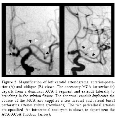
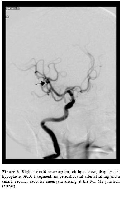
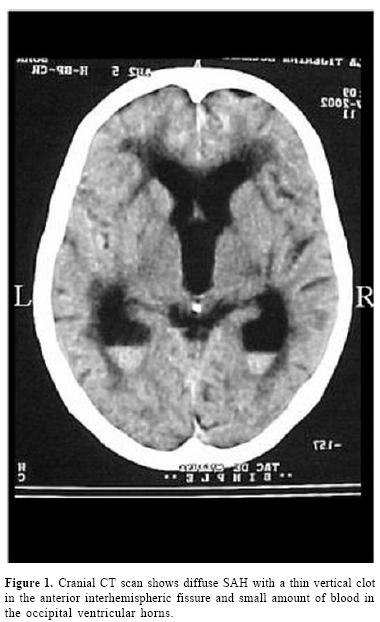
<body><![CDATA[<p align="justify"><font face="verdana" size="2">Upon admission, her blood pressure was 160/ 90 and the rest of the physiological constants, were normal. The patientwas disoriented but could attend verbal commands and move freely, if not with her left side. There was moderate neck stiffness, mild left hemiparesis and bilateral extensor plantar response. The pupils were symmetrical and reactive and the fundoscopic examination revealed slight hypertensive arteriolar changes but no subhyloid hemorrhages. A noncontrasted cranial CT scan that relatives carried along <a href="#f1">(figure 1)</a> showed diffuse SAH, a thin linear clot in the frontal interhemispheric fissure and small amount of intraventricular blood at the bottom of both occipital horns. The ventricular system was slightly enlarged due patient's age.</font></p>     <p align="center"><font face="verdana" size="2"><a name="f1" id="f1"></a></font></p>     <p align="center"><font face="verdana" size="2"><img src="/img/revistas/aneuroc/v10n1/a07f1.jpg"></font></p>     <p align="justify"><font face="verdana" size="2">Transfemoral cerebral panangiography was carried out under light sedation the following day. Left ICA injection <a href="#f2">(figure 2)</a> revealed dominance of ACA&#150;1 with opacification of both pericallosal arteries and a saccular aneurysm arising from the left corner of the anterior communicating artery (ACoA), which was recognized as the SAH's culprit. In addition, an anomalous arterial channel, roughly paralleling the course of the left MCA, was clearly observed in the anterior&#150;posterior and oblique views. As this second, "twin" like MCA departed from the proximal portion of ACA, extended laterally in the sylvian fissure to arborisation, supplying <i>pari&#150;passus </i>thin basal perforating branches, it was readily categorized as an <i>accessory </i>MCA. The right ICA injection <a href="#f3">(figure 3)</a> demonstrated an hypoplastic A1 segment, 1 no pericallosal filling and asecondaneurysm, emerging at the M1&#150;M2 junction. The proposed micro&#150;neurosurgical or endovascular treatment was refused.</font></p>     <p align="center"><font face="verdana" size="2"><a name="f2"></a></font></p>     <p align="center"><font face="verdana" size="2"><img src="/img/revistas/aneuroc/v10n1/a07f2.jpg"></font></p>     <p align="center"><font face="verdana" size="2"><a name="f3"></a></font></p>     <p align="center"><font face="verdana" size="2"><img src="/img/revistas/aneuroc/v10n1/a07f3.jpg"></font></p>     <p align="justify"><font face="verdana" size="2">The patient, however, remained hospitalized.</font></p>     <p align="justify"><font face="verdana" size="2">Sudden deterioration occurred two weeks later, the Glasgow coma scale grading declining to 7. After immediate endotracheal intubation and stabilization in the Intensive Care Unit, repeat CT scan of the head disclosed acute hydrocephalus with no trace of recurrent SAH. Actually, the subarachnoid and intraventricular blood observed in the previous CT scan, had vanished. A system for continuous external ventricular drainage was inserted. Regretfully, the patient expired on december 17, 2002, due to nosocomial pneumonia with no neurological improvement. Autopsy request was not granted.</font></p>     ]]></body>
<body><![CDATA[<p align="justify"><font face="verdana" size="2">&nbsp;</font></p>     <p align="justify"><font face="verdana" size="2"><b>DISCUSSION</b></font></p>     <p align="justify"><font face="verdana" size="2">An abnormal arterial conduit    running back from ACA into the sylvian fissure to replace the orbital branch    of the MCA, was observed in a <i>posmortem </i>investigation by Blackburn, as    early as 1908<sup>2</sup>. The nomination, however, of such irregular recurrent    arterial conduits as <i>accessory </i>MCA, took place in 1962, by Crompton<sup>3</sup>,    following the examination of 347 cadaveric brains. In his investigation, 11    examples of double MCA were found, an incidence of 3.2%. The circumstance that    10 of the anomalous arteries departed from ICA and one from ACA did not preclude    him from characterizing them all with the same name, attending more to its course    than to its different arterial origin. Two years later, Jain<sup>4</sup> published    10 examples of, again, <i>accessory </i>MCA, discovered among 300 <i>postmortem    </i>sylvian arteries dissected, a rate of 3.3%. But at this time, eight of the    variants sprang from ACA, and only two from ICA. Finally, in 1973, Teal <i>et    al.</i><sup>5</sup>, in reporting five additional angiographic examples of double    MCA, clarified the confusion by restricting the designation of <i>accessory    </i>MCA to the anomalous channel departing from ACA, and to term the one deriving    from ICA as <i>duplicate </i>MCA.</font></p>     <p align="justify"><font face="verdana" size="2">Hence, by universal acceptance, all <i>accessory </i>MCAs arise from ACA, most often from ACA&#150;1<sup>1,4,6,8</sup>, but they will at times depart from ACA&#150;2<sup>6,9,10</sup> or ACoA<sup>10,11</sup>. The <i>duplicate </i>MCA, in turn, will develop directly from ICA, either at the level of the anterior choroidal artery (AChA)<sup>6</sup>, between AChA and ICA end<sup>7,10</sup>, or opposite the origin of ACA, as an early ICA bifurcation<sup>1</sup>. As rather unique necropsy findings, an accessory MCA and a duplicate MCA, both, concurred in one occasion in the same cerebral hemisphere<sup>6</sup>; in one other, the accessory MCA presented bilaterally<sup>4</sup>, and in a third instance, an ICA&#150;originated repeat MCA, was associated with reduplication of the circle of Willis<sup>12</sup>. A bilateral accessory MCA has been diagnosed also in a few patients by means of cerebral arteriography<sup>11,13</sup>.</font></p>     <p align="justify"><font face="verdana" size="2">More recent <i>posfmortem</i>    microanatomical studies of the sylvian arteries upon unfixed adult brains<sup>16</sup>    have validated MCA duplication at a rate of 2.9% to 4.0%. In 1968, Handa <i>et    al.</i><sup>13</sup> and Krayenbh&uuml;l and Yasargil<sup>14</sup> first showed    such MCA anomalies by <i>in&#150;vivo </i>cerebral arteriography. The reported    angiographical incidence of accessory and duplicate MCAs combinedly ranges from    0.32%<sup>8</sup> and 1.6% <sup>7</sup>, to as high as 4.6% <sup>15</sup> but    some of these anomalies will pass clinically unrecognized or unpublished<sup>16</sup>.    Proximal <i>fenestration </i>(short, partial duplication) is a third MCA anomaly    disclosed at autopsy<sup>1,3,6,</sup><sup>11</sup> or    observed in cerebral angiograms at a rate of 0.26%<sup>1</sup><i>.</i></font></p>     <p align="justify"><font face="verdana" size="2">MCA abnormalities like these have been described as fortuitous angiographic findings in the investigation of patients suffering from head injury or ischemic stroke<sup>7,</sup> <sup>8,16</sup>, craniopharyngioma or paraganglioma<sup>7</sup>, moyamoya disease<sup>10,17</sup> or cerebral arteriovenous malformation<sup>18</sup>. Yet, their most frequent associations are intracranial aneurysms, usually single<sup>1,3,9,14,19,20</sup>, at times originating from the abnormal arterial conduit itself<sup>3,15,21</sup>. In a series of 24 patients with SAH and MCA variants drawn from the literature by Umanski <i>et al.1 </i>there were 35 associated intracranial aneurysms diagnosed. In five occasions, an aneurysm arose at the site of the anomaly (20.8%) and in five patients, the aneurysms were multiple (three patients with two, one with four, and one with six aneurysms). Our patient had two aneurysms. Nevertheless, a proclivity for aneurysmal development greater in these patients than in the general population, there seems not to be<sup>1</sup>.</font></p>     <p align="justify"><font face="verdana" size="2">The embryological significance of the MCA irregularities is not completely dilucidated as to whether they are the result of early blood flow dynamics or represent a persistent fetal vascular supply. Marubayashi<sup>18 </sup>suggests the <i>accessory MCA </i>is a residual congenital artery and the <i>duplicate </i>MCA represents an early ICA bifurcation. Handa et al<sup>19</sup>. See the <i>accessory </i>MCA as an hypertrophic recurrent Heubner artery (RHA) for it originates from ACA and supplies basal perforating branches. Yet, the accessory MCA and the RHA may coexist, usually with independent departure. Takahashi <i>et al.</i><sup>22 </sup>consider both, the accessory MCA and the RHA, resulting from a primitive anastomotic vascular network supplying the pyriform cortex, with final predominance of one or the other, usually the latter. Furtheremore, Komiyama <i>et al.</i><sup>7</sup> believe the duplicating MCA is a precocious ramification of early MCA branching while Yasargil and Smith<sup>10</sup> prefer not to derive any definitive conclusions in this regard.</font></p>     <p align="justify"><font face="verdana" size="2">In any case, their existence in the brain, in particular the accessory MCA, may serve as significant alternative channels for collateral blood supply reducing a potentially devastating neurological deficit in cases of acute or chronic, embolic or surgical ICA and/ or MCA occlusion<sup>16,17,20</sup>. In this eventuality, not rarely, the <i>accessory </i>MCA will be demonstrated to fill angiographically from the opposite ICA, across the midline. The preoperatory definition of this type of abnormalities will help the microvascular or endovascular surgeon toward the best treatment of intracranial aneurysms and occlusive cerebral disease.</font></p>     <p align="justify"><font face="verdana" size="2">&nbsp;</font></p>     <p align="justify"><font face="verdana" size="2"><b>CONCLUSION</b></font></p>     ]]></body>
<body><![CDATA[<p align="justify"><font face="verdana" size="2">One additional angiographic example of an <i>accessory </i>MCA, in this case associated with two supratentorial intracranial aneurysms, is presented. The anomalous MCA in our patient departed from ACA and extended laterally in a recurrent fashion paralleling the course of MCA, providing a few basal perforating arteries before branching.</font></p>     <p align="justify"><font face="verdana" size="2">&nbsp;</font></p>     <p align="justify"><font face="verdana" size="2"><b>REFERENCES</b></font></p>     <!-- ref --><p align="justify"><font face="verdana" size="2">1. Umanski F, Dujovny M, Ausman    Jl, Diaz FG, Mirchandani HG. Anomalies and variations of the middle cerebral    artery: a microanatomical study. <i>Neurosurgery </i>1998; 22:1023&#150;7.</font>&nbsp;&nbsp;&nbsp;&nbsp;&nbsp;&nbsp;&nbsp;&nbsp;[&#160;<a href="javascript:void(0);" onclick="javascript: window.open('/scielo.php?script=sci_nlinks&ref=1174172&pid=S0187-4705200500010000700001&lng=','','width=640,height=500,resizable=yes,scrollbars=1,menubar=yes,');">Links</a>&#160;]<!-- end-ref --><!-- ref --><p align="justify"><font face="verdana" size="2">2. Blackburn IW. Anomalies of    the encephalic arteries among the insane: A study of the arteries at the base    of the encephalon in two hundred and twenty consecutive cases of mental disease,    with special reference to anomalies of the circle of Willis. <i>J Comp Neurol    Psychol </i>1908; 17:493&#150;517.</font>&nbsp;&nbsp;&nbsp;&nbsp;&nbsp;&nbsp;&nbsp;&nbsp;[&#160;<a href="javascript:void(0);" onclick="javascript: window.open('/scielo.php?script=sci_nlinks&ref=1174173&pid=S0187-4705200500010000700002&lng=','','width=640,height=500,resizable=yes,scrollbars=1,menubar=yes,');">Links</a>&#160;]<!-- end-ref --><!-- ref --><p align="justify"><font face="verdana" size="2">3. Crompton MR. The pathology    of ruptured middle&#150;cerebral aneurysms with special reference to the differences    between the sexes. <i>Lancet </i>1962; 2:421&#150;5.</font>&nbsp;&nbsp;&nbsp;&nbsp;&nbsp;&nbsp;&nbsp;&nbsp;[&#160;<a href="javascript:void(0);" onclick="javascript: window.open('/scielo.php?script=sci_nlinks&ref=1174174&pid=S0187-4705200500010000700003&lng=','','width=640,height=500,resizable=yes,scrollbars=1,menubar=yes,');">Links</a>&#160;]<!-- end-ref --><!-- ref --><p align="justify"><font face="verdana" size="2">4. Jain KK. Some observations    on the anatomy of the middle cerebral artery. <i>Can J Surg </i>1964; 7:134&#150;9.</font>&nbsp;&nbsp;&nbsp;&nbsp;&nbsp;&nbsp;&nbsp;&nbsp;[&#160;<a href="javascript:void(0);" onclick="javascript: window.open('/scielo.php?script=sci_nlinks&ref=1174175&pid=S0187-4705200500010000700004&lng=','','width=640,height=500,resizable=yes,scrollbars=1,menubar=yes,');">Links</a>&#160;]<!-- end-ref --><!-- ref --><p align="justify"><font face="verdana" size="2">5. Teal JS, Rumbaugh CL, Bergeron    RT, Segall HD. Anomalies of the middle cerebral artery: accessory artery, duplication    and early bifurcation. <i>Am J Roentgenol Radium Therapy Nucl Med </i>1973;    118:567&#150;75.</font>&nbsp;&nbsp;&nbsp;&nbsp;&nbsp;&nbsp;&nbsp;&nbsp;[&#160;<a href="javascript:void(0);" onclick="javascript: window.open('/scielo.php?script=sci_nlinks&ref=1174176&pid=S0187-4705200500010000700005&lng=','','width=640,height=500,resizable=yes,scrollbars=1,menubar=yes,');">Links</a>&#160;]<!-- end-ref --><!-- ref --><p align="justify"><font face="verdana" size="2">6. Gibo H, Carver CC, Rhoton AL, Lenkey C, Mitchell RJ. Microsurgical anatomy of the middle cerebral artery. <i>J Neurosurg </i>1981:54:151&#150;69.</font>&nbsp;&nbsp;&nbsp;&nbsp;&nbsp;&nbsp;&nbsp;&nbsp;[&#160;<a href="javascript:void(0);" onclick="javascript: window.open('/scielo.php?script=sci_nlinks&ref=1174177&pid=S0187-4705200500010000700006&lng=','','width=640,height=500,resizable=yes,scrollbars=1,menubar=yes,');">Links</a>&#160;]<!-- end-ref --><!-- ref --><p align="justify"><font face="verdana" size="2">7. Komiyama M, Nakajima H, Nishikawa M, Yasui T. Middle cerebral artery variations: duplicated and accessory arteries. <i>AJNR </i>1998:19:45&#150;9.</font>&nbsp;&nbsp;&nbsp;&nbsp;&nbsp;&nbsp;&nbsp;&nbsp;[&#160;<a href="javascript:void(0);" onclick="javascript: window.open('/scielo.php?script=sci_nlinks&ref=1174178&pid=S0187-4705200500010000700007&lng=','','width=640,height=500,resizable=yes,scrollbars=1,menubar=yes,');">Links</a>&#160;]<!-- end-ref --><!-- ref --><p align="justify"><font face="verdana" size="2">8. Watanabe T, Togo M. Accessory middle cerebral artery. <i>J Neurosurg </i>1974:41:248&#150;51.</font>&nbsp;&nbsp;&nbsp;&nbsp;&nbsp;&nbsp;&nbsp;&nbsp;[&#160;<a href="javascript:void(0);" onclick="javascript: window.open('/scielo.php?script=sci_nlinks&ref=1174179&pid=S0187-4705200500010000700008&lng=','','width=640,height=500,resizable=yes,scrollbars=1,menubar=yes,');">Links</a>&#160;]<!-- end-ref --><!-- ref --><p align="justify"><font face="verdana" size="2">9. Tacconi L, Johnston FG, Symon L. Accessory middle cerebral artery. Case report. <i>J Neurosurg </i>1995:83:916&#150;8.</font>&nbsp;&nbsp;&nbsp;&nbsp;&nbsp;&nbsp;&nbsp;&nbsp;[&#160;<a href="javascript:void(0);" onclick="javascript: window.open('/scielo.php?script=sci_nlinks&ref=1174180&pid=S0187-4705200500010000700009&lng=','','width=640,height=500,resizable=yes,scrollbars=1,menubar=yes,');">Links</a>&#160;]<!-- end-ref --><!-- ref --><p align="justify"><font face="verdana" size="2">10. Yasargil GM, Smith RD. Association of middle cerebral artery anomalies with saccular aneurysms and moyamoya disease.<i>   Surg Neurol </i>1976; 6:39&#150;43.</font>&nbsp;&nbsp;&nbsp;&nbsp;&nbsp;&nbsp;&nbsp;&nbsp;[&#160;<a href="javascript:void(0);" onclick="javascript: window.open('/scielo.php?script=sci_nlinks&ref=1174181&pid=S0187-4705200500010000700010&lng=','','width=640,height=500,resizable=yes,scrollbars=1,menubar=yes,');">Links</a>&#160;]<!-- end-ref --><!-- ref --><p align="justify"><font face="verdana" size="2">11.Yasargil MG. Middle cerebral artery, in <i>Microneurosurgery. Stuttgart, <i>Georg Thieme Verlag </i>1984, vol I, 86&#150;91.&nbsp;</i></font>&nbsp;&nbsp;&nbsp;&nbsp;&nbsp;&nbsp;&nbsp;&nbsp;[&#160;<a href="javascript:void(0);" onclick="javascript: window.open('/scielo.php?script=sci_nlinks&ref=1174182&pid=S0187-4705200500010000700011&lng=','','width=640,height=500,resizable=yes,scrollbars=1,menubar=yes,');">Links</a>&#160;]<!-- end-ref --><!-- ref --><p align="justify"><font face="verdana" size="2">12. Fisher ER, Davis JS, Lemmen    MJ. Anomalous reduplication of&nbsp;the circle of Willis. <i>J Neurosurg </i>1959;    6:331&#150;6.</font>&nbsp;&nbsp;&nbsp;&nbsp;&nbsp;&nbsp;&nbsp;&nbsp;[&#160;<a href="javascript:void(0);" onclick="javascript: window.open('/scielo.php?script=sci_nlinks&ref=1174183&pid=S0187-4705200500010000700012&lng=','','width=640,height=500,resizable=yes,scrollbars=1,menubar=yes,');">Links</a>&#160;]<!-- end-ref --><!-- ref --><p align="justify"><font face="verdana" size="2">13. Handa J, Seta K, Handa H.    Die akzessorische A cerebri media. <i>Fortschr Roentgenstr </i>1968; 108:539&#150;41.</font>&nbsp;&nbsp;&nbsp;&nbsp;&nbsp;&nbsp;&nbsp;&nbsp;[&#160;<a href="javascript:void(0);" onclick="javascript: window.open('/scielo.php?script=sci_nlinks&ref=1174184&pid=S0187-4705200500010000700013&lng=','','width=640,height=500,resizable=yes,scrollbars=1,menubar=yes,');">Links</a>&#160;]<!-- end-ref --><!-- ref --><p align="justify"><font face="verdana" size="2">14. Krayenb&uuml;hl HA, Yasargil MG. <i>Cerebral Angiography, </i>ed 2. London, <i>Butterworth </i>1968.</font>&nbsp;&nbsp;&nbsp;&nbsp;&nbsp;&nbsp;&nbsp;&nbsp;[&#160;<a href="javascript:void(0);" onclick="javascript: window.open('/scielo.php?script=sci_nlinks&ref=1174185&pid=S0187-4705200500010000700014&lng=','','width=640,height=500,resizable=yes,scrollbars=1,menubar=yes,');">Links</a>&#160;]<!-- end-ref --><!-- ref --><p align="justify"><font face="verdana" size="2">15. Miyazaki S, Ito K, Ishii    S. Aneurysm at the origin of the accessory middle cerebral artery. <i>Surg Neurol    </i>1984; 22:292&#150;4.&nbsp;&nbsp;</font>&nbsp;&nbsp;&nbsp;&nbsp;&nbsp;&nbsp;&nbsp;&nbsp;[&#160;<a href="javascript:void(0);" onclick="javascript: window.open('/scielo.php?script=sci_nlinks&ref=1174186&pid=S0187-4705200500010000700015&lng=','','width=640,height=500,resizable=yes,scrollbars=1,menubar=yes,');">Links</a>&#160;]<!-- end-ref --><!-- ref --><p align="justify"><font face="verdana" size="2">16.&nbsp;Komiyama M, Nishikawa M, Yasui T. The accessory middle cerebral artery as a collateral blood supply. <i>AJNR </i>1997;18:587&#150;90.</font>&nbsp;&nbsp;&nbsp;&nbsp;&nbsp;&nbsp;&nbsp;&nbsp;[&#160;<a href="javascript:void(0);" onclick="javascript: window.open('/scielo.php?script=sci_nlinks&ref=1174187&pid=S0187-4705200500010000700016&lng=','','width=640,height=500,resizable=yes,scrollbars=1,menubar=yes,');">Links</a>&#160;]<!-- end-ref --><!-- ref --><p align="justify"><font face="verdana" size="2">17.&nbsp;Komiyama M, Yasui T. Accessory middle cerebral artery and moyamoya disease. <i>J Neurol Neurosurg Psychiatry </i>2001; 71:129&#150;30 (letter).</font>&nbsp;&nbsp;&nbsp;&nbsp;&nbsp;&nbsp;&nbsp;&nbsp;[&#160;<a href="javascript:void(0);" onclick="javascript: window.open('/scielo.php?script=sci_nlinks&ref=1174188&pid=S0187-4705200500010000700017&lng=','','width=640,height=500,resizable=yes,scrollbars=1,menubar=yes,');">Links</a>&#160;]<!-- end-ref --><!-- ref --><p align="justify"><font face="verdana" size="2">18. Marubayashi T. Accessory    middle cerebral artery and duplication of middle cerebral artery&#150;terminology,    incidence, vascular etiology and developmental significance. <i>Neurolog&iacute;a    <i>Medico Chirurgica </i>1992; 32:262&#150;7.</i></font>&nbsp;&nbsp;&nbsp;&nbsp;&nbsp;&nbsp;&nbsp;&nbsp;[&#160;<a href="javascript:void(0);" onclick="javascript: window.open('/scielo.php?script=sci_nlinks&ref=1174189&pid=S0187-4705200500010000700018&lng=','','width=640,height=500,resizable=yes,scrollbars=1,menubar=yes,');">Links</a>&#160;]<!-- end-ref --><!-- ref --><p align="justify"><font face="verdana" size="2">19. Handa J, Shimizu Y, Matsuda M, Handa H. The accessory &nbsp;middle cerebral artery: Report of further two cases. <i>Clin Radiol 1970;21:415&#150;6.</i></font>&nbsp;&nbsp;&nbsp;&nbsp;&nbsp;&nbsp;&nbsp;&nbsp;[&#160;<a href="javascript:void(0);" onclick="javascript: window.open('/scielo.php?script=sci_nlinks&ref=1174190&pid=S0187-4705200500010000700019&lng=','','width=640,height=500,resizable=yes,scrollbars=1,menubar=yes,');">Links</a>&#160;]<!-- end-ref --><!-- ref --><p align="justify"><font face="verdana" size="2">20. Mueller DP, Sato Y, Yuh WTC. Accessory middle cerebral artery  as a source of collateral blood flow. <i>AJNR </i>1991; 12:1223&#150;4.</font>&nbsp;&nbsp;&nbsp;&nbsp;&nbsp;&nbsp;&nbsp;&nbsp;[&#160;<a href="javascript:void(0);" onclick="javascript: window.open('/scielo.php?script=sci_nlinks&ref=1174191&pid=S0187-4705200500010000700020&lng=','','width=640,height=500,resizable=yes,scrollbars=1,menubar=yes,');">Links</a>&#160;]<!-- end-ref --><!-- ref --><p align="justify"><font face="verdana" size="2">21.Waga S, Kojima T, Morooka    Y, Sakakura M. Aneurysm of the accessory middle cerebral artery. <i>Surg Neurol    </i>1977; 8:359&#150;60.</font>&nbsp;&nbsp;&nbsp;&nbsp;&nbsp;&nbsp;&nbsp;&nbsp;[&#160;<a href="javascript:void(0);" onclick="javascript: window.open('/scielo.php?script=sci_nlinks&ref=1174192&pid=S0187-4705200500010000700021&lng=','','width=640,height=500,resizable=yes,scrollbars=1,menubar=yes,');">Links</a>&#160;]<!-- end-ref --><!-- ref --><p align="justify"><font face="verdana" size="2">22. Takahashi S, Hoshino F, Uemura    K, Takahashi A, Sakamoto K. Accessory middle cerebral artery: is it a variant    form of the recurrent artery of Heubner? <i>AJNR </i>1989; 10:563&#150;8 (abstr).</font>&nbsp;&nbsp;&nbsp;&nbsp;&nbsp;&nbsp;&nbsp;&nbsp;[&#160;<a href="javascript:void(0);" onclick="javascript: window.open('/scielo.php?script=sci_nlinks&ref=1174193&pid=S0187-4705200500010000700022&lng=','','width=640,height=500,resizable=yes,scrollbars=1,menubar=yes,');">Links</a>&#160;]<!-- end-ref --> ]]></body><back>
<ref-list>
<ref id="B1">
<label>1</label><nlm-citation citation-type="journal">
<person-group person-group-type="author">
<name>
<surname><![CDATA[Umanski]]></surname>
<given-names><![CDATA[F]]></given-names>
</name>
<name>
<surname><![CDATA[Dujovny]]></surname>
<given-names><![CDATA[M]]></given-names>
</name>
<name>
<surname><![CDATA[Ausman]]></surname>
<given-names><![CDATA[Jl]]></given-names>
</name>
<name>
<surname><![CDATA[Diaz]]></surname>
<given-names><![CDATA[FG]]></given-names>
</name>
<name>
<surname><![CDATA[Mirchandani]]></surname>
<given-names><![CDATA[HG]]></given-names>
</name>
</person-group>
<article-title xml:lang="en"><![CDATA[Anomalies and variations of the middle cerebral artery: a microanatomical study]]></article-title>
<source><![CDATA[Neurosurgery]]></source>
<year>1998</year>
<volume>22</volume>
<page-range>1023-7</page-range></nlm-citation>
</ref>
<ref id="B2">
<label>2</label><nlm-citation citation-type="journal">
<person-group person-group-type="author">
<name>
<surname><![CDATA[Blackburn]]></surname>
<given-names><![CDATA[IW]]></given-names>
</name>
</person-group>
<article-title xml:lang="en"><![CDATA[Anomalies of the encephalic arteries among the insane: A study of the arteries at the base of the encephalon in two hundred and twenty consecutive cases of mental disease, with special reference to anomalies of the circle of Willis]]></article-title>
<source><![CDATA[J Comp Neurol Psychol]]></source>
<year>1908</year>
<volume>17</volume>
<page-range>493-517</page-range></nlm-citation>
</ref>
<ref id="B3">
<label>3</label><nlm-citation citation-type="journal">
<person-group person-group-type="author">
<name>
<surname><![CDATA[Crompton]]></surname>
<given-names><![CDATA[MR]]></given-names>
</name>
</person-group>
<article-title xml:lang="en"><![CDATA[The pathology of ruptured middle-cerebral aneurysms with special reference to the differences between the sexes]]></article-title>
<source><![CDATA[Lancet]]></source>
<year>1962</year>
<numero>2</numero>
<issue>2</issue>
<page-range>421-5</page-range></nlm-citation>
</ref>
<ref id="B4">
<label>4</label><nlm-citation citation-type="journal">
<person-group person-group-type="author">
<name>
<surname><![CDATA[Jain]]></surname>
<given-names><![CDATA[KK]]></given-names>
</name>
</person-group>
<article-title xml:lang="en"><![CDATA[Some observations on the anatomy of the middle cerebral artery]]></article-title>
<source><![CDATA[Can J Surg]]></source>
<year>1964</year>
<volume>7</volume>
<page-range>134-9</page-range></nlm-citation>
</ref>
<ref id="B5">
<label>5</label><nlm-citation citation-type="journal">
<person-group person-group-type="author">
<name>
<surname><![CDATA[Teal]]></surname>
<given-names><![CDATA[JS]]></given-names>
</name>
<name>
<surname><![CDATA[Rumbaugh]]></surname>
<given-names><![CDATA[CL]]></given-names>
</name>
<name>
<surname><![CDATA[Bergeron]]></surname>
<given-names><![CDATA[RT]]></given-names>
</name>
<name>
<surname><![CDATA[Segall]]></surname>
<given-names><![CDATA[HD]]></given-names>
</name>
</person-group>
<article-title xml:lang="en"><![CDATA[Anomalies of the middle cerebral artery: accessory artery, duplication and early bifurcation]]></article-title>
<source><![CDATA[Am J Roentgenol Radium Therapy Nucl Med]]></source>
<year>1973</year>
<numero>118</numero>
<issue>118</issue>
<page-range>567-75</page-range></nlm-citation>
</ref>
<ref id="B6">
<label>6</label><nlm-citation citation-type="journal">
<person-group person-group-type="author">
<name>
<surname><![CDATA[Gibo]]></surname>
<given-names><![CDATA[H]]></given-names>
</name>
<name>
<surname><![CDATA[Carver]]></surname>
<given-names><![CDATA[CC]]></given-names>
</name>
<name>
<surname><![CDATA[Rhoton]]></surname>
<given-names><![CDATA[AL]]></given-names>
</name>
<name>
<surname><![CDATA[Lenkey]]></surname>
<given-names><![CDATA[C]]></given-names>
</name>
<name>
<surname><![CDATA[Mitchell]]></surname>
<given-names><![CDATA[RJ]]></given-names>
</name>
</person-group>
<article-title xml:lang="en"><![CDATA[Microsurgical anatomy of the middle cerebral artery]]></article-title>
<source><![CDATA[J Neurosurg]]></source>
<year>1981</year>
<volume>54</volume>
<page-range>151-69</page-range></nlm-citation>
</ref>
<ref id="B7">
<label>7</label><nlm-citation citation-type="journal">
<person-group person-group-type="author">
<name>
<surname><![CDATA[Komiyama]]></surname>
<given-names><![CDATA[M]]></given-names>
</name>
<name>
<surname><![CDATA[Nakajima]]></surname>
<given-names><![CDATA[H]]></given-names>
</name>
<name>
<surname><![CDATA[Nishikawa]]></surname>
<given-names><![CDATA[M]]></given-names>
</name>
<name>
<surname><![CDATA[Yasui]]></surname>
<given-names><![CDATA[T]]></given-names>
</name>
</person-group>
<article-title xml:lang="en"><![CDATA[Middle cerebral artery variations: duplicated and accessory arteries]]></article-title>
<source><![CDATA[AJNR]]></source>
<year>1998</year>
<volume>19</volume>
<page-range>45-9</page-range></nlm-citation>
</ref>
<ref id="B8">
<label>8</label><nlm-citation citation-type="journal">
<person-group person-group-type="author">
<name>
<surname><![CDATA[Watanabe]]></surname>
<given-names><![CDATA[T]]></given-names>
</name>
<name>
<surname><![CDATA[Togo]]></surname>
<given-names><![CDATA[M]]></given-names>
</name>
</person-group>
<article-title xml:lang="en"><![CDATA[Accessory middle cerebral artery]]></article-title>
<source><![CDATA[J Neurosurg]]></source>
<year>1974</year>
<volume>41</volume>
<page-range>248-51</page-range></nlm-citation>
</ref>
<ref id="B9">
<label>9</label><nlm-citation citation-type="journal">
<person-group person-group-type="author">
<name>
<surname><![CDATA[Tacconi]]></surname>
<given-names><![CDATA[L]]></given-names>
</name>
<name>
<surname><![CDATA[Johnston]]></surname>
<given-names><![CDATA[FG]]></given-names>
</name>
<name>
<surname><![CDATA[Symon]]></surname>
<given-names><![CDATA[L]]></given-names>
</name>
</person-group>
<article-title xml:lang="en"><![CDATA[Accessory middle cerebral artery: Case report]]></article-title>
<source><![CDATA[J Neurosurg]]></source>
<year>1995</year>
<volume>83</volume>
<page-range>916-8</page-range></nlm-citation>
</ref>
<ref id="B10">
<label>10</label><nlm-citation citation-type="journal">
<person-group person-group-type="author">
<name>
<surname><![CDATA[Yasargil]]></surname>
<given-names><![CDATA[GM]]></given-names>
</name>
<name>
<surname><![CDATA[Smith]]></surname>
<given-names><![CDATA[RD]]></given-names>
</name>
</person-group>
<article-title xml:lang="en"><![CDATA[Association of middle cerebral artery anomalies with saccular aneurysms and moyamoya disease]]></article-title>
<source><![CDATA[Surg Neurol]]></source>
<year>1976</year>
<volume>6</volume>
<page-range>39-43</page-range></nlm-citation>
</ref>
<ref id="B11">
<label>11</label><nlm-citation citation-type="book">
<person-group person-group-type="author">
<name>
<surname><![CDATA[Yasargil]]></surname>
<given-names><![CDATA[MG]]></given-names>
</name>
</person-group>
<article-title xml:lang="en"><![CDATA[Middle cerebral artery]]></article-title>
<source><![CDATA[Microneurosurgery]]></source>
<year>1984</year>
<volume>I</volume>
<page-range>86-91</page-range><publisher-loc><![CDATA[Stuttgart ]]></publisher-loc>
<publisher-name><![CDATA[Georg Thieme Verlag]]></publisher-name>
</nlm-citation>
</ref>
<ref id="B12">
<label>12</label><nlm-citation citation-type="journal">
<person-group person-group-type="author">
<name>
<surname><![CDATA[Fisher]]></surname>
<given-names><![CDATA[ER]]></given-names>
</name>
<name>
<surname><![CDATA[Davis]]></surname>
<given-names><![CDATA[JS]]></given-names>
</name>
<name>
<surname><![CDATA[Lemmen]]></surname>
<given-names><![CDATA[MJ]]></given-names>
</name>
</person-group>
<article-title xml:lang="en"><![CDATA[Anomalous reduplication of the circle of Willis]]></article-title>
<source><![CDATA[J Neurosurg]]></source>
<year>1959</year>
<volume>6</volume>
<page-range>331-6</page-range></nlm-citation>
</ref>
<ref id="B13">
<label>13</label><nlm-citation citation-type="journal">
<person-group person-group-type="author">
<name>
<surname><![CDATA[Handa]]></surname>
<given-names><![CDATA[J]]></given-names>
</name>
<name>
<surname><![CDATA[Seta]]></surname>
<given-names><![CDATA[K]]></given-names>
</name>
<name>
<surname><![CDATA[Handa]]></surname>
<given-names><![CDATA[H]]></given-names>
</name>
</person-group>
<article-title xml:lang="de"><![CDATA[Die akzessorische A cerebri media]]></article-title>
<source><![CDATA[Fortschr Roentgenstr]]></source>
<year>1968</year>
<volume>108</volume>
<page-range>539-41</page-range></nlm-citation>
</ref>
<ref id="B14">
<label>14</label><nlm-citation citation-type="book">
<person-group person-group-type="author">
<name>
<surname><![CDATA[Krayenbühl]]></surname>
<given-names><![CDATA[HA]]></given-names>
</name>
<name>
<surname><![CDATA[Yasargil]]></surname>
<given-names><![CDATA[MG]]></given-names>
</name>
</person-group>
<source><![CDATA[Cerebral Angiography]]></source>
<year>1968</year>
<edition>2</edition>
<publisher-loc><![CDATA[London ]]></publisher-loc>
<publisher-name><![CDATA[Butterworth]]></publisher-name>
</nlm-citation>
</ref>
<ref id="B15">
<label>15</label><nlm-citation citation-type="journal">
<person-group person-group-type="author">
<name>
<surname><![CDATA[Miyazaki]]></surname>
<given-names><![CDATA[S]]></given-names>
</name>
<name>
<surname><![CDATA[Ito]]></surname>
<given-names><![CDATA[K]]></given-names>
</name>
<name>
<surname><![CDATA[Ishii]]></surname>
<given-names><![CDATA[S]]></given-names>
</name>
</person-group>
<article-title xml:lang="en"><![CDATA[Aneurysm at the origin of the accessory middle cerebral artery]]></article-title>
<source><![CDATA[Surg Neurol]]></source>
<year>1984</year>
<volume>22</volume>
<page-range>292-4</page-range></nlm-citation>
</ref>
<ref id="B16">
<label>16</label><nlm-citation citation-type="journal">
<person-group person-group-type="author">
<name>
<surname><![CDATA[Komiyama]]></surname>
<given-names><![CDATA[M]]></given-names>
</name>
<name>
<surname><![CDATA[Nishikawa]]></surname>
<given-names><![CDATA[M]]></given-names>
</name>
<name>
<surname><![CDATA[Yasui]]></surname>
<given-names><![CDATA[T]]></given-names>
</name>
</person-group>
<article-title xml:lang="en"><![CDATA[The accessory middle cerebral artery as a collateral blood supply]]></article-title>
<source><![CDATA[AJNR]]></source>
<year>1997</year>
<volume>18</volume>
<page-range>587-90</page-range></nlm-citation>
</ref>
<ref id="B17">
<label>17</label><nlm-citation citation-type="journal">
<person-group person-group-type="author">
<name>
<surname><![CDATA[Komiyama]]></surname>
<given-names><![CDATA[M]]></given-names>
</name>
<name>
<surname><![CDATA[Yasui]]></surname>
<given-names><![CDATA[T]]></given-names>
</name>
</person-group>
<article-title xml:lang="en"><![CDATA[Accessory middle cerebral artery and moyamoya disease]]></article-title>
<source><![CDATA[J Neurol Neurosurg Psychiatry]]></source>
<year>2001</year>
<volume>71</volume>
<page-range>129-30</page-range></nlm-citation>
</ref>
<ref id="B18">
<label>18</label><nlm-citation citation-type="journal">
<person-group person-group-type="author">
<name>
<surname><![CDATA[Marubayashi]]></surname>
<given-names><![CDATA[T]]></given-names>
</name>
</person-group>
<article-title xml:lang="en"><![CDATA[Accessory middle cerebral artery and duplication of middle cerebral artery-terminology, incidence, vascular etiology and developmental significance]]></article-title>
<source><![CDATA[Neurología Medico Chirurgica]]></source>
<year>1992</year>
<volume>32</volume>
<page-range>262-7</page-range></nlm-citation>
</ref>
<ref id="B19">
<label>19</label><nlm-citation citation-type="journal">
<person-group person-group-type="author">
<name>
<surname><![CDATA[Handa]]></surname>
<given-names><![CDATA[J]]></given-names>
</name>
<name>
<surname><![CDATA[Shimizu]]></surname>
<given-names><![CDATA[Y]]></given-names>
</name>
<name>
<surname><![CDATA[Matsuda]]></surname>
<given-names><![CDATA[M]]></given-names>
</name>
<name>
<surname><![CDATA[Handa]]></surname>
<given-names><![CDATA[H]]></given-names>
</name>
</person-group>
<article-title xml:lang="en"><![CDATA[The accessory middle cerebral artery: Report of further two cases]]></article-title>
<source><![CDATA[Clin Radiol]]></source>
<year>1970</year>
<volume>21</volume>
<page-range>415-6</page-range></nlm-citation>
</ref>
<ref id="B20">
<label>20</label><nlm-citation citation-type="journal">
<person-group person-group-type="author">
<name>
<surname><![CDATA[Mueller]]></surname>
<given-names><![CDATA[DP]]></given-names>
</name>
<name>
<surname><![CDATA[Sato]]></surname>
<given-names><![CDATA[Y]]></given-names>
</name>
<name>
<surname><![CDATA[Yuh]]></surname>
<given-names><![CDATA[WTC]]></given-names>
</name>
</person-group>
<article-title xml:lang="en"><![CDATA[Accessory middle cerebral artery as a source of collateral blood flow]]></article-title>
<source><![CDATA[AJNR]]></source>
<year>1991</year>
<volume>12</volume>
<page-range>1223-4</page-range></nlm-citation>
</ref>
<ref id="B21">
<label>21</label><nlm-citation citation-type="journal">
<person-group person-group-type="author">
<name>
<surname><![CDATA[Waga]]></surname>
<given-names><![CDATA[S]]></given-names>
</name>
<name>
<surname><![CDATA[Kojima]]></surname>
<given-names><![CDATA[T]]></given-names>
</name>
<name>
<surname><![CDATA[Morooka]]></surname>
<given-names><![CDATA[Y]]></given-names>
</name>
<name>
<surname><![CDATA[Sakakura]]></surname>
<given-names><![CDATA[M]]></given-names>
</name>
</person-group>
<article-title xml:lang="en"><![CDATA[Aneurysm of the accessory middle cerebral artery]]></article-title>
<source><![CDATA[Surg Neurol]]></source>
<year>1977</year>
<volume>8</volume>
<page-range>359-60</page-range></nlm-citation>
</ref>
<ref id="B22">
<label>22</label><nlm-citation citation-type="journal">
<person-group person-group-type="author">
<name>
<surname><![CDATA[Takahashi]]></surname>
<given-names><![CDATA[S]]></given-names>
</name>
<name>
<surname><![CDATA[Hoshino]]></surname>
<given-names><![CDATA[F]]></given-names>
</name>
<name>
<surname><![CDATA[Uemura]]></surname>
<given-names><![CDATA[K]]></given-names>
</name>
<name>
<surname><![CDATA[Takahashi]]></surname>
<given-names><![CDATA[A]]></given-names>
</name>
<name>
<surname><![CDATA[Sakamoto]]></surname>
<given-names><![CDATA[K]]></given-names>
</name>
</person-group>
<article-title xml:lang="en"><![CDATA[Accessory middle cerebral artery: is it a variant form of the recurrent artery of Heubner?]]></article-title>
<source><![CDATA[AJNR]]></source>
<year>1989</year>
<volume>10</volume>
<page-range>563-8</page-range></nlm-citation>
</ref>
</ref-list>
</back>
</article>