<?xml version="1.0" encoding="ISO-8859-1"?><article xmlns:mml="http://www.w3.org/1998/Math/MathML" xmlns:xlink="http://www.w3.org/1999/xlink" xmlns:xsi="http://www.w3.org/2001/XMLSchema-instance">
<front>
<journal-meta>
<journal-id>0036-3634</journal-id>
<journal-title><![CDATA[Salud Pública de México]]></journal-title>
<abbrev-journal-title><![CDATA[Salud pública Méx]]></abbrev-journal-title>
<issn>0036-3634</issn>
<publisher>
<publisher-name><![CDATA[Instituto Nacional de Salud Pública]]></publisher-name>
</publisher>
</journal-meta>
<article-meta>
<article-id>S0036-36342009000700007</article-id>
<title-group>
<article-title xml:lang="en"><![CDATA[The impact of methods for estimating bone health and the global burden of bone disease]]></article-title>
<article-title xml:lang="es"><![CDATA[Impacto de los métodos para medir la salud ósea y la carga global de enfermedades óseas]]></article-title>
</title-group>
<contrib-group>
<contrib contrib-type="author">
<name>
<surname><![CDATA[Cole]]></surname>
<given-names><![CDATA[Zoë A]]></given-names>
</name>
<xref ref-type="aff" rid="A01"/>
</contrib>
<contrib contrib-type="author">
<name>
<surname><![CDATA[Dennison]]></surname>
<given-names><![CDATA[Elaine M]]></given-names>
</name>
<xref ref-type="aff" rid="A01"/>
</contrib>
<contrib contrib-type="author">
<name>
<surname><![CDATA[Cooper]]></surname>
<given-names><![CDATA[Cyrus]]></given-names>
</name>
<xref ref-type="aff" rid="A01"/>
</contrib>
</contrib-group>
<aff id="A01">
<institution><![CDATA[,University of Southampton MRC Epidemiology Resource Centre ]]></institution>
<addr-line><![CDATA[ ]]></addr-line>
</aff>
<pub-date pub-type="pub">
<day>00</day>
<month>00</month>
<year>2009</year>
</pub-date>
<pub-date pub-type="epub">
<day>00</day>
<month>00</month>
<year>2009</year>
</pub-date>
<volume>51</volume>
<fpage>s38</fpage>
<lpage>s45</lpage>
<copyright-statement/>
<copyright-year/>
<self-uri xlink:href="http://www.scielo.org.mx/scielo.php?script=sci_arttext&amp;pid=S0036-36342009000700007&amp;lng=en&amp;nrm=iso"></self-uri><self-uri xlink:href="http://www.scielo.org.mx/scielo.php?script=sci_abstract&amp;pid=S0036-36342009000700007&amp;lng=en&amp;nrm=iso"></self-uri><self-uri xlink:href="http://www.scielo.org.mx/scielo.php?script=sci_pdf&amp;pid=S0036-36342009000700007&amp;lng=en&amp;nrm=iso"></self-uri><abstract abstract-type="short" xml:lang="en"><p><![CDATA[Osteoporosis constitutes a major public health problem through its association with age related fractures. Fracture rates are generally higher in caucasian women than in other populations. Important determinants include estrogen deficiency in women, low body mass index, cigarette smoking, alcohol consumption, poor dietary calcium intake, physical inactivity, certain drugs and illnesses. Thus, modification of physical activity and dietary calcium/vitamin D nutrition should complement high risk approaches. In addition, the recently developed WHO algorithm for evaluation of 10-year absolute risk of fracture provides a means whereby various therapies can be targeted cost-effectively to those at risk. Risk factors, together with bone mineral density (BMD) and biochemical indices of bone turnover, can be utilised to derive absolute risks of fracture and cost-utility thresholds at which treatment is justified. These data will provide the basis for translation into coherent public health strategies aiming to prevent osteoporosis both in individuals and in the general population.]]></p></abstract>
<abstract abstract-type="short" xml:lang="es"><p><![CDATA[La osteoporosis constituye un importante problema de salud pública debido a su asociación con fracturas relacionadas con la edad. Las tasas de fractura generalmente son más altas en mujeres caucásicas que en otros grupos poblacionales. Los principales determinantes incluyen deficiencia de estrógeno en mujeres, bajo índice de masa corporal, consumo de tabaco y alcohol, escaso consumo de calcio, inactividad física y algunas drogas y enfermedades. De este modo, la modificación de la actividad física y el consumo de nutrimentos con calcio y vitamina D deben complementar los tratamientos en alto riesgo. Además, el recientemente desarrollado algoritmo de la OMS para la evaluación de riesgo de fractura absoluto a 10 años constituye una herramienta que permite plantear eficientemente diversas terapias a aquellos que están en riesgo. Los factores de riesgo, junto con la densidad mineral ósea y los índices bioquímicos de regeneración ósea pueden utilizarse para obtener riesgos de fractura absolutos así como umbrales costo-utilidad que justifiquen el tratamiento. Estos datos proveerán una base para su traducción en estrategias de salud pública con la finalidad de prevenir la osteoporosis tanto en los individuos como en la población en general.]]></p></abstract>
<kwd-group>
<kwd lng="en"><![CDATA[osteoporosis]]></kwd>
<kwd lng="en"><![CDATA[fracture risk]]></kwd>
<kwd lng="en"><![CDATA[prevention]]></kwd>
<kwd lng="en"><![CDATA[bone density]]></kwd>
<kwd lng="en"><![CDATA[risk factors]]></kwd>
<kwd lng="en"><![CDATA[WHO algorithm]]></kwd>
<kwd lng="en"><![CDATA[absolute risk of fracture]]></kwd>
<kwd lng="en"><![CDATA[public health]]></kwd>
<kwd lng="es"><![CDATA[osteoporosis]]></kwd>
<kwd lng="es"><![CDATA[riesgo de fractura]]></kwd>
<kwd lng="es"><![CDATA[prevención]]></kwd>
<kwd lng="es"><![CDATA[densidad ósea]]></kwd>
<kwd lng="es"><![CDATA[factores de riesgo]]></kwd>
<kwd lng="es"><![CDATA[algoritmo de la OMS]]></kwd>
<kwd lng="es"><![CDATA[riesgo de fractura absoluto]]></kwd>
<kwd lng="es"><![CDATA[salud pública]]></kwd>
</kwd-group>
</article-meta>
</front><body><![CDATA[ <p align="right"><font size="2" face="Verdana"><b>ART&Iacute;CULO DE REVISI&Oacute;N</b></font></p>     <p>&nbsp;</p>     <p><font size="4" face="verdana"><b>The impact of methods for estimating bone    health and the global burden of bone disease</b></font></p>     <p>&nbsp;</p>     <p><b><font size="3" face="verdana">Impacto de los m&eacute;todos para medir la salud &oacute;sea y la carga global de enfermedades &oacute;seas</font></b></p>     <p>&nbsp;</p>     <p>&nbsp;</p>     <p><font size="2" face="Verdana"><b>Zo&euml; A Cole, MRCP; Elaine M Dennison;    PhD; Cyrus Cooper, FMedSci</b></font></p>     <p><font size="2" face="Verdana">MRC Epidemiology Resource Centre, University    of Southampton</font></p>     <p>&nbsp;</p>     ]]></body>
<body><![CDATA[<p>&nbsp;</p> <hr size="1" noshade>     <p><font size="2" face="VERDANA"><b>ABSTRACT</b></font></p>     <p><font size="2" face="Verdana">Osteoporosis constitutes a major public health    problem through its association with age related fractures. Fracture rates are    generally higher in caucasian women than in other populations. Important determinants    include estrogen deficiency in women, low body mass index, cigarette smoking,    alcohol consumption, poor dietary calcium intake, physical inactivity, certain    drugs and illnesses. Thus, modification of physical activity and dietary calcium/vitamin    D nutrition should complement high risk approaches. In addition, the recently    developed WHO algorithm for evaluation of 10&#45;year absolute risk of fracture    provides a means whereby various therapies can be targeted cost&#45;effectively    to those at risk. Risk factors, together with bone mineral density (BMD) and    biochemical indices of bone turnover, can be utilised to derive absolute risks    of fracture and cost&#45;utility thresholds at which treatment is justified. These    data will provide the basis for translation into coherent public health strategies    aiming to prevent osteoporosis both in individuals and in the general population.</font></p>     <p><font size="2" face="Verdana"><b>Key words:</b> osteoporosis; fracture risk;    prevention; bone density; risk factors; WHO algorithm; absolute risk of fracture;    public health</font></p> <hr size="1" noshade>     <p><font size="2" face="Verdana"><b>RESUMEN</b></font></p>     <p><font size="2" face="Verdana">La osteoporosis constituye un importante problema    de salud p&uacute;blica debido a su asociaci&oacute;n con fracturas relacionadas    con la edad. Las tasas de fractura generalmente son m&aacute;s altas en mujeres    cauc&aacute;sicas que en otros grupos poblacionales. Los principales determinantes    incluyen deficiencia de estr&oacute;geno en mujeres, bajo &iacute;ndice de masa    corporal, consumo de tabaco y alcohol, escaso consumo de calcio, inactividad    f&iacute;sica y algunas drogas y enfermedades. De este modo, la modificaci&oacute;n    de la actividad f&iacute;sica y el consumo de nutrimentos con calcio y vitamina    D deben complementar los tratamientos en alto riesgo. Adem&aacute;s, el recientemente    desarrollado algoritmo de la OMS para la evaluaci&oacute;n de riesgo de fractura    absoluto a 10 a&ntilde;os constituye una herramienta que permite plantear eficientemente    diversas terapias a aquellos que est&aacute;n en riesgo. Los factores de riesgo,    junto con la densidad mineral &oacute;sea y los &iacute;ndices bioqu&iacute;micos    de regeneraci&oacute;n &oacute;sea pueden utilizarse para obtener riesgos de    fractura absolutos as&iacute; como umbrales costo&#45;utilidad que justifiquen el    tratamiento. Estos datos proveer&aacute;n una base para su traducci&oacute;n    en estrategias de salud p&uacute;blica con la finalidad de prevenir la osteoporosis    tanto en los individuos como en la poblaci&oacute;n en general.</font></p>     <p><font size="2" face="Verdana"><b>Palabras clave:</b> osteoporosis; riesgo de    fractura; prevenci&oacute;n; densidad &oacute;sea; factores de riesgo; algoritmo    de la OMS; riesgo de fractura absoluto; salud p&uacute;blica</font></p> <hr size="1" noshade>     <p>&nbsp;</p>     <p>&nbsp;</p>     <p><font size="2" face="Verdana">Osteoporosis is a systemic disorder characterised    by low bone mass and micro&#45;architectural deterioration of bone tissue with a    consequent increase in bone fragility and susceptibility to fracture.<SUP>1</SUP>    It has a huge impact on public health through the increased morbidity, mortality    and economic costs associated with fractures. Historically the definition of    osteoporosis has been difficult. A definition based on bone mineral density    (BMD) may not encompass all the risk factors for fracture, whereas a fracture    based definition will not enable identification of at risk populations. In 1994,    the World Health Organisation (WHO) convened to resolve this issue, defining    osteoporosis in terms of fracture and BMD.<SUP>2</SUP> It is important to note    that this only takes into account the deterioration in mineralisation, and does    not reflect the decline in micro architecture. More recently, there has been    a move towards assessment of individualised 5 or 10 year absolute risk.<SUP>3</SUP>    This has the advantage of incorporating risk factors that are partly independent    of BMD, such as age and previous fracture, and thus allows decisions regarding    commencement of therapy to be made more readily. This article reviews the global    epidemiology of osteoporosis as well as methods to diagnose this disabling condition.</font></p>     ]]></body>
<body><![CDATA[<p>&nbsp;</p>     <p><font size="3" face="verdana"><b>Fracture epidemiology</b></font></p>     <p><font size="2" face="Verdana">
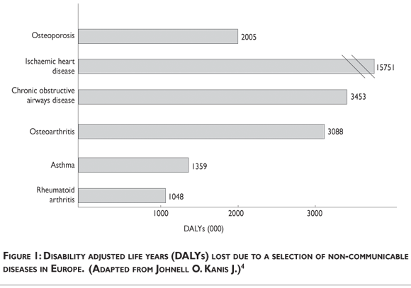
Fractures of the hip, vertebral body and distal    forearm have long been regarded as the typical osteoporotic fractures. Prospective    studies have shown that there is a heightened risk of almost all types of fracture    in individuals with low bone density irrespective of fracture site. In the year    2000 there were an estimated 9 million osteoporotic fractures of which 1.6 million    were at the hip, 1.7 million at the forearm and 1.4 million were clinical vertebral    fractures. Worldwide, osteoporotic fractures accounted for 0.83% of the global    burden of non&#45;communicable disease and 1.75% in Europe. In Europe, osteoporotic    fractures accounted for more disability adjusted life years (DALYs) than many    other chronic non communicable diseases<SUP>4</SUP> (<a href="#fig01">figure    1</a>). </font></p>     <p><a name="fig01"></a></p>     <p>&nbsp;</p>     <p align="center"><img src="/img/revistas/spm/v51s1/a07fig01.gif"></p>     <p>&nbsp;</p>     <p><font size="2" face="Verdana">
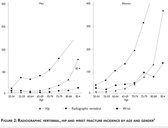
A study of British fracture occurrence indicates    that the population risk is similar in the UK. Thus one in two women that are    50 years of age will have an osteoporotic fracture in their remaining lifetime;    the figure for men is one in five.<SUP>5</SUP> The combined annual costs of    all osteoporotic fractures have been estimated to be $20 billion in the USA    and $30 billion in the European Union.<SUP>6</SUP> Fracture incidence in the    community is bimodal, showing peaks in youth and the very elderly (<a href="#fig02">figure    2</a>). In young people, fractures of the long bones predominate, usually after    substantial trauma, and they are more frequent in males than females. Over the    age of 35 years fracture incidence in women rises steeply so that rates become    twice those in men. Worldwide elderly people represent the fastest growing age    group, and the yearly number of fractures is likely to rise substantially with    continued aging of the population.</font></p>     <p><a name="fig02"></a></p>     <p>&nbsp;</p>     ]]></body>
<body><![CDATA[<p align="center"><img src="/img/revistas/spm/v51s1/a07fig02.gif"></p>     <p>&nbsp;</p>     <p><font size="2" face="Verdana"><i>Hip fracture</i></font></p>     <p><font size="2" face="Verdana">Hip fractures are the most devastating result    of osteoporosis: they require the patient to be admitted to hospital and cause    significant morbidity and mortality. Most hip fractures take place after a fall    from standing height, 80% occur in women and 90% in people older than 50 years.    The incidence increases exponentially with age (<a href="#fig01">figure 1</a>)    as a result of age related decrease in bone mass at the proximal femur and the    age related increase in falls. Worldwide there were an estimated 1.66 million    hip fractures in 1990. This has been estimated to rise to 6.3 million by 2050    with the increasing number of elderly people in the population.<SUP>7</SUP>    The lifetime risk of hip fracture for 50 year olds in the UK is 11.4% and 3.1%    for women and men respectively. Most of this risk is accrued in old age, such    that a 50 year old woman's 10 year risk of hip fracture is 0.3% rising to 8.7%    when she is 80 years old. The corresponding figures for men are 0.2 and 2.9%    respectively.<SUP>5</sup></font></p>     <p><font size="2" face="Verdana"> Hip fracture mortality is higher in men than    women and increases with age.<SUP>8</SUP> It is greatest in those with co&#45; existing    illnesses and poor pre&#45;fracture function. The risk of death is greatest after    fracture and decreases gradually with time. In the United Kingdom the 12 month    survival for hip fracture for men is 63.3% <I>versus</I> 90.0% expected and    for women 74.9% <I>versus</I> 91.1% expected.<SUP>5</SUP> Hip fractures result    in significant morbidity. Patients are prone to developing complications such    as pressure sores and bronchopneumonia. Fifty percent of those ambulatory before    the fracture are unable to walk independently afterward. Age is an important    determinant of outcome, with 14% of 50&#45;55 year old hip fracture patients being    discharged to nursing homes versus 55% of those aged &gt;90 years old.<SUP>9</sup></font></p>     <p><font size="2" face="Verdana"><i>Vertebral fracture</i></font></p>     <p><font size="2" face="Verdana">Only about a third of all radiographically identified    vertebral deformities come to specialist attention. There is also disagreement    about the radiographic definition of deformities in those that do present. Thus    in studies using radiographic screening of populations, vertebral deformities    have been estimated to be three times that of hip fracture. Data from the prospective    European Vertebral Osteoporosis Study (EVOS) have allowed accurate assessment    of radiographically determined vertebral factures in a large population.<SUP>10</SUP>    The overall age adjusted incidence of vertebral fractures was 5.7/1000 person    years for men and 10.7/1000 person years for women. The age standardised prevalence    was 12.2% for men and 12.0% for women 50&#45;79 years old. Historically it was believed    that vertebral fractures were more common in women, but the EVOS data suggest    that this is not the case at younger ages, possibly because of a higher incidence    of trauma in men (<a href="#fig03">figure 3</a>). Only around a quarter of vertebral    fractures result from falls, most result from routine activities such as bending    or lifting light objects.<SUP>11</SUP> In contrast to hip fractures, excess    mortality after vertebral fracture seems to increase progressively after diagnosis    of fracture. In the UK the observed survival in women 12 months after fracture    was 86.5% versus 93.6% expected. At 5 years it was 56.5% observed and 69.9%    expected.<SUP>5</SUP> The impact of a single vertebral fracture may be low but    multiple fractures can cause progressive loss of height and kyphosis and severe    back pain in the acute stages. Quality of life scores decrease as the number    of vertebral fractures increases.<SUP>12</sup></font></p>     <p><a name="fig03"></a></p>     <p>&nbsp;</p>     <p align="center"><img src="/img/revistas/spm/v51s1/a07fig03.gif"></p>     ]]></body>
<body><![CDATA[<p>&nbsp;</p>     <p><font size="2" face="Verdana"><i>Distal forearm fracture</i></font></p>     <p><font size="2" face="Verdana">Wrist fractures show a different pattern of occurrence    to hip and vertebral fractures. Incidence increases in white women between the    ages of 45 and 60 years, followed by a plateau. This may relate to altered neuromuscular    reflexes with ageing, and as a result, a tendency to fall sideways or backwards,    and thus not to break the fall with an outstretched arm. Most wrist fractures    occur in women and 50% occur in women over 65 years old. Data from the GPRD    show that a woman's lifetime risk of wrist fracture at 50 years old is 16.6%,    falling to 10.4% at 70 years. The incidence in men is low and does not rise    much with ageing (lifetime risk 2.9% at age 50 years and 1.4% at age 70 years).<SUP>5</SUP>    Wrist fractures do not seem to increase mortality although wrist fractures may    impact on some activities such as writing or meal preparation.</font></p>     <p><font size="2" face="Verdana"><i>Clustering of fractures in individuals</i></font></p>     <p><font size="2" face="Verdana">Epidemiological studies suggest that patients    with different types of fragility fractures are at increased risk of developing    other types of fracture. For example, the presence of a previous vertebral deformity    leads to a 7 to 10 fold increase in the risk of subsequent vertebral deformities.<SUP>13</SUP>    This is a comparable level of increased risk to that seen for individuals who    have sustained one hip fracture to then sustain a second. Furthermore, data    from Rochester, Minnesota, suggest that the risk of a hip fracture is increased    1.4 fold in women and 2.7 fold in men after the occurrence of a distal forearm    fracture.<SUP>14</SUP> The corresponding figures for subsequent vertebral fracture    are 5.2 and 10.7.</font></p>     <p><font size="2" face="Verdana"><i>Time trends and future projections</i></font></p>     <p><font size="2" face="Verdana">Life expectancy is increasing around the globe    and the number of elderly individuals is rising in every geographic region.    The world population is expected to rise from the current 323 million individuals    aged 65 years or over, to 1555 million by the year 2050. These demographic changes    alone can be expected to increase the number of hip fractures occurring among    people aged 35 years and over worldwide: the incidence is estimated to rise    from 1.66 million in 1990 to 6.26 million in 2050.<SUP>7</SUP> Assuming a constant    age&#45;specific rate of fracture, as the number of over 65s increases from 32 million    in 1990 to 69 million in 2050, the number of hip fractures in the US will increase    threefold.<SUP>7</SUP> In the UK, the number of hip fractures may increase from    46000 in 1985 to 117000 in 2016.<SUP>15</SUP> An increasingly elderly population    in Latin America and Asia could lead to a shift in the geographical distribution    of hip fractures, with only a quarter occurring in Europe and North America<SUP>7</SUP>    (<a href="#fig04">figure 4</a>). Such projections are almost certainly optimistic    considering that increases in the incidence of hip fractures have been observed    even after adjusting for the growth in the elderly population. Age&#45;adjusted    rate of hip fracture appears to have levelled off in the northern regions of    the USA, in parts of Sweden, and the UK, there is some evidence to suggest that    since implementation of osteoporosis screening and treatment programs, the incidence    of age adjusted hip fractures are now starting to fall.<SUP>16,17</SUP> However    fracture rates are still rising in some locales<SUP>18</SUP> and subsets of    the population.<SUP>19</SUP> These discrepancies in secular trends by race,    gender and age might help identify novel public health measures. </font></p>     <p><a name="fig04"></a></p>     <p>&nbsp;</p>     <p align="center"><img src="/img/revistas/spm/v51s1/a07fig04.gif"></p>     ]]></body>
<body><![CDATA[<p>&nbsp;</p>     <p><font size="2" face="Verdana"><i>Geography</i></font></p>     <p><font size="2" face="Verdana">There is a substantial variation in hip fracture    rates between populations of a given race or gender. Age adjusted rates are    highest in Scandinavian and North American populations, with almost seven fold    lower rates in southern European countries.<SUP>20</SUP> Hip fracture rates    are also lower in Asian and Latin American populations<SUP>21,22</SUP> and rates    seem to be lower in rural areas rather than urban areas in any country.<SUP>23,24</SUP>    These findings suggest an important role for environmental factors in the aetiology    of hip fracture. However, factors studied so far including smoking, alcohol    consumption, activity levels, obesity and migration status have failed to explain    these trends. The EVOS study demonstrated a threefold difference in the prevalence    of vertebral deformities between countries, with the highest rates in Scandinavia.    The prevalence range between centres was 7.5&#45;19.8% for men and 6.2&#45;20.7% for    women. The differences were not as great as those seen for hip fracture in Europe,    some of the differences could be explained by levels of physical activity and    body mass index.<SUP>8</SUP> </font></p>     <p>&nbsp;</p>     <p><font size="3" face="verdana"><b>Methods to assess bone health</b></font></p>     <p><font size="2" face="Verdana"><i>Dual energy X ray absorptiometry</i></font></p>     <p><font size="2" face="Verdana">Dual energy X ray absorptiometry (DXA) employs    X rays of two energies to produce a projection image of the region of interest.    From these projection images two measures are calculated to determine bone mass:    areal bone mineral density (g/cm<SUP>2</SUP>) and bone mineral content (BMC)    (g). Studies of DXA measures have confirmed that BMC and to a lesser degree    BMD is strongly correlated to ash weight.<SUP>25</SUP> DXA is commonly performed    at the lumbar spine and proximal femur. However it can also be measured at the    forearm, the calcaneus and the total body. The choice of the appropriate site    is based on purpose of the measurement and the technology of the equipment used.    Commercial DXA devices are either general purpose instruments for bone densitometry    examinations of the axial and peripheral skeleton or peripheral units for scans    of the forearm and /or calcaneus. The advantages of DXA include low radiation    dose, low cost, ease of use and rapidity of measurement. Peripheral DXA devices    are being increasingly utilized for BMD measurements at the forearm and calcaneus.<SUP>26</SUP>    They can be used as a pre&#45;selection tool to identify women with low BMD at the    axial skeleton, thus enabling reduction of the number of individuals who need    evaluation with axial DXA.<SUP>27</SUP> These peripheral devices have the advantages    of low cost and portability. There are however limitations inherent to DXA measurements.    This two dimensional technique can not determine between cortical and trabecular    bone, and cannot discriminate changes due to bone geometry (e.g. increases in    the third dimension) from those purely due to increased bone density (within    a fixed volume of bone). Falsely elevated BMD values at the spine can occur    in patients with degenerative spinal changes, fractures in lumbar vertebrae,    scoliosis or aortic calcification. There is still controversy as to which site    could better estimate the risk of low energy fracture. Meta&#45;analysis results    indicate that BMD measurements at the hip are superior in predicting hip fractures    compared to BMD measurements at other sites.<SUP>28</SUP> The World Health Organisation    defines osteoporosis by a T score more than 2SD below the peak bone mass; these    WHO criteria were developed for DXA measurements at the lumbar spine hip or    forearm.</font></p>     <p><font size="2" face="Verdana"><i>Quantitative computed tomography</i></font></p>     <p><font size="2" face="Verdana"> Quantitative computed tomography (QCT) provides    an alternative to DXA for the measurement of spine, hip and wrist BMD. Quantification    of BMD by QCT, like DXA, is based on the attenuation of the X ray beam as it    passes through the body. Each slice in CT represents tissue volume and thus    a volumetric BMD can be attained. This is an advantage in studies with children    and adolescents where DXA may underestimate BMD due to the growth&#45;related variations    in bone size. The other main advantage of QCT is its ability to assess separately    cortical and trabecular bone. Compared to DXA, QCT provides similar or better    results in prediction of spinal fracture in that the effects of degenerative    disease or aortic calcification can be excluded.<SUP>29</SUP> QCT has not been    used widely in clinical practice because of its high cost hardware, the increased    demand for CT whole body examinations in a clinical environment and the higher    radiation dose relative to DXA.</font></p>     <p><font size="2" face="Verdana"><i>Peripheral quantitative computed tomography</i></font></p>     ]]></body>
<body><![CDATA[<p><font size="2" face="Verdana">A recent development in QCT technology is the    availability of peripheral scanners (pQCT). These scanners provide three dimensional    mineralisation data in appendicular sites, commonly the distal radius and distal    tibia. As with traditional QCT, trabecular and cortical compartments can be    isolated and studied individually. Additionally they present a lower radiation    dose to the central body scans. The main advantage is the low investment and    running costs. PQCT measurement at the distal radius showed moderate correlation    with BMD at the hip and spine.<SUP>30,31</SUP> The greatest advantage of the    machines is the ability to image at a resolution that allows for trabecular    visualisation allowing for differences to be picked up in osteopenic women that    have fractured at the wrist compared to those without,<SUP>32</SUP> DXA is unable    to differentiate between these groups.</font></p>     <p><font size="2" face="Verdana"><i>Quantitative ultrasound</i></font></p>     <p><font size="2" face="Verdana">Quantitative ultrasound(QUS) has been introduced    recently for measuring bone density in the appendicular skeleton. Measurements    are performed primarily at the calcaneus. It is a technique that uses non&#45;ionising    sound waves to detect mineral density. Transducers transmit ultrasound energy    that travels through the bone to the receiving transducer. Two measures are    produced: Broadband ultrasound attenuation (BUA) and speed of sound (SOS). QUS    measures are well correlated with BMD derived from calcaneal DXA scans, and    can discriminate healthy from osteoporotic groups.<SUP>33</SUP> However a significant    false negative rate has been detected. Several studies have documented the ability    of QUS measurements to estimate fracture risk.<SUP>34,35</SUP> A recent study    has suggested that BUA predicts osteoporotic fractures better than DXA and independently    of BMD.<SUP>36</SUP> Interest in QUS methods can be attributed primarily to    the fact that they involve no radiation exposure. Additionally they are inexpensive    and portable. The main disadvantage is lack of sensitivity,<SUP>37</SUP> making    it inappropriate for long term monitoring of osteoporosis. Currently ultrasound    assessment is used as a screening tool, with confirmation of diagnosis via DXA    evaluation.</font></p>     <p><font size="2" face="Verdana"><i>Biochemical markers</i></font></p>     <p><font size="2" face="Verdana">Several serum and urine biochemical markers of    bone turnover have been developed. These provide non&#45;invasive and fairly inexpensive    methods for assessing rates of bone formation and resorption<I> in vivo</I>.    The most widely available markers include serum bone specific alkaline phosphatase    and the amino terminal propeptide of type 1 procollagen, which are markers of    bone formation, and urine or serum telopeptides of collagen crosslinks, which    are the markers for bone resorption. Disadvantages include their indication    of whole body bone turnover (mainly cortical bone) and day&#45;to&#45;day variability.    Despite drawbacks these markers can be used to study the pathogenesis of osteoporosis,    predict the risk of future fracture and predict and monitor response to therapy.    Prospective studies have shown an association of osteoporotic fracture with    indices of bone turnover independent of BMD in menopausal and elderly women.<SUP>38</SUP>    In elderly women with values for resorption markers exceeding the reference    range for premenopausal women, fracture risk is increased two fold after adjustment    for BMD, thus a combined approach could improve fracture prediction.<SUP>39</sup></font></p>     <p>&nbsp;</p>     <p><font size="3" face="verdana"><b>Assessment of fracture risk</b></font></p>     <p><font size="2" face="Verdana">The WHO scientific group recently convened to    develop more ways to assess fracture risk. The rationale for this initiative    is that T scores are insufficient to predict fracture risk. Although the T score    has many good attributes: it is simple and widely used, it has good correlation    with fracture risk and it can detect some high risk patients.<SUP>2</SUP> Its    shortcomings include lack of standardisation regarding which skeletal sites    to evaluate, lack of generalisation to non Caucasian groups and use of BMD as    the only risk factor evaluated. Inappropriate T score thresholds for osteoporosis    treatment have incorrectly identified patients who are not at high risk of fractures    whilst missing many who are at increased risk.<SUP>40</SUP> Several other clinical    risk factors contribute to fracture risk, in part independently of BMD. These    include age, prior fragility fracture, premature menopause, a family history    of hip fracture and the use of corticosteroids. Since several of these risk    factors are partly independent on BMD, their use in conjunction with BMD improves    sensitivity of fracture prediction without adverse effects on specificity. The    combined use of these risk factors along with age and BMD can be used in multivariate    models to estimate the 10 year probability of hip and other fractures.<SUP>41</SUP>    Thus a woman at age 60 years with an average BMD (about &#150;1.4) has an average    10 year probability of hip fracture at around 2.4%. If she has previously had    a fragility fracture this risk increased to 4.8%. Intervention thresholds will    be set by the global cost effectiveness model allowing individual countries    to determine how to utilise these interventions. The WHO initiative have concluded    from recent population based studies that clinical risk factors can be used    to enhance the performance characteristics of BMD. However application of these    models will require the incorporation of hazard functions of death and calibration    to the epidemiology of specific countries.<SUP>42</SUP> </font></p>     <p>&nbsp;</p>     <p><font size="3" face="verdana"><b>Conclusion</b></font></p>     ]]></body>
<body><![CDATA[<p><font size="2" face="Verdana">Osteoporosis is a disease that has a huge impact    on global public health. The impact of osteoporotic fracture is massive, not    just for individuals, but for the health service economy, and population as    a whole. The characterisation of some of the risk factors for osteoporotic fractures    have been elucidated, as a result the assessment of patients at high risk of    fracture can be identified by use of algorithms and appropriate preventative    and therapeutic strategies implemented. </font></p>     <p>&nbsp;</p>     <p><font size="3" face="verdana"><b>References </b></font></p>     <!-- ref --><p><font size="2" face="Verdana">1. Consensus development conference: prophylaxis    and treatment of osteoporosis. Osteoporos Int 1991;1:114&#45;117.1991.</font>&nbsp;&nbsp;&nbsp;&nbsp;&nbsp;&nbsp;&nbsp;&nbsp;[&#160;<a href="javascript:void(0);" onclick="javascript: window.open('/scielo.php?script=sci_nlinks&ref=9339556&pid=S0036-3634200900070000700001&lng=','','width=640,height=500,resizable=yes,scrollbars=1,menubar=yes,');">Links</a>&#160;]<!-- end-ref --><!-- ref --><p><font size="2" face="Verdana">2. Assessment of fracture risk and its application    to screening for postmenopausal osteoporosis. Report of a WHO Study Group. World    Health Organ Tech Rep Ser 1994; 843:1&#45;129.</font>&nbsp;&nbsp;&nbsp;&nbsp;&nbsp;&nbsp;&nbsp;&nbsp;[&#160;<a href="javascript:void(0);" onclick="javascript: window.open('/scielo.php?script=sci_nlinks&ref=9339557&pid=S0036-3634200900070000700002&lng=','','width=640,height=500,resizable=yes,scrollbars=1,menubar=yes,');">Links</a>&#160;]<!-- end-ref --><!-- ref --><p><font size="2" face="Verdana">3. Kanis JA, Johnell O, Oden A, Dawson A, De    Laet C, Jonsson B. Ten year probabilities of osteoporotic fractures according    to BMD and diagnostic thresholds. Osteoporos Int 2001; 12(12):989&#45;995.</font>&nbsp;&nbsp;&nbsp;&nbsp;&nbsp;&nbsp;&nbsp;&nbsp;[&#160;<a href="javascript:void(0);" onclick="javascript: window.open('/scielo.php?script=sci_nlinks&ref=9339558&pid=S0036-3634200900070000700003&lng=','','width=640,height=500,resizable=yes,scrollbars=1,menubar=yes,');">Links</a>&#160;]<!-- end-ref --><!-- ref --><p><font size="2" face="Verdana">4. Johnell O, Kanis JA. An estimate of the worldwide    prevalence and disability associated with osteoporotic fractures. Osteoporos    Int 2006; 17(12):1726&#45;1733.</font>&nbsp;&nbsp;&nbsp;&nbsp;&nbsp;&nbsp;&nbsp;&nbsp;[&#160;<a href="javascript:void(0);" onclick="javascript: window.open('/scielo.php?script=sci_nlinks&ref=9339559&pid=S0036-3634200900070000700004&lng=','','width=640,height=500,resizable=yes,scrollbars=1,menubar=yes,');">Links</a>&#160;]<!-- end-ref --><!-- ref --><p><font size="2" face="Verdana">5. van Staa TP, Dennison EM, Leufkens HG, Cooper    C. Epidemiology of fractures in England and Wales. Bone 2001; 29(6):517&#45;522.</font>&nbsp;&nbsp;&nbsp;&nbsp;&nbsp;&nbsp;&nbsp;&nbsp;[&#160;<a href="javascript:void(0);" onclick="javascript: window.open('/scielo.php?script=sci_nlinks&ref=9339560&pid=S0036-3634200900070000700005&lng=','','width=640,height=500,resizable=yes,scrollbars=1,menubar=yes,');">Links</a>&#160;]<!-- end-ref --><!-- ref --><p><font size="2" face="Verdana">6. Cummings SR, Melton LJ. Epidemiology and outcomes    of osteoporotic fractures. Lancet 2002; 359(9319):1761&#45;1767.</font>&nbsp;&nbsp;&nbsp;&nbsp;&nbsp;&nbsp;&nbsp;&nbsp;[&#160;<a href="javascript:void(0);" onclick="javascript: window.open('/scielo.php?script=sci_nlinks&ref=9339561&pid=S0036-3634200900070000700006&lng=','','width=640,height=500,resizable=yes,scrollbars=1,menubar=yes,');">Links</a>&#160;]<!-- end-ref --><!-- ref --><p><font size="2" face="Verdana">7. Cooper C, Campion G, Melton LJ, III. Hip fractures    in the elderly: a world&#45;wide projection. Osteoporos Int 1992; 2(6):285&#45;289.</font>&nbsp;&nbsp;&nbsp;&nbsp;&nbsp;&nbsp;&nbsp;&nbsp;[&#160;<a href="javascript:void(0);" onclick="javascript: window.open('/scielo.php?script=sci_nlinks&ref=9339562&pid=S0036-3634200900070000700007&lng=','','width=640,height=500,resizable=yes,scrollbars=1,menubar=yes,');">Links</a>&#160;]<!-- end-ref --><!-- ref --><p><font size="2" face="Verdana">8. Cooper C, Atkinson EJ, Jacobsen SJ, O'Fallon    WM, Melton LJ, III. Population&#45;based study of survival after osteoporotic fractures.    Am J Epidemiol 1993; 137(9):1001&#45;1005.</font>&nbsp;&nbsp;&nbsp;&nbsp;&nbsp;&nbsp;&nbsp;&nbsp;[&#160;<a href="javascript:void(0);" onclick="javascript: window.open('/scielo.php?script=sci_nlinks&ref=9339563&pid=S0036-3634200900070000700008&lng=','','width=640,height=500,resizable=yes,scrollbars=1,menubar=yes,');">Links</a>&#160;]<!-- end-ref --><!-- ref --><p><font size="2" face="Verdana">9. Chrischilles EA, Butler CD, Davis CS, Wallace    RB. A model of lifetime osteoporosis impact. Arch Intern Med 1991; 151(10):2026&#45;2032.</font>&nbsp;&nbsp;&nbsp;&nbsp;&nbsp;&nbsp;&nbsp;&nbsp;[&#160;<a href="javascript:void(0);" onclick="javascript: window.open('/scielo.php?script=sci_nlinks&ref=9339564&pid=S0036-3634200900070000700009&lng=','','width=640,height=500,resizable=yes,scrollbars=1,menubar=yes,');">Links</a>&#160;]<!-- end-ref --><!-- ref --><p><font size="2" face="Verdana">10. O'Neill TW, Felsenberg D, Varlow J, Cooper    C, Kanis JA, Silman AJ. The prevalence of vertebral deformity in european men    and women: the European Vertebral Osteoporosis Study. J Bone Miner Res 1996;    11(7):1010&#45;1018.</font>&nbsp;&nbsp;&nbsp;&nbsp;&nbsp;&nbsp;&nbsp;&nbsp;[&#160;<a href="javascript:void(0);" onclick="javascript: window.open('/scielo.php?script=sci_nlinks&ref=9339565&pid=S0036-3634200900070000700010&lng=','','width=640,height=500,resizable=yes,scrollbars=1,menubar=yes,');">Links</a>&#160;]<!-- end-ref --><!-- ref --><p><font size="2" face="Verdana">11. Cooper C, Atkinson EJ, O'Fallon WM, Melton    LJ, III. Incidence of clinically diagnosed vertebral fractures: a population&#45;based    study in Rochester, Minnesota, 1985&#45;1989. J Bone Miner Res 1992; 7(2):221&#45;227.</font>&nbsp;&nbsp;&nbsp;&nbsp;&nbsp;&nbsp;&nbsp;&nbsp;[&#160;<a href="javascript:void(0);" onclick="javascript: window.open('/scielo.php?script=sci_nlinks&ref=9339566&pid=S0036-3634200900070000700011&lng=','','width=640,height=500,resizable=yes,scrollbars=1,menubar=yes,');">Links</a>&#160;]<!-- end-ref --><!-- ref --><p><font size="2" face="Verdana">12. Oleksik A, Lips P, Dawson A, Minshall ME,    Shen W, Cooper C <I>et al.</I> Health&#45;related quality of life in postmenopausal    women with low BMD with or without prevalent vertebral fractures. J Bone Miner    Res 2000; 15(7):1384&#45;1392.</font>&nbsp;&nbsp;&nbsp;&nbsp;&nbsp;&nbsp;&nbsp;&nbsp;[&#160;<a href="javascript:void(0);" onclick="javascript: window.open('/scielo.php?script=sci_nlinks&ref=9339567&pid=S0036-3634200900070000700012&lng=','','width=640,height=500,resizable=yes,scrollbars=1,menubar=yes,');">Links</a>&#160;]<!-- end-ref --><!-- ref --><p><font size="2" face="Verdana">13. Ross PD, Davis JW, Epstein RS, Wasnich RD.    Pre&#45;existing fractures and bone mass predict vertebral fracture incidence in    women. Ann Intern Med 1991; 114(11):919&#45;923.</font>&nbsp;&nbsp;&nbsp;&nbsp;&nbsp;&nbsp;&nbsp;&nbsp;[&#160;<a href="javascript:void(0);" onclick="javascript: window.open('/scielo.php?script=sci_nlinks&ref=9339568&pid=S0036-3634200900070000700013&lng=','','width=640,height=500,resizable=yes,scrollbars=1,menubar=yes,');">Links</a>&#160;]<!-- end-ref --><!-- ref --><p><font size="2" face="Verdana">14. Cuddihy MT, Gabriel SE, Crowson CS, O'Fallon    WM, Melton LJ, III. Forearm fractures as predictors of subsequent osteoporotic    fractures. Osteoporos Int 1999; 9(6):469&#45;475.</font>&nbsp;&nbsp;&nbsp;&nbsp;&nbsp;&nbsp;&nbsp;&nbsp;[&#160;<a href="javascript:void(0);" onclick="javascript: window.open('/scielo.php?script=sci_nlinks&ref=9339569&pid=S0036-3634200900070000700014&lng=','','width=640,height=500,resizable=yes,scrollbars=1,menubar=yes,');">Links</a>&#160;]<!-- end-ref --><!-- ref --><p><font size="2" face="Verdana">15. Royal College of Physicians. Fractured neck    of femur: prevention and management. Summary and report of the Royal College    of Physicians. J R Coll Physicians Lond 1989; 23:8&#45;12.</font>&nbsp;&nbsp;&nbsp;&nbsp;&nbsp;&nbsp;&nbsp;&nbsp;[&#160;<a href="javascript:void(0);" onclick="javascript: window.open('/scielo.php?script=sci_nlinks&ref=9339570&pid=S0036-3634200900070000700015&lng=','','width=640,height=500,resizable=yes,scrollbars=1,menubar=yes,');">Links</a>&#160;]<!-- end-ref --><!-- ref --><p><font size="2" face="Verdana">16. Kannus P, Niemi S, Parkkari J, Palvanen M,    Vuori I, Jarvinen M. Nationwide decline in incidence of hip fracture. J Bone    Miner Res 2006; 21(12):1836&#45;1838.</font>&nbsp;&nbsp;&nbsp;&nbsp;&nbsp;&nbsp;&nbsp;&nbsp;[&#160;<a href="javascript:void(0);" onclick="javascript: window.open('/scielo.php?script=sci_nlinks&ref=9339571&pid=S0036-3634200900070000700016&lng=','','width=640,height=500,resizable=yes,scrollbars=1,menubar=yes,');">Links</a>&#160;]<!-- end-ref --><!-- ref --><p><font size="2" face="Verdana">17. Jaglal SB, Weller I, Mamdani M, Hawker G,    Kreder H, Jaakkimainen L, <I>et al. </I>Population trends in BMD testing, treatment,    and hip and wrist fracture rates: are the hip fracture projections wrong? J    Bone Miner Res 2005; 20(6):898&#45;905.</font>&nbsp;&nbsp;&nbsp;&nbsp;&nbsp;&nbsp;&nbsp;&nbsp;[&#160;<a href="javascript:void(0);" onclick="javascript: window.open('/scielo.php?script=sci_nlinks&ref=9339572&pid=S0036-3634200900070000700017&lng=','','width=640,height=500,resizable=yes,scrollbars=1,menubar=yes,');">Links</a>&#160;]<!-- end-ref --><!-- ref --><p><font size="2" face="Verdana">18. Hagino H, Katagiri H, Okano T, Yamamoto K,    Teshima R. Increasing incidence of hip fracture in Tottori Prefecture, Japan:    trend from 1986 to 2001. Osteoporos Int 2005; 16(12):1963&#45;1968.</font>&nbsp;&nbsp;&nbsp;&nbsp;&nbsp;&nbsp;&nbsp;&nbsp;[&#160;<a href="javascript:void(0);" onclick="javascript: window.open('/scielo.php?script=sci_nlinks&ref=9339573&pid=S0036-3634200900070000700018&lng=','','width=640,height=500,resizable=yes,scrollbars=1,menubar=yes,');">Links</a>&#160;]<!-- end-ref --><!-- ref --><p><font size="2" face="Verdana">19. Zingmond DS, Melton LJ, III, Silverman SL.    Increasing hip fracture incidence in California Hispanics, 1983 to 2000. Osteoporos    Int 2004; 15(8):603&#45;610.</font>&nbsp;&nbsp;&nbsp;&nbsp;&nbsp;&nbsp;&nbsp;&nbsp;[&#160;<a href="javascript:void(0);" onclick="javascript: window.open('/scielo.php?script=sci_nlinks&ref=9339574&pid=S0036-3634200900070000700019&lng=','','width=640,height=500,resizable=yes,scrollbars=1,menubar=yes,');">Links</a>&#160;]<!-- end-ref --><!-- ref --><p><font size="2" face="Verdana">20. Johnell O, Gullberg B, Allander E, Kanis    JA. The apparent incidence of hip fracture in Europe: a study of national register    sources. MEDOS Study Group. Osteoporos Int 1992; 2(6):298&#45;302.</font>&nbsp;&nbsp;&nbsp;&nbsp;&nbsp;&nbsp;&nbsp;&nbsp;[&#160;<a href="javascript:void(0);" onclick="javascript: window.open('/scielo.php?script=sci_nlinks&ref=9339575&pid=S0036-3634200900070000700020&lng=','','width=640,height=500,resizable=yes,scrollbars=1,menubar=yes,');">Links</a>&#160;]<!-- end-ref --><!-- ref --><p><font size="2" face="Verdana">21. Yan L, Zhou B, Prentice A, Wang X, Golden    MH. Epidemiological study of hip fracture in Shenyang, People's Republic of    China. Bone 1999; 24(2):151&#45;155.</font>&nbsp;&nbsp;&nbsp;&nbsp;&nbsp;&nbsp;&nbsp;&nbsp;[&#160;<a href="javascript:void(0);" onclick="javascript: window.open('/scielo.php?script=sci_nlinks&ref=9339576&pid=S0036-3634200900070000700021&lng=','','width=640,height=500,resizable=yes,scrollbars=1,menubar=yes,');">Links</a>&#160;]<!-- end-ref --><!-- ref --><p><font size="2" face="Verdana">22. Morales&#45;Torres J, Gutierrez&#45;Urena S. The    burden of osteoporosis in Latin America. Osteoporos Int 2004; 15(8):625&#45;632.</font>&nbsp;&nbsp;&nbsp;&nbsp;&nbsp;&nbsp;&nbsp;&nbsp;[&#160;<a href="javascript:void(0);" onclick="javascript: window.open('/scielo.php?script=sci_nlinks&ref=9339577&pid=S0036-3634200900070000700022&lng=','','width=640,height=500,resizable=yes,scrollbars=1,menubar=yes,');">Links</a>&#160;]<!-- end-ref --><!-- ref --><p><font size="2" face="Verdana">23. Kaastad TS, Meyer HE, Falch JA. Incidence    of hip fracture in Oslo, Norway: differences within the city. Bone 1998; 22(2):175&#45;178.</font>&nbsp;&nbsp;&nbsp;&nbsp;&nbsp;&nbsp;&nbsp;&nbsp;[&#160;<a href="javascript:void(0);" onclick="javascript: window.open('/scielo.php?script=sci_nlinks&ref=9339578&pid=S0036-3634200900070000700023&lng=','','width=640,height=500,resizable=yes,scrollbars=1,menubar=yes,');">Links</a>&#160;]<!-- end-ref --><!-- ref --><p><font size="2" face="Verdana">24. Sanders KM, Nicholson GC, Ugoni AM, Seeman    E, Pasco JA, Kotowicz MA. Fracture rates lower in rural than urban communities:    the Geelong Osteoporosis Study. J Epidemiol Community Health 2002; 56(6):466&#45;470.</font>&nbsp;&nbsp;&nbsp;&nbsp;&nbsp;&nbsp;&nbsp;&nbsp;[&#160;<a href="javascript:void(0);" onclick="javascript: window.open('/scielo.php?script=sci_nlinks&ref=9339579&pid=S0036-3634200900070000700024&lng=','','width=640,height=500,resizable=yes,scrollbars=1,menubar=yes,');">Links</a>&#160;]<!-- end-ref --><!-- ref --><p><font size="2" face="Verdana">25. Sran MM, Khan KM, Keiver K, Chew JB, McKay    HA, Oxland TR. Accuracy of DXA scanning of the thoracic spine: cadaveric studies    comparing BMC, areal BMD and geometric estimates of volumetric BMD against ash    weight and CT measures of bone volume. Eur Spine J 2005; 14(10):971&#45;976.</font>&nbsp;&nbsp;&nbsp;&nbsp;&nbsp;&nbsp;&nbsp;&nbsp;[&#160;<a href="javascript:void(0);" onclick="javascript: window.open('/scielo.php?script=sci_nlinks&ref=9339580&pid=S0036-3634200900070000700025&lng=','','width=640,height=500,resizable=yes,scrollbars=1,menubar=yes,');">Links</a>&#160;]<!-- end-ref --><!-- ref --><p><font size="2" face="Verdana">26. Adams JE. Dual&#45;energy X ray absorptiometry.    In: Gramp S, editor. Radiology of Osteoporosis. New York: Springer, 2003.</font>&nbsp;&nbsp;&nbsp;&nbsp;&nbsp;&nbsp;&nbsp;&nbsp;[&#160;<a href="javascript:void(0);" onclick="javascript: window.open('/scielo.php?script=sci_nlinks&ref=9339581&pid=S0036-3634200900070000700026&lng=','','width=640,height=500,resizable=yes,scrollbars=1,menubar=yes,');">Links</a>&#160;]<!-- end-ref --><!-- ref --><p><font size="2" face="Verdana">27. Damilakis J, Papadokostakis G, Perisinakis    K, Hadjipavlou A, Gourtsoyiannis N. Can radial bone mineral density and quantitative    ultrasound measurements reduce the number of women who need axial density skeletal    assessment? Osteoporos Int 2003; 14(8):688&#45;693.</font>&nbsp;&nbsp;&nbsp;&nbsp;&nbsp;&nbsp;&nbsp;&nbsp;[&#160;<a href="javascript:void(0);" onclick="javascript: window.open('/scielo.php?script=sci_nlinks&ref=9339582&pid=S0036-3634200900070000700027&lng=','','width=640,height=500,resizable=yes,scrollbars=1,menubar=yes,');">Links</a>&#160;]<!-- end-ref --><!-- ref --><p><font size="2" face="Verdana">28. Marshall D, Johnell O, Wedel H. Meta&#45;analysis    of how well measures of bone mineral density predict occurrence of osteoporotic    fractures. BMJ 1996; 312(7041):1254&#45;1259.</font>&nbsp;&nbsp;&nbsp;&nbsp;&nbsp;&nbsp;&nbsp;&nbsp;[&#160;<a href="javascript:void(0);" onclick="javascript: window.open('/scielo.php?script=sci_nlinks&ref=9339583&pid=S0036-3634200900070000700028&lng=','','width=640,height=500,resizable=yes,scrollbars=1,menubar=yes,');">Links</a>&#160;]<!-- end-ref --><!-- ref --><p><font size="2" face="Verdana">29. Lang TF, Guglielmi G, van KC, De SA, Cammisa    M, Genant HK. Measurement of bone mineral density at the spine and proximal    femur by volumetric quantitative computed tomography and dual&#45;energy X&#45;ray absorptiometry    in elderly women with and without vertebral fractures. Bone 2002; 30(1):247&#45;250.</font>&nbsp;&nbsp;&nbsp;&nbsp;&nbsp;&nbsp;&nbsp;&nbsp;[&#160;<a href="javascript:void(0);" onclick="javascript: window.open('/scielo.php?script=sci_nlinks&ref=9339584&pid=S0036-3634200900070000700029&lng=','','width=640,height=500,resizable=yes,scrollbars=1,menubar=yes,');">Links</a>&#160;]<!-- end-ref --><!-- ref --><p><font size="2" face="Verdana">30. Gatti D, Rossini M, Zamberlan N, Braga V,    Fracassi E, Adami S. Effect of aging on trabecular and compact bone components    of proximal and ultradistal radius. Osteoporos Int 1996; 6(5):355&#45;360.</font>&nbsp;&nbsp;&nbsp;&nbsp;&nbsp;&nbsp;&nbsp;&nbsp;[&#160;<a href="javascript:void(0);" onclick="javascript: window.open('/scielo.php?script=sci_nlinks&ref=9339585&pid=S0036-3634200900070000700030&lng=','','width=640,height=500,resizable=yes,scrollbars=1,menubar=yes,');">Links</a>&#160;]<!-- end-ref --><!-- ref --><p><font size="2" face="Verdana">31. Grampp S, Lang P, Jergas M, Gluer CC, Mathur    A, Engelke K <I>et al.</I> Assessment of the skeletal status by peripheral quantitative    computed tomography of the forearm: short&#45;term precision in vivo and comparison    to dual X&#45;ray absorptiometry. J Bone Miner Res 1995; 10(10):1566&#45;1576.</font>&nbsp;&nbsp;&nbsp;&nbsp;&nbsp;&nbsp;&nbsp;&nbsp;[&#160;<a href="javascript:void(0);" onclick="javascript: window.open('/scielo.php?script=sci_nlinks&ref=9339586&pid=S0036-3634200900070000700031&lng=','','width=640,height=500,resizable=yes,scrollbars=1,menubar=yes,');">Links</a>&#160;]<!-- end-ref --><!-- ref --><p><font size="2" face="Verdana">32. Boutroy S, Bouxsein ML, Munoz F, Delmas PD.    In vivo assessment of trabecular bone microarchitecture by high&#45;resolution peripheral    quantitative computed tomography. J Clin Endocrinol Metab 2005; 90(12):6508&#45;6515.</font>&nbsp;&nbsp;&nbsp;&nbsp;&nbsp;&nbsp;&nbsp;&nbsp;[&#160;<a href="javascript:void(0);" onclick="javascript: window.open('/scielo.php?script=sci_nlinks&ref=9339587&pid=S0036-3634200900070000700032&lng=','','width=640,height=500,resizable=yes,scrollbars=1,menubar=yes,');">Links</a>&#160;]<!-- end-ref --><!-- ref --><p><font size="2" face="Verdana">33. Hans D, Molina P, Schott AM, Sebert JL, Cormier    C, Kotzki PO <I>et al. </i>Ultrasonographic heel measurements to predict hip    fracture in elderly women: the EPIDOS prospective study. Lancet 1996;348(9026):    511&#45;514.</font>&nbsp;&nbsp;&nbsp;&nbsp;&nbsp;&nbsp;&nbsp;&nbsp;[&#160;<a href="javascript:void(0);" onclick="javascript: window.open('/scielo.php?script=sci_nlinks&ref=9339588&pid=S0036-3634200900070000700033&lng=','','width=640,height=500,resizable=yes,scrollbars=1,menubar=yes,');">Links</a>&#160;]<!-- end-ref --><!-- ref --><p><font size="2" face="Verdana">34. Gluer CC, Eastell R, Reid DM, Felsenberg    D, Roux C, Barkmann R <I>et al. </I>Association of five quantitative ultrasound    devices and bone densitometry with osteoporotic vertebral fractures in a population&#45;based    sample: the OPUS Study. J Bone Miner Res 2004; 19(5):782&#45;793.</font>&nbsp;&nbsp;&nbsp;&nbsp;&nbsp;&nbsp;&nbsp;&nbsp;[&#160;<a href="javascript:void(0);" onclick="javascript: window.open('/scielo.php?script=sci_nlinks&ref=9339589&pid=S0036-3634200900070000700034&lng=','','width=640,height=500,resizable=yes,scrollbars=1,menubar=yes,');">Links</a>&#160;]<!-- end-ref --><!-- ref --><p><font size="2" face="Verdana">35. Gluer CC, Hans D. How to use ultrasound for    risk assessment: a need for defining strategies. Osteoporos Int 1999; 9(3):193&#45;195.</font>&nbsp;&nbsp;&nbsp;&nbsp;&nbsp;&nbsp;&nbsp;&nbsp;[&#160;<a href="javascript:void(0);" onclick="javascript: window.open('/scielo.php?script=sci_nlinks&ref=9339590&pid=S0036-3634200900070000700035&lng=','','width=640,height=500,resizable=yes,scrollbars=1,menubar=yes,');">Links</a>&#160;]<!-- end-ref --><!-- ref --><p><font size="2" face="Verdana">36. Stewart A, Kumar V, Reid DM. Long&#45;term fracture    prediction by DXA and QUS: a 10&#45;year prospective study. J Bone Miner Res 2006;    21(3): 413&#45;418.</font>&nbsp;&nbsp;&nbsp;&nbsp;&nbsp;&nbsp;&nbsp;&nbsp;[&#160;<a href="javascript:void(0);" onclick="javascript: window.open('/scielo.php?script=sci_nlinks&ref=9339591&pid=S0036-3634200900070000700036&lng=','','width=640,height=500,resizable=yes,scrollbars=1,menubar=yes,');">Links</a>&#160;]<!-- end-ref --><!-- ref --><p><font size="2" face="Verdana">37. Moyad MA. Osteoporosis: a rapid review of    risk factors and screening methods. Urol Oncol 2003; 21(5):375&#45;379.</font>&nbsp;&nbsp;&nbsp;&nbsp;&nbsp;&nbsp;&nbsp;&nbsp;[&#160;<a href="javascript:void(0);" onclick="javascript: window.open('/scielo.php?script=sci_nlinks&ref=9339592&pid=S0036-3634200900070000700037&lng=','','width=640,height=500,resizable=yes,scrollbars=1,menubar=yes,');">Links</a>&#160;]<!-- end-ref --><!-- ref --><p><font size="2" face="Verdana">38. Garnero P, Sornay&#45;Rendu E, Claustrat B, Delmas    PD. Biochemical markers of bone turnover, endogenous hormones and the risk of    fractures in postmenopausal women: the OFELY study. J Bone Miner Res 2000; 15(8):1526&#45;1536.</font>&nbsp;&nbsp;&nbsp;&nbsp;&nbsp;&nbsp;&nbsp;&nbsp;[&#160;<a href="javascript:void(0);" onclick="javascript: window.open('/scielo.php?script=sci_nlinks&ref=9339593&pid=S0036-3634200900070000700038&lng=','','width=640,height=500,resizable=yes,scrollbars=1,menubar=yes,');">Links</a>&#160;]<!-- end-ref --><!-- ref --><p><font size="2" face="Verdana">39. Johnell O, Oden A, De Laet C, Garnero P,    Delmas PD, Kanis JA. Biochemical indices of bone turnover and the assessment    of fracture probability. Osteoporos Int 2002; 13(7):523&#45;526.</font>&nbsp;&nbsp;&nbsp;&nbsp;&nbsp;&nbsp;&nbsp;&nbsp;[&#160;<a href="javascript:void(0);" onclick="javascript: window.open('/scielo.php?script=sci_nlinks&ref=9339594&pid=S0036-3634200900070000700039&lng=','','width=640,height=500,resizable=yes,scrollbars=1,menubar=yes,');">Links</a>&#160;]<!-- end-ref --><!-- ref --><p><font size="2" face="Verdana">40. Richards JB, Leslie WD, Joseph L, Siminoski    K, Hanley DA, Adachi JD <I>et al. </I>Changes to osteoporosis prevalence according    to method of risk assessment. J Bone Miner Res 2007; 22(2):228&#45;234.</font>&nbsp;&nbsp;&nbsp;&nbsp;&nbsp;&nbsp;&nbsp;&nbsp;[&#160;<a href="javascript:void(0);" onclick="javascript: window.open('/scielo.php?script=sci_nlinks&ref=9339595&pid=S0036-3634200900070000700040&lng=','','width=640,height=500,resizable=yes,scrollbars=1,menubar=yes,');">Links</a>&#160;]<!-- end-ref --><!-- ref --><p><font size="2" face="Verdana">41. Kanis JA, Borgstrom F, De Laet C, Johansson    H, Johnell O, Jonsson B <I>et al.</i> Assessment of fracture risk. Osteoporos    Int 2005; 16(6):581&#45;589.</font>&nbsp;&nbsp;&nbsp;&nbsp;&nbsp;&nbsp;&nbsp;&nbsp;[&#160;<a href="javascript:void(0);" onclick="javascript: window.open('/scielo.php?script=sci_nlinks&ref=9339596&pid=S0036-3634200900070000700041&lng=','','width=640,height=500,resizable=yes,scrollbars=1,menubar=yes,');">Links</a>&#160;]<!-- end-ref --><!-- ref --><p><font size="2" face="Verdana">42. Kanis JA, Oden A, Johnell O, Johansson H,    De Laet C, Brown J<I> et al. </I>The use of clinical risk factors enhances the    performance of BMD in the prediction of hip and osteoporotic fractures in men    and women. Osteoporos Int 2007.</font>&nbsp;&nbsp;&nbsp;&nbsp;&nbsp;&nbsp;&nbsp;&nbsp;[&#160;<a href="javascript:void(0);" onclick="javascript: window.open('/scielo.php?script=sci_nlinks&ref=9339597&pid=S0036-3634200900070000700042&lng=','','width=640,height=500,resizable=yes,scrollbars=1,menubar=yes,');">Links</a>&#160;]<!-- end-ref --><p>&nbsp;</p>     <p>&nbsp;</p>     <p><font size="2" face="Verdana">Received on: January 7, 2008    <br>   Accepted on: March 7, 2008</font></p>     <p>&nbsp;</p>     ]]></body>
<body><![CDATA[<p>&nbsp;</p>     <p><font size="2" face="Verdana">Address reprint requests to: Professor Cyrus    Cooper. MRC Epidemiology Resource Centre, University of Southampton,Southampton    General Hospital. Southampton, SO16 6YD, UK.    <br>   E&#45;mail: <a href="mailto:cc@mrc.soton.ac.uk">cc@mrc.soton.ac.uk</a></font></p>      ]]></body><back>
<ref-list>
<ref id="B1">
<label>1</label><nlm-citation citation-type="journal">
<article-title xml:lang="en"><![CDATA[Consensus development conference: prophylaxis and treatment of osteoporosis]]></article-title>
<source><![CDATA[Osteoporos Int]]></source>
<year>1991</year>
<volume>1</volume>
<numero>114</numero>
<issue>114</issue>
<page-range>117.1991</page-range></nlm-citation>
</ref>
<ref id="B2">
<label>2</label><nlm-citation citation-type="journal">
<article-title xml:lang="en"><![CDATA[Assessment of fracture risk and its application to screening for postmenopausal osteoporosis: Report of a WHO Study Group]]></article-title>
<source><![CDATA[World Health Organ Tech Rep Ser]]></source>
<year>1994</year>
<volume>843</volume>
<page-range>1-129</page-range></nlm-citation>
</ref>
<ref id="B3">
<label>3</label><nlm-citation citation-type="journal">
<person-group person-group-type="author">
<name>
<surname><![CDATA[Kanis]]></surname>
<given-names><![CDATA[JA]]></given-names>
</name>
<name>
<surname><![CDATA[Johnell]]></surname>
<given-names><![CDATA[O]]></given-names>
</name>
<name>
<surname><![CDATA[Oden]]></surname>
<given-names><![CDATA[A]]></given-names>
</name>
<name>
<surname><![CDATA[Dawson]]></surname>
<given-names><![CDATA[A]]></given-names>
</name>
<name>
<surname><![CDATA[De Laet]]></surname>
<given-names><![CDATA[C]]></given-names>
</name>
<name>
<surname><![CDATA[Jonsson]]></surname>
<given-names><![CDATA[B]]></given-names>
</name>
</person-group>
<article-title xml:lang="en"><![CDATA[Ten year probabilities of osteoporotic fractures according to BMD and diagnostic thresholds]]></article-title>
<source><![CDATA[Osteoporos Int]]></source>
<year>2001</year>
<volume>12</volume>
<numero>12</numero>
<issue>12</issue>
<page-range>989-995</page-range></nlm-citation>
</ref>
<ref id="B4">
<label>4</label><nlm-citation citation-type="journal">
<person-group person-group-type="author">
<name>
<surname><![CDATA[Johnell]]></surname>
<given-names><![CDATA[O]]></given-names>
</name>
<name>
<surname><![CDATA[Kanis]]></surname>
<given-names><![CDATA[JA]]></given-names>
</name>
</person-group>
<article-title xml:lang="en"><![CDATA[An estimate of the worldwide prevalence and disability associated with osteoporotic fractures]]></article-title>
<source><![CDATA[Osteoporos Int]]></source>
<year>2006</year>
<volume>17</volume>
<numero>12</numero>
<issue>12</issue>
<page-range>1726-1733</page-range></nlm-citation>
</ref>
<ref id="B5">
<label>5</label><nlm-citation citation-type="journal">
<person-group person-group-type="author">
<name>
<surname><![CDATA[van Staa]]></surname>
<given-names><![CDATA[TP]]></given-names>
</name>
<name>
<surname><![CDATA[Dennison]]></surname>
<given-names><![CDATA[EM]]></given-names>
</name>
<name>
<surname><![CDATA[Leufkens]]></surname>
<given-names><![CDATA[HG]]></given-names>
</name>
<name>
<surname><![CDATA[Cooper]]></surname>
<given-names><![CDATA[C]]></given-names>
</name>
</person-group>
<article-title xml:lang="en"><![CDATA[Epidemiology of fractures in England and Wales]]></article-title>
<source><![CDATA[Bone]]></source>
<year>2001</year>
<volume>29</volume>
<numero>6</numero>
<issue>6</issue>
<page-range>517-522</page-range></nlm-citation>
</ref>
<ref id="B6">
<label>6</label><nlm-citation citation-type="journal">
<person-group person-group-type="author">
<name>
<surname><![CDATA[Cummings]]></surname>
<given-names><![CDATA[SR]]></given-names>
</name>
<name>
<surname><![CDATA[Melton]]></surname>
<given-names><![CDATA[LJ]]></given-names>
</name>
</person-group>
<article-title xml:lang="en"><![CDATA[Epidemiology and outcomes of osteoporotic fractures]]></article-title>
<source><![CDATA[Lancet]]></source>
<year>2002</year>
<volume>359</volume>
<numero>9319</numero>
<issue>9319</issue>
<page-range>1761-1767</page-range></nlm-citation>
</ref>
<ref id="B7">
<label>7</label><nlm-citation citation-type="journal">
<person-group person-group-type="author">
<name>
<surname><![CDATA[Cooper]]></surname>
<given-names><![CDATA[C]]></given-names>
</name>
<name>
<surname><![CDATA[Campion]]></surname>
<given-names><![CDATA[G]]></given-names>
</name>
<name>
<surname><![CDATA[Melton]]></surname>
<given-names><![CDATA[LJ]]></given-names>
</name>
<name>
<surname><![CDATA[III]]></surname>
</name>
</person-group>
<article-title xml:lang="en"><![CDATA[Hip fractures in the elderly: a world-wide projection]]></article-title>
<source><![CDATA[Osteoporos Int]]></source>
<year>1992</year>
<volume>2</volume>
<numero>6</numero>
<issue>6</issue>
<page-range>285-289</page-range></nlm-citation>
</ref>
<ref id="B8">
<label>8</label><nlm-citation citation-type="journal">
<person-group person-group-type="author">
<name>
<surname><![CDATA[Cooper]]></surname>
<given-names><![CDATA[C]]></given-names>
</name>
<name>
<surname><![CDATA[Atkinson]]></surname>
<given-names><![CDATA[EJ]]></given-names>
</name>
<name>
<surname><![CDATA[Jacobsen]]></surname>
<given-names><![CDATA[SJ]]></given-names>
</name>
<name>
<surname><![CDATA[O'Fallon WM]]></surname>
</name>
<name>
<surname><![CDATA[Melton]]></surname>
<given-names><![CDATA[LJ]]></given-names>
</name>
<name>
<surname><![CDATA[III]]></surname>
</name>
</person-group>
<article-title xml:lang="en"><![CDATA[Population-based study of survival after osteoporotic fractures]]></article-title>
<source><![CDATA[Am J Epidemiol]]></source>
<year>1993</year>
<volume>137</volume>
<numero>9</numero>
<issue>9</issue>
<page-range>1001-1005</page-range></nlm-citation>
</ref>
<ref id="B9">
<label>9</label><nlm-citation citation-type="journal">
<person-group person-group-type="author">
<name>
<surname><![CDATA[Chrischilles]]></surname>
<given-names><![CDATA[EA]]></given-names>
</name>
<name>
<surname><![CDATA[Butler]]></surname>
<given-names><![CDATA[CD]]></given-names>
</name>
<name>
<surname><![CDATA[Davis]]></surname>
<given-names><![CDATA[CS]]></given-names>
</name>
<name>
<surname><![CDATA[Wallace]]></surname>
<given-names><![CDATA[RB]]></given-names>
</name>
</person-group>
<article-title xml:lang="en"><![CDATA[A model of lifetime osteoporosis impact]]></article-title>
<source><![CDATA[Arch Intern Med]]></source>
<year>1991</year>
<volume>151</volume>
<numero>10</numero>
<issue>10</issue>
<page-range>2026-2032</page-range></nlm-citation>
</ref>
<ref id="B10">
<label>10</label><nlm-citation citation-type="journal">
<person-group person-group-type="author">
<name>
<surname><![CDATA[O'Neill TW]]></surname>
</name>
<name>
<surname><![CDATA[Felsenberg]]></surname>
<given-names><![CDATA[D]]></given-names>
</name>
<name>
<surname><![CDATA[Varlow]]></surname>
<given-names><![CDATA[J]]></given-names>
</name>
<name>
<surname><![CDATA[Cooper]]></surname>
<given-names><![CDATA[C]]></given-names>
</name>
<name>
<surname><![CDATA[Kanis]]></surname>
<given-names><![CDATA[JA]]></given-names>
</name>
<name>
<surname><![CDATA[Silman]]></surname>
<given-names><![CDATA[AJ]]></given-names>
</name>
</person-group>
<article-title xml:lang="en"><![CDATA[The prevalence of vertebral deformity in european men and women: the European Vertebral Osteoporosis Study]]></article-title>
<source><![CDATA[J Bone Miner Res]]></source>
<year>1996</year>
<volume>11</volume>
<numero>7</numero>
<issue>7</issue>
<page-range>1010-1018</page-range></nlm-citation>
</ref>
<ref id="B11">
<label>11</label><nlm-citation citation-type="journal">
<person-group person-group-type="author">
<name>
<surname><![CDATA[Cooper]]></surname>
<given-names><![CDATA[C]]></given-names>
</name>
<name>
<surname><![CDATA[Atkinson]]></surname>
<given-names><![CDATA[EJ]]></given-names>
</name>
<name>
<surname><![CDATA[O'Fallon WM]]></surname>
</name>
<name>
<surname><![CDATA[Melton]]></surname>
<given-names><![CDATA[LJ]]></given-names>
</name>
<name>
<surname><![CDATA[III]]></surname>
</name>
</person-group>
<article-title xml:lang="en"><![CDATA[Incidence of clinically diagnosed vertebral fractures: a population-based study in Rochester, Minnesota, 1985-1989]]></article-title>
<source><![CDATA[J Bone Miner Res]]></source>
<year>1992</year>
<volume>7</volume>
<numero>2</numero>
<issue>2</issue>
<page-range>221-227</page-range></nlm-citation>
</ref>
<ref id="B12">
<label>12</label><nlm-citation citation-type="journal">
<person-group person-group-type="author">
<name>
<surname><![CDATA[Oleksik]]></surname>
<given-names><![CDATA[A]]></given-names>
</name>
<name>
<surname><![CDATA[Lips]]></surname>
<given-names><![CDATA[P]]></given-names>
</name>
<name>
<surname><![CDATA[Dawson]]></surname>
<given-names><![CDATA[A]]></given-names>
</name>
<name>
<surname><![CDATA[Minshall]]></surname>
<given-names><![CDATA[ME]]></given-names>
</name>
<name>
<surname><![CDATA[Shen]]></surname>
<given-names><![CDATA[W]]></given-names>
</name>
</person-group>
<collab>Cooper C et al</collab>
<article-title xml:lang="en"><![CDATA[Health-related quality of life in postmenopausal women with low BMD with or without prevalent vertebral fractures]]></article-title>
<source><![CDATA[J Bone Miner Res]]></source>
<year>2000</year>
<volume>15</volume>
<numero>7</numero>
<issue>7</issue>
<page-range>1384-1392</page-range></nlm-citation>
</ref>
<ref id="B13">
<label>13</label><nlm-citation citation-type="journal">
<person-group person-group-type="author">
<name>
<surname><![CDATA[Ross]]></surname>
<given-names><![CDATA[PD]]></given-names>
</name>
<name>
<surname><![CDATA[Davis]]></surname>
<given-names><![CDATA[JW]]></given-names>
</name>
<name>
<surname><![CDATA[Epstein]]></surname>
<given-names><![CDATA[RS]]></given-names>
</name>
<name>
<surname><![CDATA[Wasnich]]></surname>
<given-names><![CDATA[RD]]></given-names>
</name>
</person-group>
<article-title xml:lang="en"><![CDATA[Pre-existing fractures and bone mass predict vertebral fracture incidence in women]]></article-title>
<source><![CDATA[Ann Intern Med]]></source>
<year>1991</year>
<volume>114</volume>
<numero>11</numero>
<issue>11</issue>
<page-range>919-923</page-range></nlm-citation>
</ref>
<ref id="B14">
<label>14</label><nlm-citation citation-type="journal">
<person-group person-group-type="author">
<name>
<surname><![CDATA[Cuddihy]]></surname>
<given-names><![CDATA[MT]]></given-names>
</name>
<name>
<surname><![CDATA[Gabriel]]></surname>
<given-names><![CDATA[SE]]></given-names>
</name>
<name>
<surname><![CDATA[Crowson]]></surname>
<given-names><![CDATA[CS]]></given-names>
</name>
<name>
<surname><![CDATA[O'Fallon WM]]></surname>
</name>
<name>
<surname><![CDATA[Melton]]></surname>
<given-names><![CDATA[LJ]]></given-names>
</name>
<name>
<surname><![CDATA[III]]></surname>
</name>
</person-group>
<article-title xml:lang="en"><![CDATA[Forearm fractures as predictors of subsequent osteoporotic fractures]]></article-title>
<source><![CDATA[Osteoporos Int]]></source>
<year>1999</year>
<volume>9</volume>
<numero>6</numero>
<issue>6</issue>
<page-range>469-475</page-range></nlm-citation>
</ref>
<ref id="B15">
<label>15</label><nlm-citation citation-type="journal">
<collab>Royal College of Physicians</collab>
<article-title xml:lang="en"><![CDATA[Fractured neck of femur: prevention and management. Summary and report of the Royal College of Physicians]]></article-title>
<source><![CDATA[J R Coll Physicians Lond]]></source>
<year>1989</year>
<volume>23</volume>
<page-range>8-12</page-range></nlm-citation>
</ref>
<ref id="B16">
<label>16</label><nlm-citation citation-type="journal">
<person-group person-group-type="author">
<name>
<surname><![CDATA[Kannus]]></surname>
<given-names><![CDATA[P]]></given-names>
</name>
<name>
<surname><![CDATA[Niemi]]></surname>
<given-names><![CDATA[S]]></given-names>
</name>
<name>
<surname><![CDATA[Parkkari]]></surname>
<given-names><![CDATA[J]]></given-names>
</name>
<name>
<surname><![CDATA[Palvanen]]></surname>
<given-names><![CDATA[M]]></given-names>
</name>
<name>
<surname><![CDATA[Vuori]]></surname>
<given-names><![CDATA[I]]></given-names>
</name>
<name>
<surname><![CDATA[Jarvinen]]></surname>
<given-names><![CDATA[M]]></given-names>
</name>
</person-group>
<article-title xml:lang="en"><![CDATA[Nationwide decline in incidence of hip fracture]]></article-title>
<source><![CDATA[J Bone Miner Res]]></source>
<year>2006</year>
<volume>21</volume>
<numero>12</numero>
<issue>12</issue>
<page-range>1836-1838</page-range></nlm-citation>
</ref>
<ref id="B17">
<label>17</label><nlm-citation citation-type="journal">
<person-group person-group-type="author">
<name>
<surname><![CDATA[Jaglal]]></surname>
<given-names><![CDATA[SB]]></given-names>
</name>
<name>
<surname><![CDATA[Weller]]></surname>
<given-names><![CDATA[I]]></given-names>
</name>
<name>
<surname><![CDATA[Mamdani]]></surname>
<given-names><![CDATA[M]]></given-names>
</name>
<name>
<surname><![CDATA[Hawker]]></surname>
<given-names><![CDATA[G]]></given-names>
</name>
<name>
<surname><![CDATA[Kreder]]></surname>
<given-names><![CDATA[H]]></given-names>
</name>
<name>
<surname><![CDATA[Jaakkimainen]]></surname>
<given-names><![CDATA[L]]></given-names>
</name>
</person-group>
<article-title xml:lang="en"><![CDATA[Population trends in BMD testing, treatment, and hip and wrist fracture rates: are the hip fracture projections wrong?]]></article-title>
<source><![CDATA[J Bone Miner Res]]></source>
<year>2005</year>
<volume>20</volume>
<numero>6</numero>
<issue>6</issue>
<page-range>898-905</page-range></nlm-citation>
</ref>
<ref id="B18">
<label>18</label><nlm-citation citation-type="journal">
<person-group person-group-type="author">
<name>
<surname><![CDATA[Hagino]]></surname>
<given-names><![CDATA[H]]></given-names>
</name>
<name>
<surname><![CDATA[Katagiri]]></surname>
<given-names><![CDATA[H]]></given-names>
</name>
<name>
<surname><![CDATA[Okano]]></surname>
<given-names><![CDATA[T]]></given-names>
</name>
<name>
<surname><![CDATA[Yamamoto]]></surname>
<given-names><![CDATA[K]]></given-names>
</name>
<name>
<surname><![CDATA[Teshima]]></surname>
<given-names><![CDATA[R]]></given-names>
</name>
</person-group>
<article-title xml:lang="en"><![CDATA[Increasing incidence of hip fracture in Tottori Prefecture, Japan: trend from 1986 to 2001]]></article-title>
<source><![CDATA[Osteoporos Int]]></source>
<year>2005</year>
<volume>16</volume>
<numero>12</numero>
<issue>12</issue>
<page-range>1963-1968</page-range></nlm-citation>
</ref>
<ref id="B19">
<label>19</label><nlm-citation citation-type="journal">
<person-group person-group-type="author">
<name>
<surname><![CDATA[Zingmond]]></surname>
<given-names><![CDATA[DS]]></given-names>
</name>
<name>
<surname><![CDATA[Melton]]></surname>
<given-names><![CDATA[LJ]]></given-names>
</name>
<name>
<surname><![CDATA[III]]></surname>
</name>
<name>
<surname><![CDATA[Silverman]]></surname>
<given-names><![CDATA[SL]]></given-names>
</name>
</person-group>
<article-title xml:lang="en"><![CDATA[Increasing hip fracture incidence in California Hispanics, 1983 to 2000]]></article-title>
<source><![CDATA[Osteoporos Int]]></source>
<year>2004</year>
<volume>15</volume>
<numero>8</numero>
<issue>8</issue>
<page-range>603-610</page-range></nlm-citation>
</ref>
<ref id="B20">
<label>20</label><nlm-citation citation-type="journal">
<person-group person-group-type="author">
<name>
<surname><![CDATA[Johnell]]></surname>
<given-names><![CDATA[O]]></given-names>
</name>
<name>
<surname><![CDATA[Gullberg]]></surname>
<given-names><![CDATA[B]]></given-names>
</name>
<name>
<surname><![CDATA[Allander]]></surname>
<given-names><![CDATA[E]]></given-names>
</name>
<name>
<surname><![CDATA[Kanis]]></surname>
<given-names><![CDATA[JA]]></given-names>
</name>
</person-group>
<article-title xml:lang="en"><![CDATA[The apparent incidence of hip fracture in Europe: a study of national register sources. MEDOS Study Group]]></article-title>
<source><![CDATA[Osteoporos Int]]></source>
<year>1992</year>
<volume>2</volume>
<numero>6</numero>
<issue>6</issue>
<page-range>298-302</page-range></nlm-citation>
</ref>
<ref id="B21">
<label>21</label><nlm-citation citation-type="journal">
<person-group person-group-type="author">
<name>
<surname><![CDATA[Yan]]></surname>
<given-names><![CDATA[L]]></given-names>
</name>
<name>
<surname><![CDATA[Zhou]]></surname>
<given-names><![CDATA[B]]></given-names>
</name>
<name>
<surname><![CDATA[Prentice]]></surname>
<given-names><![CDATA[A]]></given-names>
</name>
<name>
<surname><![CDATA[Wang]]></surname>
<given-names><![CDATA[X]]></given-names>
</name>
<name>
<surname><![CDATA[Golden]]></surname>
<given-names><![CDATA[MH]]></given-names>
</name>
</person-group>
<article-title xml:lang="en"><![CDATA[Epidemiological study of hip fracture in Shenyang, People's Republic of China]]></article-title>
<source><![CDATA[Bone]]></source>
<year>1999</year>
<volume>24</volume>
<numero>2</numero>
<issue>2</issue>
<page-range>151-155</page-range></nlm-citation>
</ref>
<ref id="B22">
<label>22</label><nlm-citation citation-type="journal">
<person-group person-group-type="author">
<name>
<surname><![CDATA[Morales-Torres]]></surname>
<given-names><![CDATA[J]]></given-names>
</name>
<name>
<surname><![CDATA[Gutierrez-Urena]]></surname>
<given-names><![CDATA[S]]></given-names>
</name>
</person-group>
<article-title xml:lang="en"><![CDATA[The burden of osteoporosis in Latin America]]></article-title>
<source><![CDATA[Osteoporos Int]]></source>
<year>2004</year>
<volume>15</volume>
<numero>8</numero>
<issue>8</issue>
<page-range>625-632</page-range></nlm-citation>
</ref>
<ref id="B23">
<label>23</label><nlm-citation citation-type="journal">
<person-group person-group-type="author">
<name>
<surname><![CDATA[Kaastad]]></surname>
<given-names><![CDATA[TS]]></given-names>
</name>
<name>
<surname><![CDATA[Meyer]]></surname>
<given-names><![CDATA[HE]]></given-names>
</name>
<name>
<surname><![CDATA[Falch]]></surname>
<given-names><![CDATA[JA]]></given-names>
</name>
</person-group>
<article-title xml:lang="en"><![CDATA[Incidence of hip fracture in Oslo, Norway: differences within the city]]></article-title>
<source><![CDATA[Bone]]></source>
<year>1998</year>
<volume>22</volume>
<numero>2</numero>
<issue>2</issue>
<page-range>175-178</page-range></nlm-citation>
</ref>
<ref id="B24">
<label>24</label><nlm-citation citation-type="journal">
<person-group person-group-type="author">
<name>
<surname><![CDATA[Sanders]]></surname>
<given-names><![CDATA[KM]]></given-names>
</name>
<name>
<surname><![CDATA[Nicholson]]></surname>
<given-names><![CDATA[GC]]></given-names>
</name>
<name>
<surname><![CDATA[Ugoni]]></surname>
<given-names><![CDATA[AM]]></given-names>
</name>
<name>
<surname><![CDATA[Seeman]]></surname>
<given-names><![CDATA[E]]></given-names>
</name>
<name>
<surname><![CDATA[Pasco]]></surname>
<given-names><![CDATA[JA]]></given-names>
</name>
<name>
<surname><![CDATA[Kotowicz]]></surname>
<given-names><![CDATA[MA]]></given-names>
</name>
</person-group>
<article-title xml:lang="en"><![CDATA[Fracture rates lower in rural than urban communities: the Geelong Osteoporosis Study]]></article-title>
<source><![CDATA[J Epidemiol Community Health]]></source>
<year>2002</year>
<volume>56</volume>
<numero>6</numero>
<issue>6</issue>
<page-range>466-470</page-range></nlm-citation>
</ref>
<ref id="B25">
<label>25</label><nlm-citation citation-type="journal">
<person-group person-group-type="author">
<name>
<surname><![CDATA[Sran]]></surname>
<given-names><![CDATA[MM]]></given-names>
</name>
<name>
<surname><![CDATA[Khan]]></surname>
<given-names><![CDATA[KM]]></given-names>
</name>
<name>
<surname><![CDATA[Keiver]]></surname>
<given-names><![CDATA[K]]></given-names>
</name>
<name>
<surname><![CDATA[Chew]]></surname>
<given-names><![CDATA[JB]]></given-names>
</name>
<name>
<surname><![CDATA[McKay]]></surname>
<given-names><![CDATA[HA]]></given-names>
</name>
<name>
<surname><![CDATA[Oxland]]></surname>
<given-names><![CDATA[TR]]></given-names>
</name>
</person-group>
<article-title xml:lang="en"><![CDATA[Accuracy of DXA scanning of the thoracic spine: cadaveric studies comparing BMC, areal BMD and geometric estimates of volumetric BMD against ash weight and CT measures of bone volume]]></article-title>
<source><![CDATA[Eur Spine J]]></source>
<year>2005</year>
<volume>14</volume>
<numero>10</numero>
<issue>10</issue>
<page-range>971-976</page-range></nlm-citation>
</ref>
<ref id="B26">
<label>26</label><nlm-citation citation-type="book">
<person-group person-group-type="author">
<name>
<surname><![CDATA[Adams]]></surname>
<given-names><![CDATA[JE]]></given-names>
</name>
</person-group>
<article-title xml:lang="en"><![CDATA[Dual-energy X ray absorptiometry]]></article-title>
<person-group person-group-type="editor">
<name>
<surname><![CDATA[Gramp]]></surname>
<given-names><![CDATA[S]]></given-names>
</name>
</person-group>
<source><![CDATA[Radiology of Osteoporosis]]></source>
<year>2003</year>
<publisher-loc><![CDATA[New York ]]></publisher-loc>
<publisher-name><![CDATA[Springer]]></publisher-name>
</nlm-citation>
</ref>
<ref id="B27">
<label>27</label><nlm-citation citation-type="journal">
<person-group person-group-type="author">
<name>
<surname><![CDATA[Damilakis]]></surname>
<given-names><![CDATA[J]]></given-names>
</name>
<name>
<surname><![CDATA[Papadokostakis]]></surname>
<given-names><![CDATA[G]]></given-names>
</name>
<name>
<surname><![CDATA[Perisinakis]]></surname>
<given-names><![CDATA[K]]></given-names>
</name>
<name>
<surname><![CDATA[Hadjipavlou]]></surname>
<given-names><![CDATA[A]]></given-names>
</name>
<name>
<surname><![CDATA[Gourtsoyiannis]]></surname>
<given-names><![CDATA[N]]></given-names>
</name>
</person-group>
<article-title xml:lang="en"><![CDATA[Can radial bone mineral density and quantitative ultrasound measurements reduce the number of women who need axial density skeletal assessment?]]></article-title>
<source><![CDATA[Osteoporos Int]]></source>
<year>2003</year>
<volume>14</volume>
<numero>8</numero>
<issue>8</issue>
<page-range>688-693</page-range></nlm-citation>
</ref>
<ref id="B28">
<label>28</label><nlm-citation citation-type="journal">
<person-group person-group-type="author">
<name>
<surname><![CDATA[Marshall]]></surname>
<given-names><![CDATA[D]]></given-names>
</name>
<name>
<surname><![CDATA[Johnell]]></surname>
<given-names><![CDATA[O]]></given-names>
</name>
<name>
<surname><![CDATA[Wedel]]></surname>
<given-names><![CDATA[H]]></given-names>
</name>
</person-group>
<article-title xml:lang="en"><![CDATA[Meta-analysis of how well measures of bone mineral density predict occurrence of osteoporotic fractures]]></article-title>
<source><![CDATA[BMJ]]></source>
<year>1996</year>
<volume>312</volume>
<numero>7041</numero>
<issue>7041</issue>
<page-range>1254-1259</page-range></nlm-citation>
</ref>
<ref id="B29">
<label>29</label><nlm-citation citation-type="journal">
<person-group person-group-type="author">
<name>
<surname><![CDATA[Lang]]></surname>
<given-names><![CDATA[TF]]></given-names>
</name>
<name>
<surname><![CDATA[Guglielmi]]></surname>
<given-names><![CDATA[G]]></given-names>
</name>
<name>
<surname><![CDATA[van KC]]></surname>
</name>
<name>
<surname><![CDATA[De]]></surname>
<given-names><![CDATA[SA]]></given-names>
</name>
<name>
<surname><![CDATA[Cammisa]]></surname>
<given-names><![CDATA[M]]></given-names>
</name>
<name>
<surname><![CDATA[Genant]]></surname>
<given-names><![CDATA[HK]]></given-names>
</name>
</person-group>
<article-title xml:lang="en"><![CDATA[Measurement of bone mineral density at the spine and proximal femur by volumetric quantitative computed tomography and dual-energy X-ray absorptiometry in elderly women with and without vertebral fractures]]></article-title>
<source><![CDATA[Bone]]></source>
<year>2002</year>
<volume>30</volume>
<numero>1</numero>
<issue>1</issue>
<page-range>247-250</page-range></nlm-citation>
</ref>
<ref id="B30">
<label>30</label><nlm-citation citation-type="journal">
<person-group person-group-type="author">
<name>
<surname><![CDATA[Gatti]]></surname>
<given-names><![CDATA[D]]></given-names>
</name>
<name>
<surname><![CDATA[Rossini]]></surname>
<given-names><![CDATA[M]]></given-names>
</name>
<name>
<surname><![CDATA[Zamberlan]]></surname>
<given-names><![CDATA[N]]></given-names>
</name>
<name>
<surname><![CDATA[Braga]]></surname>
<given-names><![CDATA[V]]></given-names>
</name>
<name>
<surname><![CDATA[Fracassi]]></surname>
<given-names><![CDATA[E]]></given-names>
</name>
<name>
<surname><![CDATA[Adami]]></surname>
<given-names><![CDATA[S]]></given-names>
</name>
</person-group>
<article-title xml:lang="en"><![CDATA[Effect of aging on trabecular and compact bone components of proximal and ultradistal radius]]></article-title>
<source><![CDATA[Osteoporos Int]]></source>
<year>1996</year>
<volume>6</volume>
<numero>5</numero>
<issue>5</issue>
<page-range>355-360</page-range></nlm-citation>
</ref>
<ref id="B31">
<label>31</label><nlm-citation citation-type="journal">
<person-group person-group-type="author">
<name>
<surname><![CDATA[Grampp]]></surname>
<given-names><![CDATA[S]]></given-names>
</name>
<name>
<surname><![CDATA[Lang]]></surname>
<given-names><![CDATA[P]]></given-names>
</name>
<name>
<surname><![CDATA[Jergas]]></surname>
<given-names><![CDATA[M]]></given-names>
</name>
<name>
<surname><![CDATA[Gluer]]></surname>
<given-names><![CDATA[CC]]></given-names>
</name>
<name>
<surname><![CDATA[Mathur]]></surname>
<given-names><![CDATA[A]]></given-names>
</name>
</person-group>
<collab>Engelke K et al</collab>
<article-title xml:lang="en"><![CDATA[Assessment of the skeletal status by peripheral quantitative computed tomography of the forearm: short-term precision in vivo and comparison to dual X-ray absorptiometry]]></article-title>
<source><![CDATA[J Bone Miner Res]]></source>
<year>1995</year>
<volume>10</volume>
<numero>10</numero>
<issue>10</issue>
<page-range>1566-1576</page-range></nlm-citation>
</ref>
<ref id="B32">
<label>32</label><nlm-citation citation-type="journal">
<person-group person-group-type="author">
<name>
<surname><![CDATA[Boutroy]]></surname>
<given-names><![CDATA[S]]></given-names>
</name>
<name>
<surname><![CDATA[Bouxsein]]></surname>
<given-names><![CDATA[ML]]></given-names>
</name>
<name>
<surname><![CDATA[Munoz]]></surname>
<given-names><![CDATA[F]]></given-names>
</name>
<name>
<surname><![CDATA[Delmas]]></surname>
<given-names><![CDATA[PD]]></given-names>
</name>
</person-group>
<article-title xml:lang="en"><![CDATA[In vivo assessment of trabecular bone microarchitecture by high-resolution peripheral quantitative computed tomography]]></article-title>
<source><![CDATA[J Clin Endocrinol Metab]]></source>
<year>2005</year>
<volume>90</volume>
<numero>12</numero>
<issue>12</issue>
<page-range>6508-6515</page-range></nlm-citation>
</ref>
<ref id="B33">
<label>33</label><nlm-citation citation-type="journal">
<person-group person-group-type="author">
<name>
<surname><![CDATA[Hans]]></surname>
<given-names><![CDATA[D]]></given-names>
</name>
<name>
<surname><![CDATA[Molina]]></surname>
<given-names><![CDATA[P]]></given-names>
</name>
<name>
<surname><![CDATA[Schott]]></surname>
<given-names><![CDATA[AM]]></given-names>
</name>
<name>
<surname><![CDATA[Sebert]]></surname>
<given-names><![CDATA[JL]]></given-names>
</name>
<name>
<surname><![CDATA[Cormier]]></surname>
<given-names><![CDATA[C]]></given-names>
</name>
</person-group>
<collab>Kotzki PO et al</collab>
<article-title xml:lang="en"><![CDATA[Ultrasonographic heel measurements to predict hip fracture in elderly women: the EPIDOS prospective study]]></article-title>
<source><![CDATA[Lancet]]></source>
<year>1996</year>
<volume>348</volume>
<numero>9026</numero>
<issue>9026</issue>
<page-range>511-514</page-range></nlm-citation>
</ref>
<ref id="B34">
<label>34</label><nlm-citation citation-type="journal">
<person-group person-group-type="author">
<name>
<surname><![CDATA[Gluer]]></surname>
<given-names><![CDATA[CC]]></given-names>
</name>
<name>
<surname><![CDATA[Eastell]]></surname>
<given-names><![CDATA[R]]></given-names>
</name>
<name>
<surname><![CDATA[Reid]]></surname>
<given-names><![CDATA[DM]]></given-names>
</name>
<name>
<surname><![CDATA[Felsenberg]]></surname>
<given-names><![CDATA[D]]></given-names>
</name>
<name>
<surname><![CDATA[Roux]]></surname>
<given-names><![CDATA[C]]></given-names>
</name>
</person-group>
<collab>Barkmann R et al</collab>
<article-title xml:lang="en"><![CDATA[Association of five quantitative ultrasound devices and bone densitometry with osteoporotic vertebral fractures in a population-based sample: the OPUS Study]]></article-title>
<source><![CDATA[J Bone Miner Res]]></source>
<year>2004</year>
<volume>19</volume>
<numero>5</numero>
<issue>5</issue>
<page-range>782-793</page-range></nlm-citation>
</ref>
<ref id="B35">
<label>35</label><nlm-citation citation-type="journal">
<person-group person-group-type="author">
<name>
<surname><![CDATA[Gluer]]></surname>
<given-names><![CDATA[CC]]></given-names>
</name>
<name>
<surname><![CDATA[Hans]]></surname>
<given-names><![CDATA[D]]></given-names>
</name>
</person-group>
<article-title xml:lang="en"><![CDATA[How to use ultrasound for risk assessment: a need for defining strategies]]></article-title>
<source><![CDATA[Osteoporos Int]]></source>
<year>1999</year>
<volume>9</volume>
<numero>3</numero>
<issue>3</issue>
<page-range>193-195</page-range></nlm-citation>
</ref>
<ref id="B36">
<label>36</label><nlm-citation citation-type="journal">
<person-group person-group-type="author">
<name>
<surname><![CDATA[Stewart]]></surname>
<given-names><![CDATA[A]]></given-names>
</name>
<name>
<surname><![CDATA[Kumar]]></surname>
<given-names><![CDATA[V]]></given-names>
</name>
<name>
<surname><![CDATA[Reid]]></surname>
<given-names><![CDATA[DM]]></given-names>
</name>
</person-group>
<article-title xml:lang="en"><![CDATA[Long-term fracture prediction by DXA and QUS: a 10-year prospective study]]></article-title>
<source><![CDATA[J Bone Miner Res]]></source>
<year>2006</year>
<volume>21</volume>
<numero>3</numero>
<issue>3</issue>
<page-range>413-418</page-range></nlm-citation>
</ref>
<ref id="B37">
<label>37</label><nlm-citation citation-type="journal">
<person-group person-group-type="author">
<name>
<surname><![CDATA[Moyad]]></surname>
<given-names><![CDATA[MA]]></given-names>
</name>
</person-group>
<article-title xml:lang="en"><![CDATA[Osteoporosis: a rapid review of risk factors and screening methods]]></article-title>
<source><![CDATA[Urol Oncol]]></source>
<year>2003</year>
<volume>21</volume>
<numero>5</numero>
<issue>5</issue>
<page-range>375-379</page-range></nlm-citation>
</ref>
<ref id="B38">
<label>38</label><nlm-citation citation-type="journal">
<person-group person-group-type="author">
<name>
<surname><![CDATA[Garnero]]></surname>
<given-names><![CDATA[P]]></given-names>
</name>
<name>
<surname><![CDATA[Sornay-Rendu]]></surname>
<given-names><![CDATA[E]]></given-names>
</name>
<name>
<surname><![CDATA[Claustrat]]></surname>
<given-names><![CDATA[B]]></given-names>
</name>
<name>
<surname><![CDATA[Delmas]]></surname>
<given-names><![CDATA[PD]]></given-names>
</name>
</person-group>
<article-title xml:lang="en"><![CDATA[Biochemical markers of bone turnover, endogenous hormones and the risk of fractures in postmenopausal women: the OFELY study]]></article-title>
<source><![CDATA[J Bone Miner Res]]></source>
<year>2000</year>
<volume>15</volume>
<numero>8</numero>
<issue>8</issue>
<page-range>1526-1536</page-range></nlm-citation>
</ref>
<ref id="B39">
<label>39</label><nlm-citation citation-type="journal">
<person-group person-group-type="author">
<name>
<surname><![CDATA[Johnell]]></surname>
<given-names><![CDATA[O]]></given-names>
</name>
<name>
<surname><![CDATA[Oden]]></surname>
<given-names><![CDATA[A]]></given-names>
</name>
<name>
<surname><![CDATA[De Laet]]></surname>
<given-names><![CDATA[C]]></given-names>
</name>
<name>
<surname><![CDATA[Garnero]]></surname>
<given-names><![CDATA[P]]></given-names>
</name>
<name>
<surname><![CDATA[Delmas]]></surname>
<given-names><![CDATA[PD]]></given-names>
</name>
<name>
<surname><![CDATA[Kanis]]></surname>
<given-names><![CDATA[JA]]></given-names>
</name>
</person-group>
<article-title xml:lang="en"><![CDATA[Biochemical indices of bone turnover and the assessment of fracture probability]]></article-title>
<source><![CDATA[Osteoporos Int]]></source>
<year>2002</year>
<volume>13</volume>
<numero>7</numero>
<issue>7</issue>
<page-range>523-526</page-range></nlm-citation>
</ref>
<ref id="B40">
<label>40</label><nlm-citation citation-type="journal">
<person-group person-group-type="author">
<name>
<surname><![CDATA[Richards]]></surname>
<given-names><![CDATA[JB]]></given-names>
</name>
<name>
<surname><![CDATA[Leslie]]></surname>
<given-names><![CDATA[WD]]></given-names>
</name>
<name>
<surname><![CDATA[Joseph]]></surname>
<given-names><![CDATA[L]]></given-names>
</name>
<name>
<surname><![CDATA[Siminoski]]></surname>
<given-names><![CDATA[K]]></given-names>
</name>
<name>
<surname><![CDATA[Hanley]]></surname>
<given-names><![CDATA[DA]]></given-names>
</name>
</person-group>
<collab>Adachi JD et al</collab>
<article-title xml:lang="en"><![CDATA[Changes to osteoporosis prevalence according to method of risk assessment]]></article-title>
<source><![CDATA[J Bone Miner Res]]></source>
<year>2007</year>
<volume>22</volume>
<numero>2</numero>
<issue>2</issue>
<page-range>228-234</page-range></nlm-citation>
</ref>
<ref id="B41">
<label>41</label><nlm-citation citation-type="journal">
<person-group person-group-type="author">
<name>
<surname><![CDATA[Kanis]]></surname>
<given-names><![CDATA[JA]]></given-names>
</name>
<name>
<surname><![CDATA[Borgstrom]]></surname>
<given-names><![CDATA[F]]></given-names>
</name>
<name>
<surname><![CDATA[De Laet]]></surname>
<given-names><![CDATA[C]]></given-names>
</name>
<name>
<surname><![CDATA[Johansson]]></surname>
<given-names><![CDATA[H]]></given-names>
</name>
<name>
<surname><![CDATA[Johnell]]></surname>
<given-names><![CDATA[O]]></given-names>
</name>
</person-group>
<collab>Jonsson B et al</collab>
<article-title xml:lang="en"><![CDATA[Assessment of fracture risk]]></article-title>
<source><![CDATA[Osteoporos Int]]></source>
<year>2005</year>
<volume>16</volume>
<numero>6</numero>
<issue>6</issue>
<page-range>581-589</page-range></nlm-citation>
</ref>
<ref id="B42">
<label>42</label><nlm-citation citation-type="journal">
<person-group person-group-type="author">
<name>
<surname><![CDATA[Kanis]]></surname>
<given-names><![CDATA[JA]]></given-names>
</name>
<name>
<surname><![CDATA[Oden]]></surname>
<given-names><![CDATA[A]]></given-names>
</name>
<name>
<surname><![CDATA[Johnell]]></surname>
<given-names><![CDATA[O]]></given-names>
</name>
<name>
<surname><![CDATA[Johansson]]></surname>
<given-names><![CDATA[H]]></given-names>
</name>
<name>
<surname><![CDATA[De Laet]]></surname>
<given-names><![CDATA[C]]></given-names>
</name>
<name>
<surname><![CDATA[Brown]]></surname>
<given-names><![CDATA[J]]></given-names>
</name>
</person-group>
<article-title xml:lang="en"><![CDATA[The use of clinical risk factors enhances the performance of BMD in the prediction of hip and osteoporotic fractures in men and women]]></article-title>
<source><![CDATA[Osteoporos Int]]></source>
<year>2007</year>
</nlm-citation>
</ref>
</ref-list>
</back>
</article>
