<?xml version="1.0" encoding="ISO-8859-1"?><article xmlns:mml="http://www.w3.org/1998/Math/MathML" xmlns:xlink="http://www.w3.org/1999/xlink" xmlns:xsi="http://www.w3.org/2001/XMLSchema-instance">
<front>
<journal-meta>
<journal-id>0036-3634</journal-id>
<journal-title><![CDATA[Salud Pública de México]]></journal-title>
<abbrev-journal-title><![CDATA[Salud pública Méx]]></abbrev-journal-title>
<issn>0036-3634</issn>
<publisher>
<publisher-name><![CDATA[Instituto Nacional de Salud Pública]]></publisher-name>
</publisher>
</journal-meta>
<article-meta>
<article-id>S0036-36342009000700006</article-id>
<title-group>
<article-title xml:lang="en"><![CDATA[Quantitative ultrasound for the detection and management of osteoporosis]]></article-title>
<article-title xml:lang="es"><![CDATA[Ultrasonido cuantitativo para la detección y manejo de osteoporosis]]></article-title>
</title-group>
<contrib-group>
<contrib contrib-type="author">
<name>
<surname><![CDATA[Hans]]></surname>
<given-names><![CDATA[Didier]]></given-names>
</name>
<xref ref-type="aff" rid="A01"/>
</contrib>
<contrib contrib-type="author">
<name>
<surname><![CDATA[Krieg]]></surname>
<given-names><![CDATA[Marc-Antoine]]></given-names>
</name>
<xref ref-type="aff" rid="A01"/>
</contrib>
</contrib-group>
<aff id="A01">
<institution><![CDATA[,Lausanne University Hospital Center of Bone Diseases ]]></institution>
<addr-line><![CDATA[Lausanne Switzerland]]></addr-line>
</aff>
<pub-date pub-type="pub">
<day>00</day>
<month>00</month>
<year>2009</year>
</pub-date>
<pub-date pub-type="epub">
<day>00</day>
<month>00</month>
<year>2009</year>
</pub-date>
<volume>51</volume>
<fpage>s25</fpage>
<lpage>s37</lpage>
<copyright-statement/>
<copyright-year/>
<self-uri xlink:href="http://www.scielo.org.mx/scielo.php?script=sci_arttext&amp;pid=S0036-36342009000700006&amp;lng=en&amp;nrm=iso"></self-uri><self-uri xlink:href="http://www.scielo.org.mx/scielo.php?script=sci_abstract&amp;pid=S0036-36342009000700006&amp;lng=en&amp;nrm=iso"></self-uri><self-uri xlink:href="http://www.scielo.org.mx/scielo.php?script=sci_pdf&amp;pid=S0036-36342009000700006&amp;lng=en&amp;nrm=iso"></self-uri><abstract abstract-type="short" xml:lang="en"><p><![CDATA[Quantitative ultrasound (QUS) appears to be developing into an acceptable, low-cost and readily-accessible alternative to dual X-ray absorptiometry (DXA) measurements of bone mineral density (BMD) in the detection and management of osteoporosis. Perhaps the major difficulty with their widespread use is that many different QUS devices exist that differ substantially from each other, in terms of the parameters they measure and the strength of empirical evidence supporting their use. But another problem is that virtually no data exist outside of Caucasian or Asian populations. In general, heel QUS appears to be most tested and most effective. Some, but not all heel QUS devices are effective assessing fracture risk in some, but not all populations, the evidence being strongest for Caucasian females > 55 years old, though some evidence exists for Asian females > 55 and for Caucasian and Asian males > 70. Certain devices may allow to estimate the likelihood of osteoporosis, but very limited evidence exists supporting QUS use during the initiation or monitoring of osteoporosis treatment. Likely, QUS is most effective when combined with an assessment of clinical risk factors (CRF); with DXA reserved for individuals who are not identified as either high or low risk using QUS and CRF. However, monitoring and maintenance of test and instrument accuracy, precision and reproducibility are essential if QUS devices are to be used in clinical practice; and further scientific research in non-Caucasian, non-Asian populations clearly is compulsory to validate this tool for more widespread use.]]></p></abstract>
<abstract abstract-type="short" xml:lang="es"><p><![CDATA[El ultrasonido cuantitativo (QUS) es una alternativa para la detección y manejo de la osteoporosis de bajo costo y uso práctico, si se compara con las densitometrías de rayos X de doble haz de baja energía (DXA) que determinan densidad mineral ósea (BMD). La mayor dificultad para el uso generalizado del QUS por un lado es que existen muchos instrumentos que son significativamente diferentes uno del otro y por otro en la calidad de la evidencia en que se justifica su empleo, que generalmente es insuficiente y/o poco sistematizada. Otro problema importante del QUS, es que prácticamente no existe información que no sea la generada en poblaciones asiáticas o caucásicas. En general, los estudios de calcáneo realizados con QUS son los más utilizados y mejor validados para evaluar el riesgo de fracturas en algunas poblaciones. La evidencia más grande de su efectividad se conoce para las mujeres caucásicas y asiáticas mayores de 55 años e incluso para los hombres asiáticos mayores de 70 años. Varios instrumentos cuentan con buen sustento científico, que los vuelve confiables para establecer un pronóstico preciso e identificar el riesgo individual de sufrir fracturas por osteoporosis, sin embargo, existe poca evidencia que respalde su uso para iniciar y monitorear el resultado del tratamiento de la osteoporosis. El QUS mejora su efectividad diagnóstica cuando se combina con los resultados de un cuestionario que identifica riesgos clínicos. En un escenario ideal, el DXA se debe reservar solo para aquellos individuos que no puedan ser identificados de manera confiable usando QUS y el cuestionario de riesgos clínicos. Si se quiere aceptar a los instrumentos QUS en la práctica clínica, para el monitoreo es indispensable asegurar y mantener la exactitud, precisión y reproducibilidad de los instrumentos y de los técnicos que los utilizan. Se requieren más estudios científicos de poblaciones no caucásicas o asiáticas para validar el uso generalizado del QUS.]]></p></abstract>
<kwd-group>
<kwd lng="en"><![CDATA[Quantitative ultrasound]]></kwd>
<kwd lng="en"><![CDATA[osteoporosis]]></kwd>
<kwd lng="en"><![CDATA[fragility fracture]]></kwd>
<kwd lng="en"><![CDATA[dual-energy X-ray absorptiometry]]></kwd>
<kwd lng="en"><![CDATA[evidence-based practice]]></kwd>
<kwd lng="es"><![CDATA[ultrasonido cuantitativo]]></kwd>
<kwd lng="es"><![CDATA[osteoporosis]]></kwd>
<kwd lng="es"><![CDATA[fractura por fragilidad]]></kwd>
<kwd lng="es"><![CDATA[densitometría de rayos X]]></kwd>
<kwd lng="es"><![CDATA[práctica basada en evidencias]]></kwd>
</kwd-group>
</article-meta>
</front><body><![CDATA[ <p align="right"><font size="2" face="Verdana"><b>ART&Iacute;CULO DE REVISI&Oacute;N</b></font></p>     <p>&nbsp;</p>     <p><font size="4" face="verdana"><b>Quantitative ultrasound for the detection    and management of osteoporosis</b></font></p>     <p>&nbsp;</p>     <p><b><font size="3" face="verdana">Ultrasonido cuantitativo para la detecci&oacute;n y manejo de osteoporosis</font></b></p>     <p>&nbsp;</p>     <p>&nbsp;</p>     <p><font size="2" face="Verdana"><b>Didier Hans, PhD, PD; Marc&#45;Antoine Krieg,    MD, PD</b></font></p>     <p><font size="2" face="Verdana">Center of Bone Diseases, Lausanne University    Hospital, Lausanne, Switzerland</font></p>     <p>&nbsp;</p>     ]]></body>
<body><![CDATA[<p>&nbsp;</p> <hr size="1" noshade>     <p><font size="2" face="VERDANA"><b>ABSTRACT</b></font></p>     <p><font size="2" face="Verdana">Quantitative ultrasound (QUS) appears to be developing    into an acceptable, low&#45;cost and readily&#45;accessible alternative to dual X&#45;ray    absorptiometry (DXA) measurements of bone mineral density (BMD) in the detection    and management of osteoporosis. Perhaps the major difficulty with their widespread    use is that many different QUS devices exist that differ substantially from    each other, in terms of the parameters they measure and the strength of empirical    evidence supporting their use. But another problem is that virtually no data    exist outside of Caucasian or Asian populations. In general, heel QUS appears    to be most tested and most effective. Some, but not all heel QUS devices are    effective assessing fracture risk in some, but not all populations, the evidence    being strongest for Caucasian females &gt; 55 years old, though some evidence    exists for Asian females &gt; 55 and for Caucasian and Asian males &gt; 70.    Certain devices may allow to estimate the likelihood of osteoporosis, but very    limited evidence exists supporting QUS use during the initiation or monitoring    of osteoporosis treatment. Likely, QUS is most effective when combined with    an assessment of clinical risk factors (CRF); with DXA reserved for individuals    who are not identified as either high or low risk using QUS and CRF. However,    monitoring and maintenance of test and instrument accuracy, precision and reproducibility    are essential if QUS devices are to be used in clinical practice; and further    scientific research in non&#45;Caucasian, non&#45;Asian populations clearly is compulsory    to validate this tool for more widespread use.</font></p>     <p><font size="2" face="Verdana"><b>Key words:</b> Quantitative ultrasound; osteoporosis;    fragility fracture; dual&#45;energy X&#45;ray absorptiometry; evidence&#45;based practice</font></p> <hr size="1" noshade>     <p><font size="2" face="Verdana"><b>RESUMEN</b></font></p>     <p><font size="2" face="Verdana">El ultrasonido cuantitativo (QUS) es una alternativa    para la detecci&oacute;n y manejo de la osteoporosis de bajo costo y uso pr&aacute;ctico,    si se compara con las densitometr&iacute;as de rayos X de doble haz de baja    energ&iacute;a (DXA) que determinan densidad mineral &oacute;sea (BMD). La mayor    dificultad para el uso generalizado del QUS por un lado es que existen muchos    instrumentos que son significativamente diferentes uno del otro y por otro en    la calidad de la evidencia en que se justifica su empleo, que generalmente es    insuficiente y/o poco sistematizada. Otro problema importante del QUS, es que    pr&aacute;cticamente no existe informaci&oacute;n que no sea la generada en    poblaciones asi&aacute;ticas o cauc&aacute;sicas. En general, los estudios de    calc&aacute;neo realizados con QUS son los m&aacute;s utilizados y mejor validados    para evaluar el riesgo de fracturas en algunas poblaciones. La evidencia m&aacute;s    grande de su efectividad se conoce para las mujeres cauc&aacute;sicas y asi&aacute;ticas    mayores de 55 a&ntilde;os e incluso para los hombres asi&aacute;ticos mayores    de 70 a&ntilde;os. Varios instrumentos cuentan con buen sustento cient&iacute;fico,    que los vuelve confiables para establecer un pron&oacute;stico preciso e identificar    el riesgo individual de sufrir fracturas por osteoporosis, sin embargo, existe    poca evidencia que respalde su uso para iniciar y monitorear el resultado del    tratamiento de la osteoporosis. El QUS mejora su efectividad diagn&oacute;stica    cuando se combina con los resultados de un cuestionario que identifica riesgos    cl&iacute;nicos. En un escenario ideal, el DXA se debe reservar solo para aquellos    individuos que no puedan ser identificados de manera confiable usando QUS y    el cuestionario de riesgos cl&iacute;nicos. Si se quiere aceptar a los instrumentos    QUS en la pr&aacute;ctica cl&iacute;nica, para el monitoreo es indispensable    asegurar y mantener la exactitud, precisi&oacute;n y reproducibilidad de los    instrumentos y de los t&eacute;cnicos que los utilizan. Se requieren m&aacute;s    estudios cient&iacute;ficos de poblaciones no cauc&aacute;sicas o asi&aacute;ticas    para validar el uso generalizado del QUS.</font></p>     <p><font size="2" face="Verdana"><b>Palabras clave:</b> ultrasonido cuantitativo;    osteoporosis; fractura por fragilidad; densitometr&iacute;a de rayos X; pr&aacute;ctica    basada en evidencias</font></p> <hr size="1" noshade>     <p>&nbsp;</p>     <p>&nbsp;</p>     <p><font size="2" face="Verdana">Osteoporosis is a "disease characterized    by low bone mass and micro&#45;architectural deterioration of bone tissue, leading    to enhanced bone fragility and a consequent increase in fracture risk".<SUP>1</SUP>    Hip fractures are especially problematic. In the US, for example, more than    250000 hip fractures occur annually,<SUP>2&#45;5</SUP> at least 90% of which are    attributed to osteoporosis; women over 70 years of age are particularly vulnerable.<SUP>6,7</SUP>    Those who sustain a hip fracture often suffer severe and prolonged physical    and social limitations:<SUP>6,8&#45;13</SUP> only 15% of patients are able to walk    without assistance 6 months after the event; 50% never return to their previous    functional state; and roughly 20% require long&#45;term care.<SUP>14,15</SUP> Hip    fracture patients also experience a 20% increase in mortality over the next    five years. Even in relatively smaller&#45;population countries like Canada, costs    to governments measure in the billions of dollars annually, related both to    direct health care costs and insurance.<SUP>14</SUP> And, as much of a problem    hip fractures currently are, the numbers almost certainly will increase dramatically    as an increasing percentage of the population achieves older age, with as many    as 6.3 million hip fractures predicted worldwide, annually, by 2050.<SUP>16</SUP>    </font></p>     ]]></body>
<body><![CDATA[<p><font size="2" face="Verdana"> Osteoporosis is a major public health concern    in Latin America as well, with vertebral osteoporosis affecting 12&#45;18% and femoral    osteoporosis 8&#45;22% of women 50 years and older.<SUP>17,18</SUP> Bone mineral    density may be lower in Latin American women over 50 than in their American    counter&#45;parts,<SUP>19</SUP> with osteopenia affecting almost 60% of women <u>&gt;</u>    50.<SUP>18</SUP> In addition, in Latin America, up to 362 osteoporosis&#45;related    hip fractures occur annually per 100000 persons 50 years and older;<SUP>17</SUP>    vertebral fractures affect almost one in five women over 50;<SUP>17,18</SUP>    and between 17 and 37% of hip fracture sufferers die within a year of their    fracture.<SUP>17</SUP> The social burden of osteoporosis also is high in South    and Central America. Across 20 Latin American countries, including Mexico, direct    costs have ranged from $4500 to $6000, which is higher than the per capita gross    incomes of many Latin American countries, which range from $410 to $7 550.<SUP>17</SUP>    </font></p>     <p><font size="2" face="Verdana"> For a variety of reasons that include the huge    impact osteoporosis&#45;related fractures have upon individuals and society, increased    health expectations among seniors, and recent advances in the prevention and    treatment of osteoporosis, the early detection of osteoporosis now is considered    essential. Traditionally, measurement of bone mineral density (BMD) via dual&#45;energy    x&#45;ray absorptiometry (DXA) has been the means by which osteoporosis is diagnosed    and fracture risk estimated.<SUP>20</SUP> In 1994, the World Health Organization    (WHO) published a set of diagnostic criteria to define osteoporosis in postmenopausal    Caucasian women,<SUP>21</SUP> using BMD values measured by DXA. These criteria    express BMD relative to the mean BMD of a healthy young&#45;adult reference population,    expressed as a T&#45;score, which represents the number of standard deviations a    measured BMD is from the reference population mean<I>. </I>These WHO criteria    commonly are applied to BMD measurements at the spine, hip, and forearm,<SUP>22</SUP>    and define osteoporosis as a T&#45;score of &#45;2.5 or less; in other words, a given    individual is said to have osteoporosis if her or his BMD is more than 2.5 standard    deviations less than the mean BMD of a healthy, young adult. </font></p>     <p><font size="2" face="Verdana"> Because of the high socio&#45;economic impact of    hip fractures and studies which demonstrate that BMD measurements at the proximal    femur are most strongly associated with hip fracture, current clinical treatment    guidelines for osteoporosis generally are based upon DXA measurements of BMD    at the hip &#150;at the femoral neck, for the hip measured in total, or using both    measurements.<SUP>23</SUP> This being said, a variety of problems exist with    DXA, which include difficulties extrapolating standards for hip fracture risk    to other skeletal sites, like the wrist and lumbar spine. Two other major problems    with DXA that are especially pertinent in South and Central America are (1)    its cost, and (2) the rarity of DXA instruments in many localities, especially    in poorer and/or rural areas and in less developed countries. These two latter    problems have led many investigators to search for some lower&#45;cost and more    readily&#45;available alternative to DXA for the diagnosis of osteoporosis and/or    the estimation of future risk of fragility fractures; and one such alternative    that has garnered considerable recent attention has been quantitative ultrasound    (QUS).</font></p>     <p>&nbsp;</p>     <p><font size="3" face="verdana"><b>Quantitative ultrasound: General principles</b></font></p>     <p><font size="2" face="Verdana">Initially used to detect enemy submarines underwater    during World War II, ultrasonic waves are sound waves outside the threshold    of human hearing, which pass easily through fluid and other tissues, and which    are altered upon contact with bone, in terms of their shape, intensity and speed.    Over the years, ultrasonic (US) devices have found a diverse array of clinical    applications in medicine, including uses in cardiology to assess cardiac size    and function and vascular flow, obstetrics to assess fetal development, general    medicine to examine for intra&#45;abdominal and intra&#45;peritoneal masses, and rheumatolology    and orthopedics to both diagnose and treat conditions like bursitis and tendonitis.    As opposed to qualitative ultrasound, which just generates pictures, quantitative    ultrasound uses ultrasonic waves at lower frequencies to generate empirical    measurements. </font></p>     <p><font size="2" face="Verdana"> With respect to the detection of osteoporosis,    QUS can be used to measure a variety of parameters that pertain to bone density,    parameters that are related to the velocity and attenuation of US waves as they    pass through bone. Advantages of QUS over DXA are that it is inexpensive, transportable,    and ionizing radiation free. The low cost and transportability could make QUS    an especially valuable osteoporosis detection tool wherever cost or instrument    inaccessibility renders DXA difficult or impossible. But does QUS work? Already,    there is evidence that QUS is as effective as axial DXA in predicting hip fractures    and all osteoporosis&#45;related fractures in elderly women.<SUP>24&#45;26</SUP> Having    said this, numerous potential problems still exist with the use of QUS for osteoporosis    detection. For example, care must be exercised interpreting US velocity and    attenuation, as they are calculated differently depending upon the manufacturer    and model of the ultrasound device. Similarly, there are significant differences    between QUS instruments from different manufacturers, differences that affect    the interpretation of results and limit comparisons between devices. </font></p>     <p>&nbsp;</p>     <p><font size="3" face="verdana"><b>Different QUS devices</b></font></p>     <p><font size="2" face="Verdana">QUS devices can be classified into three types,    related to the form of US transmission used:</font></p>     ]]></body>
<body><![CDATA[<blockquote>        <p><font size="2" face="Verdana">1. Trabecular sound transmission is the most      commonly utilized category of devices, for which the most evidence exists      supporting its use. It is best utilized measuring the heel.<SUP>24,25</SUP>      </font> </p>       <p><font size="2" face="Verdana">2. Cortical transverse transmission currently      only is used in phalanx contact devices;<SUP>27</SUP> to date, little evidence      supports the use of these devices clinically for osteoporosis.</font></p>       <p> <font size="2" face="Verdana">3. Cortical axial transmission presently is      being investigated for use in phalanges, the radius and the tibia;<SUP>27</SUP>      no clinical application have been proven, to date. </font> </p> </blockquote>     <p><font size="2" face="Verdana"> 
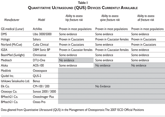
As just noted, heel devices currently appear    to have the most clinical applications, with some devices &#150;like the GE&#45;Lunar    Achilles and the Hologic Sahara&#150; better tested and more proven effective than    others (<a href="#tab01">table I</a>). For these purposes, the recommended parameter    of interest generally has been the heel stiffness index (SI) or the Quantitative    index (QUI), which is a composite score combining the results of broadband ultrasound    attenuation (BUA) and speed of sound (SOS), as measured in meters per second.</font></p>     <p><a name="tab01"></a></p>     <p>&nbsp;</p>     <p align="center"><img src="/img/revistas/spm/v51s1/a06tab01.gif"></p>     <p>&nbsp;</p>     <p><font size="2" face="Verdana">The remainder of this paper will review the clinical    use of QUS in the following settings: 1) the prediction of fracture risk; 2)    the diagnosis of osteoporosis; 3) the initiation of osteoporosis treatment or    prevention; 4) the monitoring of such treatment; and 5) osteoporosis case finding.    The paper will conclude by examining 6) quality assurance and quality control    issues pertaining to the clinical application of QUS. </font></p>     ]]></body>
<body><![CDATA[<p><font size="2" face="Verdana"><i>1) Using QUS to predict fracture risk</i></font></p>     <p><font size="2" face="Verdana">At the present time, there is satisfactory (cross&#45;sectional    and/or prospective) evidence that QUS can be used to assess fracture risk in    some, but not all populations, as defined by sex, age and ethnic background.<SUP>28&#45;96    </SUP>This is particularly true of heel QUS and for hip versus spinal fractures.    Having said this, because of various methodological issues, it is difficult    to compare studies. Nonetheless, combining the results from 13 studies involving    9561 patients,<SUP>33,39,40,43&#45;45,48,52,58&#45;60,69,79</SUP> it is reasonable to    state that the increase in relative risk observed for each standard deviation    decrease in stiffness index (SI), measured at the heel using QUS, is roughly    2.0 for the hip and spine, and approximately 1.5 for all fractures combined.    Consequently, heel QUS is much the same as DXA BMD, in terms of hip and spine    fracture risk per standard deviation decrease.<SUP>97,98</sup></font></p>     <p><font size="2" face="Verdana"> Although some differences may exist in the expression    of osteoporosis and overall fracture risk in Hispanics versus general Caucasians    and other ethnic populations, <SUP>99,100</SUP> there is ample empirical evidence    that the heel QUS stiffness index, using some but not all QUS devices, is predictive    of hip fracture risk in Caucasian and Asian women over age 55, and of any fracture    risk in Caucasian women &gt; 55 (<a href="#tab02">table II</a>). Weaker evidence    exists that the heel QUS stiffness index, again using some but not all QUS devices,    is predictive of hip fracture risk in Caucasian and Asian men over age 70; of    vertebral fracture risk in Caucasian and Asian women over age 55; and of any    fracture risk in Asian women and Caucasian or Asian men &gt; 70. With respect    to QUS devices from one of the other two categories, phalanx QUS devices utilizing    cortical transverse transmission might predict non&#45;vertebral fracture risk in    Caucasian women &gt; 70; however, to date, cortical axial transmission devices    have no proven clinical application. </font></p>     <p><a name="tab02"></a></p>     <p>&nbsp;</p>     <p align="center"><img src="/img/revistas/spm/v51s1/a06tab02.gif"></p>     <p>&nbsp;</p>     <p><font size="2" face="Verdana">Another practical question is: which QUS device    is best to use? As indicated in <a href="#tab01">table I</a>, the <I>G</I>E    Lunar Achilles and the Hologic Sahara are among the best tested devices, at    the hip, spine and overall, and both seem effective for most females; the former    may be preferable in males. Some evidence exists supporting the use of the Norland    Cuba Clinical and the IGEA DBM Sonic BP, at least among Caucasians. However    general results on the IGEA DBM Sonic device are not very impressive. Consequently,    the three heel devices appear to be the most reasonable to use, at this time,    though further testing of these and other devices clearly is necessary.</font></p>     <p><font size="2" face="Verdana"><i>2) Using QUS to diagnose osteoporosis </i></font></p>     <p><font size="2" face="Verdana">Diagnosing osteoporosis using QUS is less supported    by evidence and more complicated and problematic than assessing fracture risk    is. To start with, the T&#45;score diagnostic criteria of &#45;2.5, classically used    for DXA BMD, cannot be applied to QUS without discrepancies in the numbers of    women diagnosed with osteoporosis. This is because there are tremendous variations    in QUS measurements by skeletal site, and because different QUS devices yield    different results. If, for example, the prevalence of osteoporosis is defined    as &#45;2.5 standard deviations from the mean threshold for QUS, even within the    same sample population, different QUS instruments and different skeletal sites    generate prevalence estimates that vary as much as ten&#45;fold; for example, prevalence    estimates among Caucasian women &gt; 65 have ranged from 4 to 50%.<SUP>96,101&#45;105    </SUP>To overcome this dilemma, there is a need for pre&#45;defined, device&#45;specific    diagnostic thresholds. One recommended system suggests calibrating QUS measurements    with DXA results, the latter used as the 'gold standard', so that an upper QUS    threshold is set to identify osteoporosis with 90% sensitivity, and a lower    threshold is set to identify osteoporosis with 90% specificity.<SUP>106,107</SUP>    A similar approach already has been recommended by the UK National Osteoporosis    Society to define upper and lower thresholds for pDXA,<SUP>108,109 </SUP>the    results of which are highly correlated with QUS. Using such a system, one could    identify osteoporosis with high probability in patients whose results fall below    the lower threshold for QUS, where specificity exceeds 90%; between the upper    and lower thresholds, the diagnosis of osteoporosis would be considered quite    equivocal, so that another means of measurement, like DXA BMD, would be highly    recommended; and above the upper threshold for QUS, where the sensitivity of    a value below the threshold is 90%, osteoporosis would be deemed unlikely.
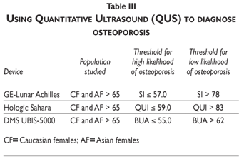
 </font></p>     ]]></body>
<body><![CDATA[<p><font size="2" face="Verdana"> We, in fact, utilized this approach in 5954    women 75 years and older who took part in the EPIDOS Study, utilizing the 90%    sensitivity threshold for the Achilles stiffness index of SI= 78%, and the 90%    specificity threshold of SI= 57%. Using these cut&#45;off points generated 11% false    positive (FP) and 13% false negative (FN) results, which are comparable to the    FP and FN rates of many other tests. Based upon these results, we believe that    device&#45;specific heel QUS thresholds for 90% sensitivity and 90% specificity    in specific populations defined by sex, age, and ethnic background, can be used    to identify individuals who have either a high or a low likelihood of osteoporosis,    even though only limited evidence exists supporting the use of any existing    QUS device for this purpose. Devices that have been evaluated include the GE&#45;Lunar    Achilles, the Hologic Sahara, and the DMS UBIS&#45;5000, each of which uses a different    measure and different upper and lower likelihood thresholds, as indicated in    <a href="#tab03">table III</a>. What can be concluded is that, regardless of    the QUS device used, values that fall between the upper and lower thresholds    strongly warrant further evaluation using DXA BMD as a more definitive test.</font></p>     <p><a name="tab03"></a></p>     <p>&nbsp;</p>     <p align="center"><img src="/img/revistas/spm/v51s1/a06tab03.gif"></p>     <p>&nbsp;</p>     <p><font size="2" face="Verdana"><i>3) Using QUS to initiate osteoporosis treatment</i></font></p>     <p><font size="2" face="Verdana">Except in patients with a low&#45;energy fracture    of the hip or spine, when the fracture alone is adequate to require treatment,    all currently&#45;published guidelines or recommendations for the initiation of    osteoporosis treatment are based upon DXA BMD values; in no instance, to date,    are the results of QUS the definitive parameter. Despite this, several studies    have demonstrated high levels of correlation (r ~ 0.90) between heel trabecular    sound transmission and BMD at matched skeletal sites.<SUP>110&#45;113 </SUP>Moreover,    both SOS and BUA, standard QUS measurements, are dependent upon overall bone    strength which, in turn, is related to bone density, architecture and turnover,    and the extent of bone mineralization.<SUP>110,112,114&#45;121</SUP> These factors    likely work together to maintain overall bone quality and strength, and to prevent    fractures and other bone failure.<SUP>110,112,114&#45;121</SUP> QUS parameters related    to heel trabecular transverse transmission are highly correlated with bone strength.<SUP>117,122&#45;129</SUP>    Consequently, it is conceivable that guidelines could be created using QUS to    guide when to initiate osteoporosis treatment, especially if combined with the    use of clinical risk factors.<SUP>130</SUP> To date, however, no randomized    clinical trails have been published examining whether individuals identified    as high risk for fracture by QUS respond to treatment. </font></p>     <p><font size="2" face="Verdana"> At the present time, published evidence does    not support using QUS device&#45;specific values to initiate osteoporosis treatment    in women younger than 65 years or in men of any age, but some evidence exists    supporting QUS use in other populations, when DXA is not available. These populations    include 1) Caucasian and 2) Asian women who are between the ages of 65 and 74    years, exhibit results below the lower specific device threshold (i.e. SI <u>&lt;</u>    57.0 with the GE&#45;Lunar Achilles device), and have at least two clinical risk    factors (<a href="#tab04">table IV</a>); and 3) Caucasian and 4) Asian women    who are 75 years or older, exhibit results below the lower specific device threshold    (i.e. SI <u>&lt;</u> 57.0 with the GE&#45;Lunar Achilles device), and have at least    one clinical risk factor besides age. <a href="#tab04">Table IV</a> lists pertinent    clinical risk factors that we have identified by examining published meta&#45;analyses,    as well as literature reviews written by Kanis<SUP>131</SUP> and Durosier.<SUP>98</SUP>    </font></p>     <p><a name="tab04"></a></p>     <p>&nbsp;</p>     ]]></body>
<body><![CDATA[<p align="center"><img src="/img/revistas/spm/v51s1/a06tab04.gif"></p>     <p>&nbsp;</p>     <p><font size="2" face="Verdana">Currently, a World Health Organization (WHO)    task force is developing a model to predict the 10&#45;year probability of osteoporosis&#45;related    fractures, combining femoral neck DXA BMD measurements and CRF. This also could    be done combining QUS and CRF, using a device&#45;specific T&#45;score. QUS&#45;generated    high and low risk probabilities &#150;for example, SI <u>&lt;</u> 57.0 and &gt; 78,    respectively, for the GE&#45;Lunar Achilles device&#150; then could be used to decide    whether treatment is warranted. Because different devices have their own high&#45;and    low&#45;probability threshold values that all correspond to roughly the same two    levels of fracture risk, this approach could be utilized independently of the    measurement instrument used. Preliminary results demonstrating the benefits    of this combined CRF plus technology approach already have been published by    Hans <I>et al</I>.<SUP>142</sup></font></p>     <p><font size="2" face="Verdana"> Unfortunately, whereas QUS and BMD are highly    correlated in trabecular bone<SUP>143</SUP> and this correlation reasonably    well understood, the situation is considerably more complex with cortical measurements.    Many properties influence these measurements, including cortical thickness,    mineralization, porosity and lamellar structure, and it is not clear to what    degree these various properties contribute to bone strength.<SUP>144&#45;147</SUP>    Consequently, QUS devices that measure cortical bone, like the cortical transverse    transmission devices currently used to assess phalanges, and the cortical axial    transmission devices being investigated for use with phalanges, the radius and    tibia, cannot be recommended as tools to determine the appropriateness of initiating    osteoporosis treatment at this time.</font></p>     <p><font size="2" face="Verdana"><i>4) Using QUS to monitor osteoporosis treatment    </i></font></p>     <p><font size="2" face="Verdana">At this time, QUS cannot be recommended for the    monitoring of treatment response in patients with osteoporosis, both due to    the absence of large&#45;scale, randomized, double&#45;blinded and placebo&#45;controlled    clinical trials (RCT) and the relatively equivocal evidence that has been generated    by the studies that have been published.<SUP>148&#45;164</SUP> It has been observed    that changes in heel QUS parameters, especially the stiffness index (SI), do    mimic the treatment response observed in BMD. In two studies involving alendronate,    for example, the Achilles SI was observed to significantly increase with treatment    over time.<SUP>149,150</SUP> Clearly, however, further RCT are needed to determine    if QUS parameters are sensitive enough to change with treatment, if the various    QUS instruments are sensitive enough to detect these changes, and if the precision    of these instruments is such that repeated measures can be performed without    excessive 'noise'. There is some evidence that instrument precision is adequate    in the short&#45;term; but what about over a longer period of time? What can be    said is that, if QUS is going to be used to monitor treatment, likely the heel    devices will be most successful, since trabecular measurements appear to be    more accurate than those that have been achieved with any of the cortical devices.</font></p>     <p><font size="2" face="Verdana"><i>5) Case finding</i></font></p>     <p><font size="2" face="Verdana">Case finding involves distinguishing subjects    at highest or lowest risk for a given disorder, who hence do not require further    investigation because their disease status is reasonably well known, from subjects    at intermediate risk whose disease status remains equivocal, thereby requiring    further evaluation. At present, there is fair empirical support for the use    of heel QUS for osteoporosis case finding among Caucasian and Asian females    who are at least 65 years old. The evidence for the use of cortical devices    is poor, and for males and other ethnic populations generally lacking altogether.    Nonetheless, we feel that the proposed case&#45;finding protocol depicted in <a href="#fig01">figure    1</a> is reasonable for clinical practice, in terms of distinguishing individuals    who require from those who do not require further evaluation of fragility fracture    risk.</font></p>     <p><a name="fig01"></a></p>     <p>&nbsp;</p>     ]]></body>
<body><![CDATA[<p align="center"><img src="/img/revistas/spm/v51s1/a06fig01.gif"></p>     <p>&nbsp;</p>     <p><font size="2" face="Verdana">To begin, each patient would undergo an assessment    to identify any clinical risk factors. Using this protocol, patients who are    suspected to be at risk for secondary osteoporosis &#150;for example, because of    prolonged systemic use of corticosteroids&#150; or who have a clinically&#45;evident    vertebral fracture would proceed directly to management deemed appropriate for    their condition. All others would undergo a heel QUS. Based upon this, those    whose QUS parameters suggest a low likelihood of future fracture would be assigned    to receive primary prevention, unless they have had a fragility fracture, in    which case they would undergo DXA BMD. Those for whom results indicate an intermediate    risk of future fracture, in that they lie between the upper and lower thresholds,    also would undergo DXA BMD. And those patients for whom the risk of future fracture    is deemed to be high, based upon the results of heel QUS, would proceed to treatment    if they are <u>&gt;</u> 75 years old and have at least one other CRF besides    age, or if they are &lt; 75 and have at least two clinical risk factors. Otherwise,    they too would proceed to DXA BMD. </font></p>     <p><font size="2" face="Verdana"> The primary advantage of this protocol, especially    in terms of cost, is that it saves performing costly and, sometimes relatively    inaccessible DXA on all patients. In particular, those with a low risk of fracture    by QUS would avoid DXA unless they have had a fragility fracture; and those    with a high risk of fracture by QUS would avoid DXA if they have at least two    CRF, counting age. As stated at the outset, this would have particular relevance    in localities in which access to DXA is scarce or too costly to be performed,    both of which likely apply to the majority of patients in many developing countries.</font></p>     <p><font size="2" face="Verdana"><i>6) QUS quality assurance and quality control</i></font></p>     <p><font size="2" face="Verdana">Technically, quality assurance primarily deals    with the performance of the equipment, whereas quality control is more heavily    grounded in theory and statistics, emphasizing the quality of the actual test.    For practical purposes, however, these two concepts often are treated as the    same,<SUP>165</SUP> and this paper will not seek to further delineate them.    The primary issues of importance are those of test/equipment accuracy, precision,    and reproducibility. Accuracy is a measure of how close a provided answer or    value is to the true answer or value.<SUP>166</SUP> If one were to visualize    a game of darts, for example, darts that hit the bull's eye are said to be accurate.    Precision, on the other hand, is a measure of consistency. If five darts are    thrown, do they all end up close to each other (high precision) or widely spread    (low precision)?<SUP>166</SUP> The two concepts, accuracy and precision, are    not the same. One could have all one's darts bunched together far from the bull's    eye (high precision, low accuracy); or one could have all one's darts widely    spread, but all equidistant from the bull's eye, so that the average of their    positions is near to the center dot (high accuracy, low precision). Reproducibility    measures how well the same test done on the same person or sample yields the    same result, whether the test is performed by the same technician (intra&#45;observer)    or a different technician (inter&#45;observer).<SUP>166</SUP> These three parameters    are important, whether a test is being performed to diagnose disease, monitor    its course, or identify potential cases. </font></p>     <p><font size="2" face="Verdana"> 
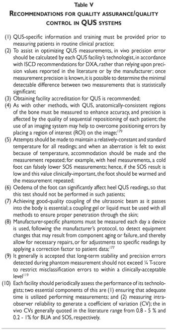
With heel QUS, there are several potential sources    of <I>in vivo</I> measurement error, which include surrounding soft tissue and    foot positioning;<SUP>167&#45;170</SUP> soft&#45;tissue thickness,<SUP>171&#45;173 </SUP>temperature<SUP>170,174</SUP>    and composition; the quality of sound transmission from the coupling medium    to the skin; and properties of the coupling medium between the transducers and    the skin, whether it be a fluid bath or sound transmitting pads.<SUP>167,170,175&#45;178</SUP>    One of the most important components of QA entails using some sort of test object,    which can be either a standard or phantom,<SUP>165</SUP> to monitor instrument    performance and make necessary calibrations when measurement accuracy begins    to stray. A standard is an object of known acoustic properties, which does not    necessarily resemble the anatomy of interest. Conversely, a phantom is designed    to emulate the anatomy and acoustic properties that exist during<I> in vivo</I>    measurements as much as possible. With respect to optimizing QUS device performance,    phantoms are more useful. Unfortunately, no universally&#45;applicable phantoms    exist. Consequently, manufacturer&#45;specific phantoms must be used, and measured    each day that the respective device is used, following the manufacturer's protocol,    to detect performance changes that may result from component aging or failure.    Detection of these changes allows both for necessary repairs, and for adjustments    to specific readings by applying a correction factor to patient data.<SUP>177    </SUP><a href="#tab05">Table V</a> provides a brief list of important guidelines    regarding quality assurance and control:</font></p>     <p><a name="tab05"></a></p>     <p>&nbsp;</p>     <p align="center"><img src="/img/revistas/spm/v51s1/a06tab05.gif"></p>     ]]></body>
<body><![CDATA[<p>&nbsp;</p>     <p><font size="2" face="Verdana"><b><font size="3">Summary</font></b></font></p>     <p><font size="2" face="Verdana">To date, no satisfactory evidence exists either    supporting or refuting the usefulness of quantitative ultrasound in Latin populations,    so that further research clearly is warranted. Nonetheless, there is enough    evidence in other populations to suggest that QUS may be an acceptable, low&#45;cost    and readily&#45;accessible alternative to DXA measurements of BMD in the management    of osteoporosis in Hispanics. Many different QUS devices exist that are quite    different in terms of the parameters they measure and the strength of empirical    evidence supporting their use. In general, heel QUS appears to be most tested    and most effective. Some, but not all heel QUS devices are effective assessing    fracture risk in some, but not all populations, the evidence being strongest    for Caucasian females &gt; 55 years old, though some evidence exists for Asian    females &gt; 55 and for Caucasian and Asian males &gt; 70. Certain devices may    allow for the accurate diagnosis of osteoporosis, but very limited evidence    exists supporting the use of QUS use during the initiation or monitoring of    osteoporosis treatment. A reasonable protocol for osteoporosis case&#45;finding    relies upon the combined assessment of clinical risk factors and heel QUS. However,    monitoring and maintenance of test and instrument accuracy, precision and reproducibility    are paramount to the effective clinical use of QUS. And, as stated earlier,    there is a huge call for further research in non&#45;Caucasian, non&#45;Asian populations.</font></p>     <p>&nbsp;</p>     <p><font size="3" face="verdana"><b>References</b></font></p>     <!-- ref --><p><font size="2" face="Verdana">1. Anonymous. Consensus development conference:    diagnosis, prophylaxis and treatment of osteoporosis. Am J Med 1993;94:646&#45;650.</font>&nbsp;&nbsp;&nbsp;&nbsp;&nbsp;&nbsp;&nbsp;&nbsp;[&#160;<a href="javascript:void(0);" onclick="javascript: window.open('/scielo.php?script=sci_nlinks&ref=9281934&pid=S0036-3634200900070000600001&lng=','','width=640,height=500,resizable=yes,scrollbars=1,menubar=yes,');">Links</a>&#160;]<!-- end-ref --><!-- ref --><p><font size="2" face="Verdana">2. Riggs, BLaM, LJ, 3rd, Medical progress series:    Involutional osteoporosis: N Engl J Med 1986;314:1676&#45;1686.</font>&nbsp;&nbsp;&nbsp;&nbsp;&nbsp;&nbsp;&nbsp;&nbsp;[&#160;<a href="javascript:void(0);" onclick="javascript: window.open('/scielo.php?script=sci_nlinks&ref=9281935&pid=S0036-3634200900070000600002&lng=','','width=640,height=500,resizable=yes,scrollbars=1,menubar=yes,');">Links</a>&#160;]<!-- end-ref --><!-- ref --><p><font size="2" face="Verdana">3. Riggs BL, LJ Melton, 3rd. The worldwide problem    of osteoporosis: insights afforded by epidemiology. Bone 1995;17(5 suppl):505S&#45;511S.</font>&nbsp;&nbsp;&nbsp;&nbsp;&nbsp;&nbsp;&nbsp;&nbsp;[&#160;<a href="javascript:void(0);" onclick="javascript: window.open('/scielo.php?script=sci_nlinks&ref=9281936&pid=S0036-3634200900070000600003&lng=','','width=640,height=500,resizable=yes,scrollbars=1,menubar=yes,');">Links</a>&#160;]<!-- end-ref --><!-- ref --><p><font size="2" face="Verdana">4. Kelsey JL, Hoffman S. Risk factors for hip    fracture. N Engl J Med 1987;316(7):404&#45;406.</font>&nbsp;&nbsp;&nbsp;&nbsp;&nbsp;&nbsp;&nbsp;&nbsp;[&#160;<a href="javascript:void(0);" onclick="javascript: window.open('/scielo.php?script=sci_nlinks&ref=9281937&pid=S0036-3634200900070000600004&lng=','','width=640,height=500,resizable=yes,scrollbars=1,menubar=yes,');">Links</a>&#160;]<!-- end-ref --><!-- ref --><p><font size="2" face="Verdana">5. Statistics NCfH. Advance data from vital and    health statistics: 1985 summary: national hospital discharge survey. 1986, US    Public Health Service (PHS): Hyattsville. p. PHS 86&#45;1250.</font>&nbsp;&nbsp;&nbsp;&nbsp;&nbsp;&nbsp;&nbsp;&nbsp;[&#160;<a href="javascript:void(0);" onclick="javascript: window.open('/scielo.php?script=sci_nlinks&ref=9281938&pid=S0036-3634200900070000600005&lng=','','width=640,height=500,resizable=yes,scrollbars=1,menubar=yes,');">Links</a>&#160;]<!-- end-ref --><!-- ref --><p><font size="2" face="Verdana">6. Cummings SR, et al. Epidemiology of osteoporosis    and osteoporotic fractures. Epidemiol Rev 1985;7:178&#45;208.</font>&nbsp;&nbsp;&nbsp;&nbsp;&nbsp;&nbsp;&nbsp;&nbsp;[&#160;<a href="javascript:void(0);" onclick="javascript: window.open('/scielo.php?script=sci_nlinks&ref=9281939&pid=S0036-3634200900070000600006&lng=','','width=640,height=500,resizable=yes,scrollbars=1,menubar=yes,');">Links</a>&#160;]<!-- end-ref --><!-- ref --><p><font size="2" face="Verdana">7. Melton LJ, 3rd, Riggs BL, Epidemiology of    age&#45;related fractures, in The osteoporotic syndrome: detection, prevention,    and treatment. Grune &amp; Stratton: New York, 1987:45&#45;72.</font>&nbsp;&nbsp;&nbsp;&nbsp;&nbsp;&nbsp;&nbsp;&nbsp;[&#160;<a href="javascript:void(0);" onclick="javascript: window.open('/scielo.php?script=sci_nlinks&ref=9281940&pid=S0036-3634200900070000600007&lng=','','width=640,height=500,resizable=yes,scrollbars=1,menubar=yes,');">Links</a>&#160;]<!-- end-ref --><p><font size="2" face="Verdana">8. Baudoin C. &#91;Fractures of the proximal femur.    Epidemiology and economic impact&#93;. Presse Med 1997;26(3):1451&#45;1456.</font></p>     <!-- ref --><p><font size="2" face="Verdana">9. Melton LJ, 3rd. Epidemiology of hip fractures:    implications of the exponential increase with age. Bone 1996;18(3 suppl):121S&#45;125S.</font>&nbsp;&nbsp;&nbsp;&nbsp;&nbsp;&nbsp;&nbsp;&nbsp;[&#160;<a href="javascript:void(0);" onclick="javascript: window.open('/scielo.php?script=sci_nlinks&ref=9281942&pid=S0036-3634200900070000600009&lng=','','width=640,height=500,resizable=yes,scrollbars=1,menubar=yes,');">Links</a>&#160;]<!-- end-ref --><!-- ref --><p><font size="2" face="Verdana">10. Fisher ES, et al. Hip fracture incidence    and mortality in New England. Epidemiology 1991;2(2):116&#45;122.</font>&nbsp;&nbsp;&nbsp;&nbsp;&nbsp;&nbsp;&nbsp;&nbsp;[&#160;<a href="javascript:void(0);" onclick="javascript: window.open('/scielo.php?script=sci_nlinks&ref=9281943&pid=S0036-3634200900070000600010&lng=','','width=640,height=500,resizable=yes,scrollbars=1,menubar=yes,');">Links</a>&#160;]<!-- end-ref --><!-- ref --><p><font size="2" face="Verdana">11. Sernbo I, Johnell O. Consequences of a hip    fracture: a prospective study over 1 year. Osteoporosis Int 1993;3(3):148&#45;153.</font>&nbsp;&nbsp;&nbsp;&nbsp;&nbsp;&nbsp;&nbsp;&nbsp;[&#160;<a href="javascript:void(0);" onclick="javascript: window.open('/scielo.php?script=sci_nlinks&ref=9281944&pid=S0036-3634200900070000600011&lng=','','width=640,height=500,resizable=yes,scrollbars=1,menubar=yes,');">Links</a>&#160;]<!-- end-ref --><!-- ref --><p><font size="2" face="Verdana">12. Wolinsky FD, Fitzgerald JF, Stump TE. The    effect of hip fracture on mortality, hospitalization, and functional status:    a prospective study. Am J Public Health 1997;87(3):398&#45;403.</font>&nbsp;&nbsp;&nbsp;&nbsp;&nbsp;&nbsp;&nbsp;&nbsp;[&#160;<a href="javascript:void(0);" onclick="javascript: window.open('/scielo.php?script=sci_nlinks&ref=9281945&pid=S0036-3634200900070000600012&lng=','','width=640,height=500,resizable=yes,scrollbars=1,menubar=yes,');">Links</a>&#160;]<!-- end-ref --><!-- ref --><p><font size="2" face="Verdana">13. Cummings SR, Rubin SM, Black D. The future    of hip fractures in the United States. Numbers, costs, and potential effects    of postmenopausal estrogen. Clin Orthop 1990(252):163&#45;166.</font>&nbsp;&nbsp;&nbsp;&nbsp;&nbsp;&nbsp;&nbsp;&nbsp;[&#160;<a href="javascript:void(0);" onclick="javascript: window.open('/scielo.php?script=sci_nlinks&ref=9281946&pid=S0036-3634200900070000600013&lng=','','width=640,height=500,resizable=yes,scrollbars=1,menubar=yes,');">Links</a>&#160;]<!-- end-ref --><!-- ref --><p><font size="2" face="Verdana">14. Brown J, Josse R. Clinical practices guidelines    for the diagnosis and management of osteoporosis in Canada. CMAJ 2002;167(10):S1&#45;S34.</font>&nbsp;&nbsp;&nbsp;&nbsp;&nbsp;&nbsp;&nbsp;&nbsp;[&#160;<a href="javascript:void(0);" onclick="javascript: window.open('/scielo.php?script=sci_nlinks&ref=9281947&pid=S0036-3634200900070000600014&lng=','','width=640,height=500,resizable=yes,scrollbars=1,menubar=yes,');">Links</a>&#160;]<!-- end-ref --><!-- ref --><p><font size="2" face="Verdana">15. Magnusson H, et al. Bone mass changes in    weight&#45;loaded and unloaded skeletal regions following a fracture of the hip.    Calcif Tiss Int 2001;69(2):78&#45;83.</font>&nbsp;&nbsp;&nbsp;&nbsp;&nbsp;&nbsp;&nbsp;&nbsp;[&#160;<a href="javascript:void(0);" onclick="javascript: window.open('/scielo.php?script=sci_nlinks&ref=9281948&pid=S0036-3634200900070000600015&lng=','','width=640,height=500,resizable=yes,scrollbars=1,menubar=yes,');">Links</a>&#160;]<!-- end-ref --><!-- ref --><p><font size="2" face="Verdana">16. Kiebzak GM, et al. Undertreatment of osteoporosis    in men with hip fracture Arch Int Med 2002;162(19):2217&#45;2222.</font>&nbsp;&nbsp;&nbsp;&nbsp;&nbsp;&nbsp;&nbsp;&nbsp;[&#160;<a href="javascript:void(0);" onclick="javascript: window.open('/scielo.php?script=sci_nlinks&ref=9281949&pid=S0036-3634200900070000600016&lng=','','width=640,height=500,resizable=yes,scrollbars=1,menubar=yes,');">Links</a>&#160;]<!-- end-ref --><!-- ref --><p><font size="2" face="Verdana">17. Morales&#45;Torres J, Guti&eacute;rrez&#45;Ure&ntilde;a    S, O.C.o.P.&#45;A.L.o.A.f. Rheumatology, The burden of osteoporosis in Latin America.    Osteoporos Int 2004;15(8):625&#45;632.</font>&nbsp;&nbsp;&nbsp;&nbsp;&nbsp;&nbsp;&nbsp;&nbsp;[&#160;<a href="javascript:void(0);" onclick="javascript: window.open('/scielo.php?script=sci_nlinks&ref=9281950&pid=S0036-3634200900070000600017&lng=','','width=640,height=500,resizable=yes,scrollbars=1,menubar=yes,');">Links</a>&#160;]<!-- end-ref --><p><font size="2" face="Verdana">18. Murrillo&#45;Uribe A, et al. &#91;Osteoporosis in    Mexican postmenopausal women. Magnitude of the problem. Multicenter study&#93; &#91;Article    in Spanish&#93;. Ginecol Obstet Mex 1999;67:227&#45;233.</font></p>     <!-- ref --><p><font size="2" face="Verdana">19. Beyene Y, Martin MC. Menopausal experiences    and bone density of Mayan women in Yucatan, Mexico. Am J Hum Biol 2001;13(4):505&#45;511.</font>&nbsp;&nbsp;&nbsp;&nbsp;&nbsp;&nbsp;&nbsp;&nbsp;[&#160;<a href="javascript:void(0);" onclick="javascript: window.open('/scielo.php?script=sci_nlinks&ref=9281952&pid=S0036-3634200900070000600019&lng=','','width=640,height=500,resizable=yes,scrollbars=1,menubar=yes,');">Links</a>&#160;]<!-- end-ref --><!-- ref --><p><font size="2" face="Verdana">20. Kanis JA, et al. Guidelines for diagnosis    and management of osteoporosis. The European Foundation for Osteoporosis and    Bone Disease. Osteoporos Int 1997; 7(4):390&#45;406.</font>&nbsp;&nbsp;&nbsp;&nbsp;&nbsp;&nbsp;&nbsp;&nbsp;[&#160;<a href="javascript:void(0);" onclick="javascript: window.open('/scielo.php?script=sci_nlinks&ref=9281953&pid=S0036-3634200900070000600020&lng=','','width=640,height=500,resizable=yes,scrollbars=1,menubar=yes,');">Links</a>&#160;]<!-- end-ref --><!-- ref --><p><font size="2" face="Verdana">21. WHO, Assessment of fracture risk and its    application to screening for postmenopausal osteoporosis. Geneva, World Health    Organization, 1994.</font>&nbsp;&nbsp;&nbsp;&nbsp;&nbsp;&nbsp;&nbsp;&nbsp;[&#160;<a href="javascript:void(0);" onclick="javascript: window.open('/scielo.php?script=sci_nlinks&ref=9281954&pid=S0036-3634200900070000600021&lng=','','width=640,height=500,resizable=yes,scrollbars=1,menubar=yes,');">Links</a>&#160;]<!-- end-ref --><!-- ref --><p><font size="2" face="Verdana">22. Hans D, et al. Skeletal sites for osteoporosis    diagnosis: the 2005 ISCD Official Positions. J Clin Densitom 2006;9(1):15&#45;21.</font>&nbsp;&nbsp;&nbsp;&nbsp;&nbsp;&nbsp;&nbsp;&nbsp;[&#160;<a href="javascript:void(0);" onclick="javascript: window.open('/scielo.php?script=sci_nlinks&ref=9281955&pid=S0036-3634200900070000600022&lng=','','width=640,height=500,resizable=yes,scrollbars=1,menubar=yes,');">Links</a>&#160;]<!-- end-ref --><!-- ref --><p><font size="2" face="Verdana">23. Kanis JA, Gluer CC. An update on the diagnosis    and assessment of osteoporosis with densitometry. Committee of Scientific Advisors,    International Osteoporosis Foundation. Osteoporos Int 2000;11(3):192&#45;202.</font>&nbsp;&nbsp;&nbsp;&nbsp;&nbsp;&nbsp;&nbsp;&nbsp;[&#160;<a href="javascript:void(0);" onclick="javascript: window.open('/scielo.php?script=sci_nlinks&ref=9281956&pid=S0036-3634200900070000600023&lng=','','width=640,height=500,resizable=yes,scrollbars=1,menubar=yes,');">Links</a>&#160;]<!-- end-ref --><!-- ref --><p><font size="2" face="Verdana">24. Njeh CF, Black DM. Calcaneal quantitative    ultrasound: Water&#45;coupled in: Quantitative Ultrasound: Assessment of Osteoporosis    and Bone Status, ed. Njeh CF, Hans D, Fuerst T, Gluer CC, Genant HK. 1999, London,    UK: Martin Dunitz:109&#45;124.</font>&nbsp;&nbsp;&nbsp;&nbsp;&nbsp;&nbsp;&nbsp;&nbsp;[&#160;<a href="javascript:void(0);" onclick="javascript: window.open('/scielo.php?script=sci_nlinks&ref=9281957&pid=S0036-3634200900070000600024&lng=','','width=640,height=500,resizable=yes,scrollbars=1,menubar=yes,');">Links</a>&#160;]<!-- end-ref --><!-- ref --><p><font size="2" face="Verdana">25. Cheng S, Hans D, Genant HK. Calcaneal quantitative    ultrasound: Gel&#45;coupled in: Quantitative Ultrasound: Assessment of Osteoporosis    and Bone Status, ed. C.F. Njeh, Hans, D., Fuerst, T., Gluer, C.C., Genant, H.K.    1999, London, UK: Martin Dunitz:125&#45;144.</font>&nbsp;&nbsp;&nbsp;&nbsp;&nbsp;&nbsp;&nbsp;&nbsp;[&#160;<a href="javascript:void(0);" onclick="javascript: window.open('/scielo.php?script=sci_nlinks&ref=9281958&pid=S0036-3634200900070000600025&lng=','','width=640,height=500,resizable=yes,scrollbars=1,menubar=yes,');">Links</a>&#160;]<!-- end-ref --><!-- ref --><p><font size="2" face="Verdana">26. Marin F, et al. Relationship between bone    quantitative ultrasound and fractures: a meta&#45;analysis. J Bone Miner Res 2006;21(7):1126&#45;1135.</font>&nbsp;&nbsp;&nbsp;&nbsp;&nbsp;&nbsp;&nbsp;&nbsp;[&#160;<a href="javascript:void(0);" onclick="javascript: window.open('/scielo.php?script=sci_nlinks&ref=9281959&pid=S0036-3634200900070000600026&lng=','','width=640,height=500,resizable=yes,scrollbars=1,menubar=yes,');">Links</a>&#160;]<!-- end-ref --><!-- ref --><p><font size="2" face="Verdana">27. Hans D, Fan B, Fuerst T. Non&#45;heel quantitative    ultrasound devices. in: Quantitative Ultrasound: Assessment of Osteoporosis    and Bone Status, ed. Njeh CF, Hans D, Fuerst T, Gluer CC. Genant, H.K. 1999,    London, UK: Martin Dunitz:145&#45;162.</font>&nbsp;&nbsp;&nbsp;&nbsp;&nbsp;&nbsp;&nbsp;&nbsp;[&#160;<a href="javascript:void(0);" onclick="javascript: window.open('/scielo.php?script=sci_nlinks&ref=9281960&pid=S0036-3634200900070000600027&lng=','','width=640,height=500,resizable=yes,scrollbars=1,menubar=yes,');">Links</a>&#160;]<!-- end-ref --><!-- ref --><p><font size="2" face="Verdana">28. Alenfeld FE, et al. Ultrasound measurements    at the proximal phalanges in healthy women and patients with hip fractures.    Osteoporos Int 1998;8(5):393&#45;398.</font>&nbsp;&nbsp;&nbsp;&nbsp;&nbsp;&nbsp;&nbsp;&nbsp;[&#160;<a href="javascript:void(0);" onclick="javascript: window.open('/scielo.php?script=sci_nlinks&ref=9281961&pid=S0036-3634200900070000600028&lng=','','width=640,height=500,resizable=yes,scrollbars=1,menubar=yes,');">Links</a>&#160;]<!-- end-ref --><!-- ref --><p><font size="2" face="Verdana">29. Augat, P., et al., Assessment of bone mineral    at appendicular sites in females with fractures of the proximal femur. Bone    1998;22(4):395&#45;402.</font>&nbsp;&nbsp;&nbsp;&nbsp;&nbsp;&nbsp;&nbsp;&nbsp;[&#160;<a href="javascript:void(0);" onclick="javascript: window.open('/scielo.php?script=sci_nlinks&ref=9281962&pid=S0036-3634200900070000600029&lng=','','width=640,height=500,resizable=yes,scrollbars=1,menubar=yes,');">Links</a>&#160;]<!-- end-ref --><!-- ref --><p><font size="2" face="Verdana">30. Barkmann R, et al. A new method for quantitative    ultrasound measurements at multiple skeletal sites: first results of precision    and fracture discrimination. J Clin Densitom 2000;3(1):1&#45;7.</font>&nbsp;&nbsp;&nbsp;&nbsp;&nbsp;&nbsp;&nbsp;&nbsp;[&#160;<a href="javascript:void(0);" onclick="javascript: window.open('/scielo.php?script=sci_nlinks&ref=9281963&pid=S0036-3634200900070000600030&lng=','','width=640,height=500,resizable=yes,scrollbars=1,menubar=yes,');">Links</a>&#160;]<!-- end-ref --><!-- ref --><p><font size="2" face="Verdana">31. Bauer DC, et al. Quantitative ultrasound    and vertebral fracture in postmenopausal women. Fracture Intervention Trial    Research Group. J Bone Miner Res 1995;10(3):353&#45;358.</font>&nbsp;&nbsp;&nbsp;&nbsp;&nbsp;&nbsp;&nbsp;&nbsp;[&#160;<a href="javascript:void(0);" onclick="javascript: window.open('/scielo.php?script=sci_nlinks&ref=9281964&pid=S0036-3634200900070000600031&lng=','','width=640,height=500,resizable=yes,scrollbars=1,menubar=yes,');">Links</a>&#160;]<!-- end-ref --><!-- ref --><p><font size="2" face="Verdana">32. Cepollaro C, et al. The combined use of ultrasound    and densitometry in the prediction of vertebral fracture. Br J Radiol 1997;70(835):691&#45;696.</font>&nbsp;&nbsp;&nbsp;&nbsp;&nbsp;&nbsp;&nbsp;&nbsp;[&#160;<a href="javascript:void(0);" onclick="javascript: window.open('/scielo.php?script=sci_nlinks&ref=9281965&pid=S0036-3634200900070000600032&lng=','','width=640,height=500,resizable=yes,scrollbars=1,menubar=yes,');">Links</a>&#160;]<!-- end-ref --><!-- ref --><p><font size="2" face="Verdana">33. Clowes JA, Eastell R, Peel NF. The discriminative    ability of peripheral and axial bone measurements to identify proximal femoral,    vertebral, distal forearm and proximal humeral fractures: a case control study.    Osteoporos Int 2005;16(12):1794&#45;1802.</font>&nbsp;&nbsp;&nbsp;&nbsp;&nbsp;&nbsp;&nbsp;&nbsp;[&#160;<a href="javascript:void(0);" onclick="javascript: window.open('/scielo.php?script=sci_nlinks&ref=9281966&pid=S0036-3634200900070000600033&lng=','','width=640,height=500,resizable=yes,scrollbars=1,menubar=yes,');">Links</a>&#160;]<!-- end-ref --><!-- ref --><p><font size="2" face="Verdana">34. Damilakis J, et al. Discrimination of hip    fractures by quantitative ultrasound of the phalanges and the calcaneus and    dual X&#45;ray absorptiometry. Eur J Radiol 2004;50(3):268&#45;272.</font>&nbsp;&nbsp;&nbsp;&nbsp;&nbsp;&nbsp;&nbsp;&nbsp;[&#160;<a href="javascript:void(0);" onclick="javascript: window.open('/scielo.php?script=sci_nlinks&ref=9281967&pid=S0036-3634200900070000600034&lng=','','width=640,height=500,resizable=yes,scrollbars=1,menubar=yes,');">Links</a>&#160;]<!-- end-ref --><!-- ref --><p><font size="2" face="Verdana">35. Damilakis J, et al. Ultrasound velocity through    the cortex of phalanges, radius, and tibia in normal and osteoporotic postmenopausal    women using a new multisite quantitative ultrasound device. Invest Radiol 2003;38(4):207&#45;211.</font>&nbsp;&nbsp;&nbsp;&nbsp;&nbsp;&nbsp;&nbsp;&nbsp;[&#160;<a href="javascript:void(0);" onclick="javascript: window.open('/scielo.php?script=sci_nlinks&ref=9281968&pid=S0036-3634200900070000600035&lng=','','width=640,height=500,resizable=yes,scrollbars=1,menubar=yes,');">Links</a>&#160;]<!-- end-ref --><!-- ref --><p><font size="2" face="Verdana">36. Donaldson MM, et al. Calcaneal ultrasound    attenuation in an elderly population: measurement position and relationships    with body size and past fractures. Osteoporos Int 1999;10(4):316&#45;324.</font>&nbsp;&nbsp;&nbsp;&nbsp;&nbsp;&nbsp;&nbsp;&nbsp;[&#160;<a href="javascript:void(0);" onclick="javascript: window.open('/scielo.php?script=sci_nlinks&ref=9281969&pid=S0036-3634200900070000600036&lng=','','width=640,height=500,resizable=yes,scrollbars=1,menubar=yes,');">Links</a>&#160;]<!-- end-ref --><!-- ref --><p><font size="2" face="Verdana">37. Drozdzowska B, Pluskiewicz W. The ability    of quantitative ultrasound at the calcaneus to identify postmenopausal women    with different types of nontraumatic fractures. Ultrasound Med Biol 2002;28(11&#45;12):1491&#45;1497.</font>&nbsp;&nbsp;&nbsp;&nbsp;&nbsp;&nbsp;&nbsp;&nbsp;[&#160;<a href="javascript:void(0);" onclick="javascript: window.open('/scielo.php?script=sci_nlinks&ref=9281970&pid=S0036-3634200900070000600037&lng=','','width=640,height=500,resizable=yes,scrollbars=1,menubar=yes,');">Links</a>&#160;]<!-- end-ref --><!-- ref --><p><font size="2" face="Verdana">38. Drozdzowska B, Pluskiewicz W, F. de Terlizzi.    The usefulness of quantitative ultrasound at the hand phalanges in the detection    of the different types of nontraumatic fractures. Ultrasound Med Biol 2003;29(11):1545&#45;1550.</font>&nbsp;&nbsp;&nbsp;&nbsp;&nbsp;&nbsp;&nbsp;&nbsp;[&#160;<a href="javascript:void(0);" onclick="javascript: window.open('/scielo.php?script=sci_nlinks&ref=9281971&pid=S0036-3634200900070000600038&lng=','','width=640,height=500,resizable=yes,scrollbars=1,menubar=yes,');">Links</a>&#160;]<!-- end-ref --><!-- ref --><p><font size="2" face="Verdana">39. Ekman A, et al. Dual X&#45;ray absorptiometry    of hip, heel ultrasound, and densitometry of fingers can discriminate male patients    with hip fracture from control subjects: a comparison of four different methods.    J Clin Densitom 2002;5(1):79&#45;85.</font>&nbsp;&nbsp;&nbsp;&nbsp;&nbsp;&nbsp;&nbsp;&nbsp;[&#160;<a href="javascript:void(0);" onclick="javascript: window.open('/scielo.php?script=sci_nlinks&ref=9281972&pid=S0036-3634200900070000600039&lng=','','width=640,height=500,resizable=yes,scrollbars=1,menubar=yes,');">Links</a>&#160;]<!-- end-ref --><!-- ref --><p><font size="2" face="Verdana">40. Ekman A, et al. DXA of the hip and heel ultrasound    but not densitometry of the fingers can discriminate female hip fracture patients    from controls: a comparison between four different methods. Osteoporos Int 2001;12(3):185&#45;191.</font>&nbsp;&nbsp;&nbsp;&nbsp;&nbsp;&nbsp;&nbsp;&nbsp;[&#160;<a href="javascript:void(0);" onclick="javascript: window.open('/scielo.php?script=sci_nlinks&ref=9281973&pid=S0036-3634200900070000600040&lng=','','width=640,height=500,resizable=yes,scrollbars=1,menubar=yes,');">Links</a>&#160;]<!-- end-ref --><!-- ref --><p><font size="2" face="Verdana">41. Frediani B, et al. Calcaneus ultrasonometry    and dual&#45;energy X&#45;ray absorptiometry for the evaluation of vertebral fracture    risk. Calcif Tissue Int 2006;79(4):223&#45;229.</font>&nbsp;&nbsp;&nbsp;&nbsp;&nbsp;&nbsp;&nbsp;&nbsp;[&#160;<a href="javascript:void(0);" onclick="javascript: window.open('/scielo.php?script=sci_nlinks&ref=9281974&pid=S0036-3634200900070000600041&lng=','','width=640,height=500,resizable=yes,scrollbars=1,menubar=yes,');">Links</a>&#160;]<!-- end-ref --><!-- ref --><p><font size="2" face="Verdana">42. Frost ML, Blake GM, Fogelman I. A comparison    of fracture discrimination using calcaneal quantitative ultrasound and dual    X&#45;ray absorptiometry in women with a history of fracture at sites other than    the spine and hip. Calcif Tissue Int 2002;71(3):207&#45;211.</font>&nbsp;&nbsp;&nbsp;&nbsp;&nbsp;&nbsp;&nbsp;&nbsp;[&#160;<a href="javascript:void(0);" onclick="javascript: window.open('/scielo.php?script=sci_nlinks&ref=9281975&pid=S0036-3634200900070000600042&lng=','','width=640,height=500,resizable=yes,scrollbars=1,menubar=yes,');">Links</a>&#160;]<!-- end-ref --><!-- ref --><p><font size="2" face="Verdana">43. Frost ML, Blake GM, Fogelman I. Does the    combination of quantitative ultrasound and dual&#45;energy X&#45;ray absorptiometry    improve fracture discrimination? Osteoporos Int 2001;12(6):471&#45;477.</font>&nbsp;&nbsp;&nbsp;&nbsp;&nbsp;&nbsp;&nbsp;&nbsp;[&#160;<a href="javascript:void(0);" onclick="javascript: window.open('/scielo.php?script=sci_nlinks&ref=9281976&pid=S0036-3634200900070000600043&lng=','','width=640,height=500,resizable=yes,scrollbars=1,menubar=yes,');">Links</a>&#160;]<!-- end-ref --><!-- ref --><p><font size="2" face="Verdana">44. Frost ML, Blake GM, Fogelman I. Does quantitative    ultrasound imaging enhance precision and discrimination? Osteoporos Int 2000;11(5):425&#45;433.</font>&nbsp;&nbsp;&nbsp;&nbsp;&nbsp;&nbsp;&nbsp;&nbsp;[&#160;<a href="javascript:void(0);" onclick="javascript: window.open('/scielo.php?script=sci_nlinks&ref=9281977&pid=S0036-3634200900070000600044&lng=','','width=640,height=500,resizable=yes,scrollbars=1,menubar=yes,');">Links</a>&#160;]<!-- end-ref --><!-- ref --><p><font size="2" face="Verdana">45. Frost ML, Blake GM, Fogelman I. Contact quantitative    ultrasound: an evaluation of precision, fracture discrimination, age&#45;related    bone loss and applicability of the WHO criteria. Osteoporos Int 1999;10(6):441&#45;449.</font>&nbsp;&nbsp;&nbsp;&nbsp;&nbsp;&nbsp;&nbsp;&nbsp;[&#160;<a href="javascript:void(0);" onclick="javascript: window.open('/scielo.php?script=sci_nlinks&ref=9281978&pid=S0036-3634200900070000600045&lng=','','width=640,height=500,resizable=yes,scrollbars=1,menubar=yes,');">Links</a>&#160;]<!-- end-ref --><!-- ref --><p><font size="2" face="Verdana">46. Gerdhem P, et al. Ultrasound of the phalanges    is not related to a previous fracture. A comparison between ultrasound of the    phalanges, calcaneus, and DXA of the spine and hip in 75&#45;year&#45;old women. J Clin    Densitom 2002;5(2):159&#45;166.</font>&nbsp;&nbsp;&nbsp;&nbsp;&nbsp;&nbsp;&nbsp;&nbsp;[&#160;<a href="javascript:void(0);" onclick="javascript: window.open('/scielo.php?script=sci_nlinks&ref=9281979&pid=S0036-3634200900070000600046&lng=','','width=640,height=500,resizable=yes,scrollbars=1,menubar=yes,');">Links</a>&#160;]<!-- end-ref --><!-- ref --><p><font size="2" face="Verdana">47. Gluer CC, et al. Osteoporosis: association    of recent fractures with quantitative US findings. Radiology 1996;199(3):725&#45;732.</font>&nbsp;&nbsp;&nbsp;&nbsp;&nbsp;&nbsp;&nbsp;&nbsp;[&#160;<a href="javascript:void(0);" onclick="javascript: window.open('/scielo.php?script=sci_nlinks&ref=9281980&pid=S0036-3634200900070000600047&lng=','','width=640,height=500,resizable=yes,scrollbars=1,menubar=yes,');">Links</a>&#160;]<!-- end-ref --><!-- ref --><p><font size="2" face="Verdana">48. Gluer CC, et al. Association of five quantitative    ultrasound devices and bone densitometry with osteoporotic vertebral fractures    in a population&#45;based sample: the OPUS Study. J Bone Miner Res 2004;19(5):782&#45;793.</font>&nbsp;&nbsp;&nbsp;&nbsp;&nbsp;&nbsp;&nbsp;&nbsp;[&#160;<a href="javascript:void(0);" onclick="javascript: window.open('/scielo.php?script=sci_nlinks&ref=9281981&pid=S0036-3634200900070000600048&lng=','','width=640,height=500,resizable=yes,scrollbars=1,menubar=yes,');">Links</a>&#160;]<!-- end-ref --><!-- ref --><p><font size="2" face="Verdana">49. Gnudi S, Gualtieri G, Malavolta N. Simultaneous    densitometry and quantitative bone sonography in the estimation of osteoporotic    fracture risk. Br J Radiol 1998;71(846):625&#45;629.</font>&nbsp;&nbsp;&nbsp;&nbsp;&nbsp;&nbsp;&nbsp;&nbsp;[&#160;<a href="javascript:void(0);" onclick="javascript: window.open('/scielo.php?script=sci_nlinks&ref=9281982&pid=S0036-3634200900070000600049&lng=','','width=640,height=500,resizable=yes,scrollbars=1,menubar=yes,');">Links</a>&#160;]<!-- end-ref --><!-- ref --><p><font size="2" face="Verdana">50. Gnudi S, Ripamonti C. Quantitative ultrasound    at the phalanxes discriminates osteoporotic women with vertebral but not with    hip fracture. Ultrasound Med Biol 2004;30(3):357&#45;361.</font>&nbsp;&nbsp;&nbsp;&nbsp;&nbsp;&nbsp;&nbsp;&nbsp;[&#160;<a href="javascript:void(0);" onclick="javascript: window.open('/scielo.php?script=sci_nlinks&ref=9281983&pid=S0036-3634200900070000600050&lng=','','width=640,height=500,resizable=yes,scrollbars=1,menubar=yes,');">Links</a>&#160;]<!-- end-ref --><!-- ref --><p><font size="2" face="Verdana">51. Gonnelli S, et al. Diagnostic value of ultrasound    analysis and bone densitometry as predictors of vertebral deformity in postmenopausal    women. Osteoporos Int 1995;5(6):413&#45;418.</font>&nbsp;&nbsp;&nbsp;&nbsp;&nbsp;&nbsp;&nbsp;&nbsp;[&#160;<a href="javascript:void(0);" onclick="javascript: window.open('/scielo.php?script=sci_nlinks&ref=9281984&pid=S0036-3634200900070000600051&lng=','','width=640,height=500,resizable=yes,scrollbars=1,menubar=yes,');">Links</a>&#160;]<!-- end-ref --><!-- ref --><p><font size="2" face="Verdana">52. Gonnelli S, et al. Quantitative ultrasound    and dual&#45;energy X&#45;ray absorptiometry in the prediction of fragility fracture    in men. Osteoporos Int 2005;16(8):963&#45;968.</font>&nbsp;&nbsp;&nbsp;&nbsp;&nbsp;&nbsp;&nbsp;&nbsp;[&#160;<a href="javascript:void(0);" onclick="javascript: window.open('/scielo.php?script=sci_nlinks&ref=9281985&pid=S0036-3634200900070000600052&lng=','','width=640,height=500,resizable=yes,scrollbars=1,menubar=yes,');">Links</a>&#160;]<!-- end-ref --><!-- ref --><p><font size="2" face="Verdana">53. Greenspan SL, et al. Precision and discriminatory    ability of calcaneal bone assessment technologies. J Bone Miner Res 1997;12(8):1303&#45;1313.</font>&nbsp;&nbsp;&nbsp;&nbsp;&nbsp;&nbsp;&nbsp;&nbsp;[&#160;<a href="javascript:void(0);" onclick="javascript: window.open('/scielo.php?script=sci_nlinks&ref=9281986&pid=S0036-3634200900070000600053&lng=','','width=640,height=500,resizable=yes,scrollbars=1,menubar=yes,');">Links</a>&#160;]<!-- end-ref --><!-- ref --><p><font size="2" face="Verdana">54. Greenspan SL, et al. Clinical performance    of a highly portable, scanning calcaneal ultrasonometer. Osteoporos Int 2001;12(5):391&#45;398.</font>&nbsp;&nbsp;&nbsp;&nbsp;&nbsp;&nbsp;&nbsp;&nbsp;[&#160;<a href="javascript:void(0);" onclick="javascript: window.open('/scielo.php?script=sci_nlinks&ref=9281987&pid=S0036-3634200900070000600054&lng=','','width=640,height=500,resizable=yes,scrollbars=1,menubar=yes,');">Links</a>&#160;]<!-- end-ref --><!-- ref --><p><font size="2" face="Verdana">55. Guglielmi G, et al. Palangeal quantitative    ultrasound, phalangeal morphometric variables, and vertebral fracture discrimination.    Calcif Tissue Int 2003;72(4):469&#45;477.</font>&nbsp;&nbsp;&nbsp;&nbsp;&nbsp;&nbsp;&nbsp;&nbsp;[&#160;<a href="javascript:void(0);" onclick="javascript: window.open('/scielo.php?script=sci_nlinks&ref=9281988&pid=S0036-3634200900070000600055&lng=','','width=640,height=500,resizable=yes,scrollbars=1,menubar=yes,');">Links</a>&#160;]<!-- end-ref --><!-- ref --><p><font size="2" face="Verdana">56. Hadji P, et al. Quantitative ultrasound of    the os calcis in postmenopausal women with spine and hip fracture. J Clin Densitom    2000;3(3):233&#45;239.</font>&nbsp;&nbsp;&nbsp;&nbsp;&nbsp;&nbsp;&nbsp;&nbsp;[&#160;<a href="javascript:void(0);" onclick="javascript: window.open('/scielo.php?script=sci_nlinks&ref=9281989&pid=S0036-3634200900070000600056&lng=','','width=640,height=500,resizable=yes,scrollbars=1,menubar=yes,');">Links</a>&#160;]<!-- end-ref --><!-- ref --><p><font size="2" face="Verdana">57. Hamanaka Y, et al. Comparison of bone mineral    density at various skeletal sites with quantitative ultrasound parameters of    the calcaneus for assessment of vertebral fractures. J Bone Miner Metab 1999;17(3):195&#45;200.</font>&nbsp;&nbsp;&nbsp;&nbsp;&nbsp;&nbsp;&nbsp;&nbsp;[&#160;<a href="javascript:void(0);" onclick="javascript: window.open('/scielo.php?script=sci_nlinks&ref=9281990&pid=S0036-3634200900070000600057&lng=','','width=640,height=500,resizable=yes,scrollbars=1,menubar=yes,');">Links</a>&#160;]<!-- end-ref --><!-- ref --><p><font size="2" face="Verdana">58. Hans D, et al. Does combining the results    from multiple bone sites measured by a new quantitative ultrasound device improve    discrimination of hip fracture? J Bone Miner Res 1999;14(4):644&#45;651.</font>&nbsp;&nbsp;&nbsp;&nbsp;&nbsp;&nbsp;&nbsp;&nbsp;[&#160;<a href="javascript:void(0);" onclick="javascript: window.open('/scielo.php?script=sci_nlinks&ref=9281991&pid=S0036-3634200900070000600058&lng=','','width=640,height=500,resizable=yes,scrollbars=1,menubar=yes,');">Links</a>&#160;]<!-- end-ref --><!-- ref --><p><font size="2" face="Verdana">59. Hans D, et al. Is time since hip fracture    influencing the discrimination between fractured and nonfractured subjects as    assessed at the calcaneum by three technologically different quantitative ultrasound    devices? Calcif Tissue Int 2002;71(6):485&#45;492.</font>&nbsp;&nbsp;&nbsp;&nbsp;&nbsp;&nbsp;&nbsp;&nbsp;[&#160;<a href="javascript:void(0);" onclick="javascript: window.open('/scielo.php?script=sci_nlinks&ref=9281992&pid=S0036-3634200900070000600059&lng=','','width=640,height=500,resizable=yes,scrollbars=1,menubar=yes,');">Links</a>&#160;]<!-- end-ref --><!-- ref --><p><font size="2" face="Verdana">60. Hans D, et al. Hip fracture discrimination    study: QUS of the radius and the calcaneum. J Clin Densitom 2003;6(2):163&#45;172.</font>&nbsp;&nbsp;&nbsp;&nbsp;&nbsp;&nbsp;&nbsp;&nbsp;[&#160;<a href="javascript:void(0);" onclick="javascript: window.open('/scielo.php?script=sci_nlinks&ref=9281993&pid=S0036-3634200900070000600060&lng=','','width=640,height=500,resizable=yes,scrollbars=1,menubar=yes,');">Links</a>&#160;]<!-- end-ref --><!-- ref --><p><font size="2" face="Verdana">61. Hartl F, et al. Discriminatory ability of    quantitative ultrasound parameters and bone mineral density in a population&#45;based    sample of postmenopausal women with vertebral fractures: results of the Basel    Osteoporosis Study. J Bone Miner Res 2002;17(2):321&#45;330.</font>&nbsp;&nbsp;&nbsp;&nbsp;&nbsp;&nbsp;&nbsp;&nbsp;[&#160;<a href="javascript:void(0);" onclick="javascript: window.open('/scielo.php?script=sci_nlinks&ref=9281994&pid=S0036-3634200900070000600061&lng=','','width=640,height=500,resizable=yes,scrollbars=1,menubar=yes,');">Links</a>&#160;]<!-- end-ref --><!-- ref --><p><font size="2" face="Verdana">62. He YQ, et al. Assessment of a new quantitative    ultrasound calcaneus measurement: precision and discrimination of hip fractures    in elderly women compared with dual X&#45;ray absorptiometry. Osteoporos Int 2000;11(4):354&#45;360.</font>&nbsp;&nbsp;&nbsp;&nbsp;&nbsp;&nbsp;&nbsp;&nbsp;[&#160;<a href="javascript:void(0);" onclick="javascript: window.open('/scielo.php?script=sci_nlinks&ref=9281995&pid=S0036-3634200900070000600062&lng=','','width=640,height=500,resizable=yes,scrollbars=1,menubar=yes,');">Links</a>&#160;]<!-- end-ref --><!-- ref --><p><font size="2" face="Verdana">63. Hernandez JL, et al. Discriminative capacity    of calcaneal quantitative ultrasound and of osteoporosis and fracture risk factors    in postmenopausal women with osteoporotic fractures. Calcif Tissue Int 2004;74(4):357&#45;365.</font>&nbsp;&nbsp;&nbsp;&nbsp;&nbsp;&nbsp;&nbsp;&nbsp;[&#160;<a href="javascript:void(0);" onclick="javascript: window.open('/scielo.php?script=sci_nlinks&ref=9281996&pid=S0036-3634200900070000600063&lng=','','width=640,height=500,resizable=yes,scrollbars=1,menubar=yes,');">Links</a>&#160;]<!-- end-ref --><!-- ref --><p><font size="2" face="Verdana">64. Hollevoet N, et al. Tibial ultrasound velocity    in women with wrist fracture. J Clin Densitom 2004;7(3):302&#45;306.</font>&nbsp;&nbsp;&nbsp;&nbsp;&nbsp;&nbsp;&nbsp;&nbsp;[&#160;<a href="javascript:void(0);" onclick="javascript: window.open('/scielo.php?script=sci_nlinks&ref=9281997&pid=S0036-3634200900070000600064&lng=','','width=640,height=500,resizable=yes,scrollbars=1,menubar=yes,');">Links</a>&#160;]<!-- end-ref --><!-- ref --><p><font size="2" face="Verdana">65. Ingle BM, Eastell R. Site&#45;specific bone measurements    in patients with ankle fracture. Osteoporos Int 2002;13(4):342&#45;347.</font>&nbsp;&nbsp;&nbsp;&nbsp;&nbsp;&nbsp;&nbsp;&nbsp;[&#160;<a href="javascript:void(0);" onclick="javascript: window.open('/scielo.php?script=sci_nlinks&ref=9281998&pid=S0036-3634200900070000600065&lng=','','width=640,height=500,resizable=yes,scrollbars=1,menubar=yes,');">Links</a>&#160;]<!-- end-ref --><!-- ref --><p><font size="2" face="Verdana">66. Karlsson MK, et al. Age, gender, and fragility    fractures are associated with differences in quantitative ultrasound independent    of bone mineral density. Bone 2001;28(1):118&#45;122.</font>&nbsp;&nbsp;&nbsp;&nbsp;&nbsp;&nbsp;&nbsp;&nbsp;[&#160;<a href="javascript:void(0);" onclick="javascript: window.open('/scielo.php?script=sci_nlinks&ref=9281999&pid=S0036-3634200900070000600066&lng=','','width=640,height=500,resizable=yes,scrollbars=1,menubar=yes,');">Links</a>&#160;]<!-- end-ref --><!-- ref --><p><font size="2" face="Verdana">67. Knapp KM, et al. Multisite quantitative ultrasound:    Colles' fracture discrimination in postmenopausal women. Osteoporos Int 2002;13(6):474&#45;479.</font>&nbsp;&nbsp;&nbsp;&nbsp;&nbsp;&nbsp;&nbsp;&nbsp;[&#160;<a href="javascript:void(0);" onclick="javascript: window.open('/scielo.php?script=sci_nlinks&ref=9282000&pid=S0036-3634200900070000600067&lng=','','width=640,height=500,resizable=yes,scrollbars=1,menubar=yes,');">Links</a>&#160;]<!-- end-ref --><!-- ref --><p><font size="2" face="Verdana">68. Knapp KM, et al. Multisite quantitative ultrasound:    precision, age&#45; and menopause&#45;related changes, fracture discrimination, and    T&#45;score equivalence with dual&#45;energy X&#45;ray absorptiometry. Osteoporos Int 2001;12(6):456&#45;464.</font>&nbsp;&nbsp;&nbsp;&nbsp;&nbsp;&nbsp;&nbsp;&nbsp;[&#160;<a href="javascript:void(0);" onclick="javascript: window.open('/scielo.php?script=sci_nlinks&ref=9282001&pid=S0036-3634200900070000600068&lng=','','width=640,height=500,resizable=yes,scrollbars=1,menubar=yes,');">Links</a>&#160;]<!-- end-ref --><!-- ref --><p><font size="2" face="Verdana">69. Krieg, M.A., et al., Comparison of three    bone ultrasounds for the discrimination of subjects with and without osteoporotic    fractures among 7562 elderly women. J Bone Miner Res 2003;18(7):1261&#45;1266.</font>&nbsp;&nbsp;&nbsp;&nbsp;&nbsp;&nbsp;&nbsp;&nbsp;[&#160;<a href="javascript:void(0);" onclick="javascript: window.open('/scielo.php?script=sci_nlinks&ref=9282002&pid=S0036-3634200900070000600069&lng=','','width=640,height=500,resizable=yes,scrollbars=1,menubar=yes,');">Links</a>&#160;]<!-- end-ref --><!-- ref --><p><font size="2" face="Verdana">70. Kung AW, et al. Quantitative ultrasound and    symptomatic vertebral fracture risk in Chinese women. Osteoporos Int 1999;10(6):456&#45;461.</font>&nbsp;&nbsp;&nbsp;&nbsp;&nbsp;&nbsp;&nbsp;&nbsp;[&#160;<a href="javascript:void(0);" onclick="javascript: window.open('/scielo.php?script=sci_nlinks&ref=9282003&pid=S0036-3634200900070000600070&lng=','','width=640,height=500,resizable=yes,scrollbars=1,menubar=yes,');">Links</a>&#160;]<!-- end-ref --><!-- ref --><p><font size="2" face="Verdana">71. Lopez&#45;Rodriguez F, et al. Performance of    quantitative ultrasound in the discrimination of prevalent osteoporotic fractures    in a bone metabolic unit. Bone 2003;32(5):571&#45;578.</font>&nbsp;&nbsp;&nbsp;&nbsp;&nbsp;&nbsp;&nbsp;&nbsp;[&#160;<a href="javascript:void(0);" onclick="javascript: window.open('/scielo.php?script=sci_nlinks&ref=9282004&pid=S0036-3634200900070000600071&lng=','','width=640,height=500,resizable=yes,scrollbars=1,menubar=yes,');">Links</a>&#160;]<!-- end-ref --><!-- ref --><p><font size="2" face="Verdana">72. Maggi S, et al. Quantitative heel ultrasound    in a population&#45;based study in Italy and its relationship with fracture history:    the ESOPO study. Osteoporos Int 2006;17(2):237&#45;244.</font>&nbsp;&nbsp;&nbsp;&nbsp;&nbsp;&nbsp;&nbsp;&nbsp;[&#160;<a href="javascript:void(0);" onclick="javascript: window.open('/scielo.php?script=sci_nlinks&ref=9282005&pid=S0036-3634200900070000600072&lng=','','width=640,height=500,resizable=yes,scrollbars=1,menubar=yes,');">Links</a>&#160;]<!-- end-ref --><!-- ref --><p><font size="2" face="Verdana">73. Matsushita R, et al. Comparison of various    biochemical measurements with bone mineral densitometry and quantitative ultrasound    for the assessment of vertebral fracture. J Bone Miner Metab 2000;18(3):158&#45;164.</font>&nbsp;&nbsp;&nbsp;&nbsp;&nbsp;&nbsp;&nbsp;&nbsp;[&#160;<a href="javascript:void(0);" onclick="javascript: window.open('/scielo.php?script=sci_nlinks&ref=9282006&pid=S0036-3634200900070000600073&lng=','','width=640,height=500,resizable=yes,scrollbars=1,menubar=yes,');">Links</a>&#160;]<!-- end-ref --><!-- ref --><p><font size="2" face="Verdana">74. Meszaros S, et al. Calcaneous quantitative    ultrasound measurements predicts vertebral fractures in idiopathic male osteoporosis.    Joint Bone Spine, 2006.</font>&nbsp;&nbsp;&nbsp;&nbsp;&nbsp;&nbsp;&nbsp;&nbsp;[&#160;<a href="javascript:void(0);" onclick="javascript: window.open('/scielo.php?script=sci_nlinks&ref=9282007&pid=S0036-3634200900070000600074&lng=','','width=640,height=500,resizable=yes,scrollbars=1,menubar=yes,');">Links</a>&#160;]<!-- end-ref --><!-- ref --><p><font size="2" face="Verdana">75. Mikhail MB, Flaster E, Aloia JF. Stiffness    in discrimination of patients with vertebral fractures. Osteoporos Int 1999;9(1):24&#45;28.</font>&nbsp;&nbsp;&nbsp;&nbsp;&nbsp;&nbsp;&nbsp;&nbsp;[&#160;<a href="javascript:void(0);" onclick="javascript: window.open('/scielo.php?script=sci_nlinks&ref=9282008&pid=S0036-3634200900070000600075&lng=','','width=640,height=500,resizable=yes,scrollbars=1,menubar=yes,');">Links</a>&#160;]<!-- end-ref --><!-- ref --><p><font size="2" face="Verdana">76. Mulleman D, et al. Quantitative ultrasound    of bone in male osteoporosis. Osteoporos Int 2002;13(5):388&#45;393.</font>&nbsp;&nbsp;&nbsp;&nbsp;&nbsp;&nbsp;&nbsp;&nbsp;[&#160;<a href="javascript:void(0);" onclick="javascript: window.open('/scielo.php?script=sci_nlinks&ref=9282009&pid=S0036-3634200900070000600076&lng=','','width=640,height=500,resizable=yes,scrollbars=1,menubar=yes,');">Links</a>&#160;]<!-- end-ref --><!-- ref --><p><font size="2" face="Verdana">77. Muraki S, Yamamoto S, Kanai H. Ultrasound    velocity in the tibia in Japanese patients with hip fracture. J Orthop Sci 2002;7(6):623&#45;628.</font>&nbsp;&nbsp;&nbsp;&nbsp;&nbsp;&nbsp;&nbsp;&nbsp;[&#160;<a href="javascript:void(0);" onclick="javascript: window.open('/scielo.php?script=sci_nlinks&ref=9282010&pid=S0036-3634200900070000600077&lng=','','width=640,height=500,resizable=yes,scrollbars=1,menubar=yes,');">Links</a>&#160;]<!-- end-ref --><!-- ref --><p><font size="2" face="Verdana">78. Nguyen TV, Center JR, Eisman JA. Bone mineral    density&#45;independent association of quantitative ultrasound measurements and    fracture risk in women. Osteoporos Int 2004;15(12):942&#45;947.</font>&nbsp;&nbsp;&nbsp;&nbsp;&nbsp;&nbsp;&nbsp;&nbsp;[&#160;<a href="javascript:void(0);" onclick="javascript: window.open('/scielo.php?script=sci_nlinks&ref=9282011&pid=S0036-3634200900070000600078&lng=','','width=640,height=500,resizable=yes,scrollbars=1,menubar=yes,');">Links</a>&#160;]<!-- end-ref --><!-- ref --><p><font size="2" face="Verdana">79. Njeh CF, et al. Comparison of six calcaneal    quantitative ultrasound devices: precision and hip fracture discrimination.    Osteoporos Int 2000;11(12):1051&#45;1062.</font>&nbsp;&nbsp;&nbsp;&nbsp;&nbsp;&nbsp;&nbsp;&nbsp;[&#160;<a href="javascript:void(0);" onclick="javascript: window.open('/scielo.php?script=sci_nlinks&ref=9282012&pid=S0036-3634200900070000600079&lng=','','width=640,height=500,resizable=yes,scrollbars=1,menubar=yes,');">Links</a>&#160;]<!-- end-ref --><!-- ref --><p><font size="2" face="Verdana">80. Ohishi T, et al. Ultrasound measurement using    CUBA clinical system can discriminate between women with and without vertebral    fractures. Contact Ultrasound Bone Analyzer. J Clin Densitom 2000;3(3):227&#45;231.</font>&nbsp;&nbsp;&nbsp;&nbsp;&nbsp;&nbsp;&nbsp;&nbsp;[&#160;<a href="javascript:void(0);" onclick="javascript: window.open('/scielo.php?script=sci_nlinks&ref=9282013&pid=S0036-3634200900070000600080&lng=','','width=640,height=500,resizable=yes,scrollbars=1,menubar=yes,');">Links</a>&#160;]<!-- end-ref --><!-- ref --><p><font size="2" face="Verdana">81. Peretz A, et al. Evaluation of quantitative    ultrasound and dual X&#45;Ray absorptiometry measurements in women with and without    fractures. J Clin Densitom 1999;2(2):127&#45;133.</font>&nbsp;&nbsp;&nbsp;&nbsp;&nbsp;&nbsp;&nbsp;&nbsp;[&#160;<a href="javascript:void(0);" onclick="javascript: window.open('/scielo.php?script=sci_nlinks&ref=9282014&pid=S0036-3634200900070000600081&lng=','','width=640,height=500,resizable=yes,scrollbars=1,menubar=yes,');">Links</a>&#160;]<!-- end-ref --><!-- ref --><p><font size="2" face="Verdana">82. Pinheiro MM, et al. Discriminatory ability    of quantitative ultrasound measurements is similar to dual&#45;energy X&#45;ray absorptiometry    in a Brazilian women population with osteoporotic fracture. Calcif Tissue Int    2003;73(6):555&#45;564.</font>&nbsp;&nbsp;&nbsp;&nbsp;&nbsp;&nbsp;&nbsp;&nbsp;[&#160;<a href="javascript:void(0);" onclick="javascript: window.open('/scielo.php?script=sci_nlinks&ref=9282015&pid=S0036-3634200900070000600082&lng=','','width=640,height=500,resizable=yes,scrollbars=1,menubar=yes,');">Links</a>&#160;]<!-- end-ref --><!-- ref --><p><font size="2" face="Verdana">83. Pluskiewicz W, Drozdzowska B. Ultrasound    measurements at the calcaneus in men: differences between healthy and fractured    persons and the influence of age and anthropometric features on ultrasound parameters.    Osteoporos Int 1999;10(1):47&#45;51.</font>&nbsp;&nbsp;&nbsp;&nbsp;&nbsp;&nbsp;&nbsp;&nbsp;[&#160;<a href="javascript:void(0);" onclick="javascript: window.open('/scielo.php?script=sci_nlinks&ref=9282016&pid=S0036-3634200900070000600083&lng=','','width=640,height=500,resizable=yes,scrollbars=1,menubar=yes,');">Links</a>&#160;]<!-- end-ref --><!-- ref --><p><font size="2" face="Verdana">84. Ross P, et al. Predicting vertebral deformity    using bone densitometry at various skeletal sites and calcaneus ultrasound.    Bone 1995;16(3):325&#45;332.</font>&nbsp;&nbsp;&nbsp;&nbsp;&nbsp;&nbsp;&nbsp;&nbsp;[&#160;<a href="javascript:void(0);" onclick="javascript: window.open('/scielo.php?script=sci_nlinks&ref=9282017&pid=S0036-3634200900070000600084&lng=','','width=640,height=500,resizable=yes,scrollbars=1,menubar=yes,');">Links</a>&#160;]<!-- end-ref --><!-- ref --><p><font size="2" face="Verdana">85. Roux C, et al. Ultrasonic backscatter and    transmission parameters at the os calcis in postmenopausal osteoporosis. J Bone    Miner Res 2001;16(7):1353&#45;1362.</font>&nbsp;&nbsp;&nbsp;&nbsp;&nbsp;&nbsp;&nbsp;&nbsp;[&#160;<a href="javascript:void(0);" onclick="javascript: window.open('/scielo.php?script=sci_nlinks&ref=9282018&pid=S0036-3634200900070000600085&lng=','','width=640,height=500,resizable=yes,scrollbars=1,menubar=yes,');">Links</a>&#160;]<!-- end-ref --><!-- ref --><p><font size="2" face="Verdana">86. Sakata S, et al. Ultrasound bone densitometry    of os calcis in elderly Japanese women with hip fracture. Calcif Tissue Int    1997;60(1):2&#45;7.</font>&nbsp;&nbsp;&nbsp;&nbsp;&nbsp;&nbsp;&nbsp;&nbsp;[&#160;<a href="javascript:void(0);" onclick="javascript: window.open('/scielo.php?script=sci_nlinks&ref=9282019&pid=S0036-3634200900070000600086&lng=','','width=640,height=500,resizable=yes,scrollbars=1,menubar=yes,');">Links</a>&#160;]<!-- end-ref --><!-- ref --><p><font size="2" face="Verdana">87. Schneider J, et al. Discrimination of patients    with and without vertebral fractures as measured by ultrasound and DXA osteodensitometry.    Calcif Tissue Int 2004;74(3):246&#45;254.</font>&nbsp;&nbsp;&nbsp;&nbsp;&nbsp;&nbsp;&nbsp;&nbsp;[&#160;<a href="javascript:void(0);" onclick="javascript: window.open('/scielo.php?script=sci_nlinks&ref=9282020&pid=S0036-3634200900070000600087&lng=','','width=640,height=500,resizable=yes,scrollbars=1,menubar=yes,');">Links</a>&#160;]<!-- end-ref --><!-- ref --><p><font size="2" face="Verdana">88. Schott AM, et al. Ultrasound discriminates    patients with hip fracture equally well as dual energy X&#45;ray absorptiometry    and independently of bone mineral density. J Bone Miner Res 1995;10(2):243&#45;249.</font>&nbsp;&nbsp;&nbsp;&nbsp;&nbsp;&nbsp;&nbsp;&nbsp;[&#160;<a href="javascript:void(0);" onclick="javascript: window.open('/scielo.php?script=sci_nlinks&ref=9282021&pid=S0036-3634200900070000600088&lng=','','width=640,height=500,resizable=yes,scrollbars=1,menubar=yes,');">Links</a>&#160;]<!-- end-ref --><!-- ref --><p><font size="2" face="Verdana">89. Stegman MR, Heaney RP, Recker RR. Comparison    of speed of sound ultrasound with single photon absorptiometry for determining    fracture odds ratios. J Bone Miner Res 1995;10(3):346&#45;352.</font>&nbsp;&nbsp;&nbsp;&nbsp;&nbsp;&nbsp;&nbsp;&nbsp;[&#160;<a href="javascript:void(0);" onclick="javascript: window.open('/scielo.php?script=sci_nlinks&ref=9282022&pid=S0036-3634200900070000600089&lng=','','width=640,height=500,resizable=yes,scrollbars=1,menubar=yes,');">Links</a>&#160;]<!-- end-ref --><!-- ref --><p><font size="2" face="Verdana">90. Travers&#45;Gustafson D, et al. Ultrasound, densitometry,    and extraskeletal appendicular fracture risk factors: a cross&#45;sectional report    on the Saunders County Bone Quality Study. Calcif Tissue Int 1995;57(4):267&#45;271.</font>&nbsp;&nbsp;&nbsp;&nbsp;&nbsp;&nbsp;&nbsp;&nbsp;[&#160;<a href="javascript:void(0);" onclick="javascript: window.open('/scielo.php?script=sci_nlinks&ref=9282023&pid=S0036-3634200900070000600090&lng=','','width=640,height=500,resizable=yes,scrollbars=1,menubar=yes,');">Links</a>&#160;]<!-- end-ref --><!-- ref --><p><font size="2" face="Verdana">91. Stewart A, et al. Vertebral fractures in    men and women: how discriminative are bone mass measurements? Br J Radiol 1995;68(810):614&#45;620.</font>&nbsp;&nbsp;&nbsp;&nbsp;&nbsp;&nbsp;&nbsp;&nbsp;[&#160;<a href="javascript:void(0);" onclick="javascript: window.open('/scielo.php?script=sci_nlinks&ref=9282024&pid=S0036-3634200900070000600091&lng=','','width=640,height=500,resizable=yes,scrollbars=1,menubar=yes,');">Links</a>&#160;]<!-- end-ref --><!-- ref --><p><font size="2" face="Verdana">92. Turner CH, et al. Calcaneal ultrasonic measurements    discriminate hip fracture independently of bone mass. Osteoporos Int 1995;5(2):130&#45;135.</font>&nbsp;&nbsp;&nbsp;&nbsp;&nbsp;&nbsp;&nbsp;&nbsp;[&#160;<a href="javascript:void(0);" onclick="javascript: window.open('/scielo.php?script=sci_nlinks&ref=9282025&pid=S0036-3634200900070000600092&lng=','','width=640,height=500,resizable=yes,scrollbars=1,menubar=yes,');">Links</a>&#160;]<!-- end-ref --><!-- ref --><p><font size="2" face="Verdana">93. Varenna M, et al. Association of quantitative    heel ultrasound with history of osteoporotic fractures in elderly men: the ESOPO    study. Osteoporos Int 2005;16(12):1749&#45;1754.</font>&nbsp;&nbsp;&nbsp;&nbsp;&nbsp;&nbsp;&nbsp;&nbsp;[&#160;<a href="javascript:void(0);" onclick="javascript: window.open('/scielo.php?script=sci_nlinks&ref=9282026&pid=S0036-3634200900070000600093&lng=','','width=640,height=500,resizable=yes,scrollbars=1,menubar=yes,');">Links</a>&#160;]<!-- end-ref --><!-- ref --><p><font size="2" face="Verdana">94. Weiss M, et al. Discrimination of proximal    hip fracture by quantitative ultrasound measurement at the radius. Osteoporos    Int 2000;11(5):411&#45;416.</font>&nbsp;&nbsp;&nbsp;&nbsp;&nbsp;&nbsp;&nbsp;&nbsp;[&#160;<a href="javascript:void(0);" onclick="javascript: window.open('/scielo.php?script=sci_nlinks&ref=9282027&pid=S0036-3634200900070000600094&lng=','','width=640,height=500,resizable=yes,scrollbars=1,menubar=yes,');">Links</a>&#160;]<!-- end-ref --><!-- ref --><p><font size="2" face="Verdana">95. Welch A, et al. Broadband ultrasound attenuation    (BUA) of the heel bone and its correlates in men and women in the EPIC&#45;Norfolk    cohort: a cross&#45;sectional population&#45;based study. Osteoporos Int 2004;15(3):217&#45;25.</font>&nbsp;&nbsp;&nbsp;&nbsp;&nbsp;&nbsp;&nbsp;&nbsp;[&#160;<a href="javascript:void(0);" onclick="javascript: window.open('/scielo.php?script=sci_nlinks&ref=9282028&pid=S0036-3634200900070000600095&lng=','','width=640,height=500,resizable=yes,scrollbars=1,menubar=yes,');">Links</a>&#160;]<!-- end-ref --><!-- ref --><p><font size="2" face="Verdana">96. Wuster C, et al. Phalangeal osteosonogrammetry    study: age&#45;related changes, diagnostic sensitivity, and discrimination power.    The Phalangeal Osteosonogrammetry Study Group. J Bone Miner Res 2000;15(8):1603&#45;1614.</font>&nbsp;&nbsp;&nbsp;&nbsp;&nbsp;&nbsp;&nbsp;&nbsp;[&#160;<a href="javascript:void(0);" onclick="javascript: window.open('/scielo.php?script=sci_nlinks&ref=9282029&pid=S0036-3634200900070000600096&lng=','','width=640,height=500,resizable=yes,scrollbars=1,menubar=yes,');">Links</a>&#160;]<!-- end-ref --><!-- ref --><p><font size="2" face="Verdana">97. Marshall D, Johnell O, Wedel H. Meta&#45;analysis    of how well measures of bone mineral density predict occurrence of osteoporotic    fractures. Bmj 1996;312(7041):1254&#45;1259.</font>&nbsp;&nbsp;&nbsp;&nbsp;&nbsp;&nbsp;&nbsp;&nbsp;[&#160;<a href="javascript:void(0);" onclick="javascript: window.open('/scielo.php?script=sci_nlinks&ref=9282030&pid=S0036-3634200900070000600097&lng=','','width=640,height=500,resizable=yes,scrollbars=1,menubar=yes,');">Links</a>&#160;]<!-- end-ref --><!-- ref --><p><font size="2" face="Verdana">98. Durosier C, et al. Prediction and discrimination    of osteoporotic hip fracture in postmenopausal women. J Clin Densitom 2006;9(4):475&#45;495.</font>&nbsp;&nbsp;&nbsp;&nbsp;&nbsp;&nbsp;&nbsp;&nbsp;[&#160;<a href="javascript:void(0);" onclick="javascript: window.open('/scielo.php?script=sci_nlinks&ref=9282031&pid=S0036-3634200900070000600098&lng=','','width=640,height=500,resizable=yes,scrollbars=1,menubar=yes,');">Links</a>&#160;]<!-- end-ref --><!-- ref --><p><font size="2" face="Verdana">99. Bauer RL. Ethnic differences in hip fracture:    a reduced incidence in Mexican Americans. Am J Epidemiol 1988;127(1):145&#45;149.</font>&nbsp;&nbsp;&nbsp;&nbsp;&nbsp;&nbsp;&nbsp;&nbsp;[&#160;<a href="javascript:void(0);" onclick="javascript: window.open('/scielo.php?script=sci_nlinks&ref=9282032&pid=S0036-3634200900070000600099&lng=','','width=640,height=500,resizable=yes,scrollbars=1,menubar=yes,');">Links</a>&#160;]<!-- end-ref --><!-- ref --><p><font size="2" face="Verdana">100. Bauer RL, Deyo RA. Low risk of vertebral    fracture in Mexican American women. Arch Intern Med 1987;147(8):1437&#45;1439.</font>&nbsp;&nbsp;&nbsp;&nbsp;&nbsp;&nbsp;&nbsp;&nbsp;[&#160;<a href="javascript:void(0);" onclick="javascript: window.open('/scielo.php?script=sci_nlinks&ref=9282033&pid=S0036-3634200900070000600100&lng=','','width=640,height=500,resizable=yes,scrollbars=1,menubar=yes,');">Links</a>&#160;]<!-- end-ref --><!-- ref --><p><font size="2" face="Verdana">101. Frost ML, Blake GM, Fogelman I. Can the    WHO criteria for diagnosing osteoporosis be applied to calcaneal quantitative    ultrasound? Osteoporos Int 2000;11(4):321&#45;330.</font>&nbsp;&nbsp;&nbsp;&nbsp;&nbsp;&nbsp;&nbsp;&nbsp;[&#160;<a href="javascript:void(0);" onclick="javascript: window.open('/scielo.php?script=sci_nlinks&ref=9282034&pid=S0036-3634200900070000600101&lng=','','width=640,height=500,resizable=yes,scrollbars=1,menubar=yes,');">Links</a>&#160;]<!-- end-ref --><!-- ref --><p><font size="2" face="Verdana">102. Faulkner KG, E. von Stetten, Miller P. Discordance    in patient classification using T&#45;scores. J Clin Densitom 1999;2(3):343&#45;350.</font>&nbsp;&nbsp;&nbsp;&nbsp;&nbsp;&nbsp;&nbsp;&nbsp;[&#160;<a href="javascript:void(0);" onclick="javascript: window.open('/scielo.php?script=sci_nlinks&ref=9282035&pid=S0036-3634200900070000600102&lng=','','width=640,height=500,resizable=yes,scrollbars=1,menubar=yes,');">Links</a>&#160;]<!-- end-ref --><!-- ref --><p><font size="2" face="Verdana">103. Damilakis J, Perisinakis K, Gourtsoyiannis    N. Imaging ultrasonometry of the calcaneus: optimum T&#45;score thresholds for the    identification of osteoporotic subjects. Calcif Tissue Int 2001;68(4):219&#45;224.</font>&nbsp;&nbsp;&nbsp;&nbsp;&nbsp;&nbsp;&nbsp;&nbsp;[&#160;<a href="javascript:void(0);" onclick="javascript: window.open('/scielo.php?script=sci_nlinks&ref=9282036&pid=S0036-3634200900070000600103&lng=','','width=640,height=500,resizable=yes,scrollbars=1,menubar=yes,');">Links</a>&#160;]<!-- end-ref --><!-- ref --><p><font size="2" face="Verdana">104. Knapp KM, et al. Can the WHO definition    of osteoporosis be applied to multi&#45;site axial transmission quantitative ultrasound?    Osteoporos Int 2004;15(5):367&#45;374.</font>&nbsp;&nbsp;&nbsp;&nbsp;&nbsp;&nbsp;&nbsp;&nbsp;[&#160;<a href="javascript:void(0);" onclick="javascript: window.open('/scielo.php?script=sci_nlinks&ref=9282037&pid=S0036-3634200900070000600104&lng=','','width=640,height=500,resizable=yes,scrollbars=1,menubar=yes,');">Links</a>&#160;]<!-- end-ref --><!-- ref --><p><font size="2" face="Verdana">105. Hans D, et al. Reference data in a Swiss    population. Discordance in patient classification using T&#45;scores among calcaneum,    spine, and femur. J Clin Densitom 2001;4(4):291&#45;298.</font>&nbsp;&nbsp;&nbsp;&nbsp;&nbsp;&nbsp;&nbsp;&nbsp;[&#160;<a href="javascript:void(0);" onclick="javascript: window.open('/scielo.php?script=sci_nlinks&ref=9282038&pid=S0036-3634200900070000600105&lng=','','width=640,height=500,resizable=yes,scrollbars=1,menubar=yes,');">Links</a>&#160;]<!-- end-ref --><!-- ref --><p><font size="2" face="Verdana">106. Hans D, Hartl F, Krieg MA. Device&#45;specific    weighted T&#45;score for two quantitative ultrasounds: operational propositions    for the management of osteoporosis for 65 years and older women in Switzerland.    Osteoporos Int 2003;14(3):251&#45;258.</font>&nbsp;&nbsp;&nbsp;&nbsp;&nbsp;&nbsp;&nbsp;&nbsp;[&#160;<a href="javascript:void(0);" onclick="javascript: window.open('/scielo.php?script=sci_nlinks&ref=9282039&pid=S0036-3634200900070000600106&lng=','','width=640,height=500,resizable=yes,scrollbars=1,menubar=yes,');">Links</a>&#160;]<!-- end-ref --><!-- ref --><p><font size="2" face="Verdana">107. Clowes JA, Peel NF, Eastell R. Device&#45;specific    thresholds to diagnose osteoporosis at the proximal femur: an approach to interpreting    peripheral bone measurements in clinical practice. Osteoporos Int 2006;17(9):1293&#45;1302.</font>&nbsp;&nbsp;&nbsp;&nbsp;&nbsp;&nbsp;&nbsp;&nbsp;[&#160;<a href="javascript:void(0);" onclick="javascript: window.open('/scielo.php?script=sci_nlinks&ref=9282040&pid=S0036-3634200900070000600107&lng=','','width=640,height=500,resizable=yes,scrollbars=1,menubar=yes,');">Links</a>&#160;]<!-- end-ref --><!-- ref --><p><font size="2" face="Verdana">108. Black DM, Fordham JN, McCrea JD, Ryan PJ,    and the NOS Bone Densitometry Forum and the NOS Scien tific Advisory Group,    Position statement on the use of peripheral x&#45;ray absorptiometry in the management    of osteoporosis, ed. N.O. Society, 2004.</font>&nbsp;&nbsp;&nbsp;&nbsp;&nbsp;&nbsp;&nbsp;&nbsp;[&#160;<a href="javascript:void(0);" onclick="javascript: window.open('/scielo.php?script=sci_nlinks&ref=9282041&pid=S0036-3634200900070000600108&lng=','','width=640,height=500,resizable=yes,scrollbars=1,menubar=yes,');">Links</a>&#160;]<!-- end-ref --><!-- ref --><p><font size="2" face="Verdana">109. Black DM, Chinn D, Steel S, Patel R, Panayiotou    E, Thorpe J, Fordham J. The revised NOS position statement on peripheral x&#45;ray    absorptiometry: a listing of device specific T&#45;score thresholds for the clinical    interpretation of pDXA examinations, ed. N.O. Society, 2007.</font>&nbsp;&nbsp;&nbsp;&nbsp;&nbsp;&nbsp;&nbsp;&nbsp;[&#160;<a href="javascript:void(0);" onclick="javascript: window.open('/scielo.php?script=sci_nlinks&ref=9282042&pid=S0036-3634200900070000600109&lng=','','width=640,height=500,resizable=yes,scrollbars=1,menubar=yes,');">Links</a>&#160;]<!-- end-ref --><!-- ref --><p><font size="2" face="Verdana">110. Siffert RS. Kaufman, J.J., Ultrasonic bone    assessment: "The time has come". Bone 2007;40:5&#45;8.</font>&nbsp;&nbsp;&nbsp;&nbsp;&nbsp;&nbsp;&nbsp;&nbsp;[&#160;<a href="javascript:void(0);" onclick="javascript: window.open('/scielo.php?script=sci_nlinks&ref=9282043&pid=S0036-3634200900070000600110&lng=','','width=640,height=500,resizable=yes,scrollbars=1,menubar=yes,');">Links</a>&#160;]<!-- end-ref --><!-- ref --><p><font size="2" face="Verdana">111. Roux C, et al. Broadband ultrasound attenuation    imaging: a new imaging method in osteoporosis. J Bone Miner Res 1996;11(8):1112&#45;1118.</font>&nbsp;&nbsp;&nbsp;&nbsp;&nbsp;&nbsp;&nbsp;&nbsp;[&#160;<a href="javascript:void(0);" onclick="javascript: window.open('/scielo.php?script=sci_nlinks&ref=9282044&pid=S0036-3634200900070000600111&lng=','','width=640,height=500,resizable=yes,scrollbars=1,menubar=yes,');">Links</a>&#160;]<!-- end-ref --><!-- ref --><p><font size="2" face="Verdana">112. Hans D, et al. Do ultrasound measurements    on the os calcis reflect more the bone microarchitecture than the bone mass?:    a two&#45;dimensional histomorphometric study. Bone 1995;16(3):295&#45;300.</font>&nbsp;&nbsp;&nbsp;&nbsp;&nbsp;&nbsp;&nbsp;&nbsp;[&#160;<a href="javascript:void(0);" onclick="javascript: window.open('/scielo.php?script=sci_nlinks&ref=9282045&pid=S0036-3634200900070000600112&lng=','','width=640,height=500,resizable=yes,scrollbars=1,menubar=yes,');">Links</a>&#160;]<!-- end-ref --><p><font size="2" face="Verdana">113. Krieg MA, et al. &#91;Evaluation of bones using    quantitative ultrasonography&#93;. Schweiz Med Wochenschr 1996;126(5):159&#45;163.</font></p>     <!-- ref --><p><font size="2" face="Verdana">114. Seeman, E. and P.D. Delmas, Bone quality&#45;&#45;the    material and structural basis of bone strength and fragility. N Engl J Med 2006;354(21):2250&#45;261.</font>&nbsp;&nbsp;&nbsp;&nbsp;&nbsp;&nbsp;&nbsp;&nbsp;[&#160;<a href="javascript:void(0);" onclick="javascript: window.open('/scielo.php?script=sci_nlinks&ref=9282047&pid=S0036-3634200900070000600114&lng=','','width=640,height=500,resizable=yes,scrollbars=1,menubar=yes,');">Links</a>&#160;]<!-- end-ref --><p><font size="2" face="Verdana">115. Bouxsein ML. Mechanisms of osteoporosis    therapy: a bone strength perspective. Clin Cornerstone 2003;(Suppl 2):S13&#45;S21.</font></p>     ]]></body>
<body><![CDATA[<p><font size="2" face="Verdana">116. Ammann P, Rizzoli R. Bone strength and its    determinants. Osteoporos Int 2003;14(suppl 3):S13&#45;S18.</font></p>     <!-- ref --><p><font size="2" face="Verdana">117. Hans D, et al. Ultrasound velocity of trabecular    cubes reflects mainly bone density and elasticity. Calcif Tissue Int 1999;64(1):18&#45;23.</font>&nbsp;&nbsp;&nbsp;&nbsp;&nbsp;&nbsp;&nbsp;&nbsp;[&#160;<a href="javascript:void(0);" onclick="javascript: window.open('/scielo.php?script=sci_nlinks&ref=9282050&pid=S0036-3634200900070000600117&lng=','','width=640,height=500,resizable=yes,scrollbars=1,menubar=yes,');">Links</a>&#160;]<!-- end-ref --><!-- ref --><p><font size="2" face="Verdana">118. Hans D, Fuerst T, Uffmann T. Bone density    and quality measurement using ultrasound. Curr Opin Rheumatol 1996;8(4):370&#45;375.</font>&nbsp;&nbsp;&nbsp;&nbsp;&nbsp;&nbsp;&nbsp;&nbsp;[&#160;<a href="javascript:void(0);" onclick="javascript: window.open('/scielo.php?script=sci_nlinks&ref=9282051&pid=S0036-3634200900070000600118&lng=','','width=640,height=500,resizable=yes,scrollbars=1,menubar=yes,');">Links</a>&#160;]<!-- end-ref --><!-- ref --><p><font size="2" face="Verdana">119. Gluer CC. Quantitative Ultrasound&#151;it is    time to focus research efforts. Bone 2007;40(1):9&#45;13.</font>&nbsp;&nbsp;&nbsp;&nbsp;&nbsp;&nbsp;&nbsp;&nbsp;[&#160;<a href="javascript:void(0);" onclick="javascript: window.open('/scielo.php?script=sci_nlinks&ref=9282052&pid=S0036-3634200900070000600119&lng=','','width=640,height=500,resizable=yes,scrollbars=1,menubar=yes,');">Links</a>&#160;]<!-- end-ref --><!-- ref --><p><font size="2" face="Verdana">120. Gluer CC, Wu CY, Genant HK. Broadband ultrasound    attenuation signals depend on trabecular orientation: an in vitro study. Osteoporos    Int 1993;3(4):185&#45;191.</font>&nbsp;&nbsp;&nbsp;&nbsp;&nbsp;&nbsp;&nbsp;&nbsp;[&#160;<a href="javascript:void(0);" onclick="javascript: window.open('/scielo.php?script=sci_nlinks&ref=9282053&pid=S0036-3634200900070000600120&lng=','','width=640,height=500,resizable=yes,scrollbars=1,menubar=yes,');">Links</a>&#160;]<!-- end-ref --><!-- ref --><p><font size="2" face="Verdana">121. Gluer CC, et al. Three quantitative ultrasound    parameters reflect bone structure. Calcif Tissue Int 1994;55(1):46&#45;52.</font>&nbsp;&nbsp;&nbsp;&nbsp;&nbsp;&nbsp;&nbsp;&nbsp;[&#160;<a href="javascript:void(0);" onclick="javascript: window.open('/scielo.php?script=sci_nlinks&ref=9282054&pid=S0036-3634200900070000600121&lng=','','width=640,height=500,resizable=yes,scrollbars=1,menubar=yes,');">Links</a>&#160;]<!-- end-ref --><!-- ref --><p><font size="2" face="Verdana">122. Bouxsein ML, Coan BS, Lee SC. Prediction    of the strength of the elderly proximal femur by bone mineral density and quantitative    ultrasound measurements of the heel and tibia. Bone 1999;25(1):49&#45;54.</font>&nbsp;&nbsp;&nbsp;&nbsp;&nbsp;&nbsp;&nbsp;&nbsp;[&#160;<a href="javascript:void(0);" onclick="javascript: window.open('/scielo.php?script=sci_nlinks&ref=9282055&pid=S0036-3634200900070000600122&lng=','','width=640,height=500,resizable=yes,scrollbars=1,menubar=yes,');">Links</a>&#160;]<!-- end-ref --><!-- ref --><p><font size="2" face="Verdana">123. Bouxsein ML, and S.E. Radloff, Quantitative    ultrasound of the calcaneus reflects the mechanical properties of calcaneal    trabecular bone. J Bone Miner Res 1997;12(5):839&#45;846.</font>&nbsp;&nbsp;&nbsp;&nbsp;&nbsp;&nbsp;&nbsp;&nbsp;[&#160;<a href="javascript:void(0);" onclick="javascript: window.open('/scielo.php?script=sci_nlinks&ref=9282056&pid=S0036-3634200900070000600123&lng=','','width=640,height=500,resizable=yes,scrollbars=1,menubar=yes,');">Links</a>&#160;]<!-- end-ref --><!-- ref --><p><font size="2" face="Verdana">124. Cheng XG, et al. Prediction of vertebral    strength in vitro by spinal bone densitometry and calcaneal ultrasound. J Bone    Miner Res 1997;12(10):1721&#45;1728.</font>&nbsp;&nbsp;&nbsp;&nbsp;&nbsp;&nbsp;&nbsp;&nbsp;[&#160;<a href="javascript:void(0);" onclick="javascript: window.open('/scielo.php?script=sci_nlinks&ref=9282057&pid=S0036-3634200900070000600124&lng=','','width=640,height=500,resizable=yes,scrollbars=1,menubar=yes,');">Links</a>&#160;]<!-- end-ref --><!-- ref --><p><font size="2" face="Verdana">125. Lochmuller EM, et al. Mechanical strength    of the thoracolumbar spine in the elderly: prediction from in situ dual&#45;energy    X&#45;ray absorptiometry, quantitative computed tomography (QCT), upper and lower    limb peripheral QCT, and quantitative ultrasound. Bone 2002;31(1):77&#45;84.</font>&nbsp;&nbsp;&nbsp;&nbsp;&nbsp;&nbsp;&nbsp;&nbsp;[&#160;<a href="javascript:void(0);" onclick="javascript: window.open('/scielo.php?script=sci_nlinks&ref=9282058&pid=S0036-3634200900070000600125&lng=','','width=640,height=500,resizable=yes,scrollbars=1,menubar=yes,');">Links</a>&#160;]<!-- end-ref --><!-- ref --><p><font size="2" face="Verdana">126. Lochmuller EM, et al. Radius bone strength    in bending, compression, and falling and its correlation with clinical densitometry    at multiple sites. J Bone Miner Res 2002;17(9):1629&#45;1638.</font>&nbsp;&nbsp;&nbsp;&nbsp;&nbsp;&nbsp;&nbsp;&nbsp;[&#160;<a href="javascript:void(0);" onclick="javascript: window.open('/scielo.php?script=sci_nlinks&ref=9282059&pid=S0036-3634200900070000600126&lng=','','width=640,height=500,resizable=yes,scrollbars=1,menubar=yes,');">Links</a>&#160;]<!-- end-ref --><!-- ref --><p><font size="2" face="Verdana">127. Hakulinen MA, et al. Ability of ultrasound    backscattering to predict mechanical properties of bovine trabecular bone. Ultrasound    Med Biol 2004;30(7):919&#45;27.</font>&nbsp;&nbsp;&nbsp;&nbsp;&nbsp;&nbsp;&nbsp;&nbsp;[&#160;<a href="javascript:void(0);" onclick="javascript: window.open('/scielo.php?script=sci_nlinks&ref=9282060&pid=S0036-3634200900070000600127&lng=','','width=640,height=500,resizable=yes,scrollbars=1,menubar=yes,');">Links</a>&#160;]<!-- end-ref --><!-- ref --><p><font size="2" face="Verdana">128. Han S, et al. The ability of quantitative    ultrasound to predict the mechanical properties of trabecular bone under different    strain rates. Med Eng Phys 1997; 19(8):742&#45;747.</font>&nbsp;&nbsp;&nbsp;&nbsp;&nbsp;&nbsp;&nbsp;&nbsp;[&#160;<a href="javascript:void(0);" onclick="javascript: window.open('/scielo.php?script=sci_nlinks&ref=9282061&pid=S0036-3634200900070000600128&lng=','','width=640,height=500,resizable=yes,scrollbars=1,menubar=yes,');">Links</a>&#160;]<!-- end-ref --><!-- ref --><p><font size="2" face="Verdana">129. Njeh CF, et al. Prediction of human femoral    bone strength using ultrasound velocity and BMD: an in vitro study. Osteoporos    Int 1997;7(5):471&#45;477.</font>&nbsp;&nbsp;&nbsp;&nbsp;&nbsp;&nbsp;&nbsp;&nbsp;[&#160;<a href="javascript:void(0);" onclick="javascript: window.open('/scielo.php?script=sci_nlinks&ref=9282062&pid=S0036-3634200900070000600129&lng=','','width=640,height=500,resizable=yes,scrollbars=1,menubar=yes,');">Links</a>&#160;]<!-- end-ref --><!-- ref --><p><font size="2" face="Verdana">130. Durosier C, et al. Combining clinical factors    and QUS improves the detection of women both at low and high risk for hip fracture.    &#91;submitted&#93;.</font>&nbsp;&nbsp;&nbsp;&nbsp;&nbsp;&nbsp;&nbsp;&nbsp;[&#160;<a href="javascript:void(0);" onclick="javascript: window.open('/scielo.php?script=sci_nlinks&ref=9282063&pid=S0036-3634200900070000600130&lng=','','width=640,height=500,resizable=yes,scrollbars=1,menubar=yes,');">Links</a>&#160;]<!-- end-ref --><!-- ref --><p><font size="2" face="Verdana">131. Kanis JA, et al. The use of clinical risk    factors enhances the performance of BMD in the prediction of hip and osteoporotic    fractures in men and women. Osteoporos Int, 2007.</font>&nbsp;&nbsp;&nbsp;&nbsp;&nbsp;&nbsp;&nbsp;&nbsp;[&#160;<a href="javascript:void(0);" onclick="javascript: window.open('/scielo.php?script=sci_nlinks&ref=9282064&pid=S0036-3634200900070000600131&lng=','','width=640,height=500,resizable=yes,scrollbars=1,menubar=yes,');">Links</a>&#160;]<!-- end-ref --><!-- ref --><p><font size="2" face="Verdana">132. Dargent&#45;Molina P, Piault S, Breart G. A    triage strategy based on clinical risk factors for selecting elderly women for    treatment or bone densitometry: the EPIDOS prospective study. Osteoporos Int    2005;16(8):898&#45;906.</font>&nbsp;&nbsp;&nbsp;&nbsp;&nbsp;&nbsp;&nbsp;&nbsp;[&#160;<a href="javascript:void(0);" onclick="javascript: window.open('/scielo.php?script=sci_nlinks&ref=9282065&pid=S0036-3634200900070000600132&lng=','','width=640,height=500,resizable=yes,scrollbars=1,menubar=yes,');">Links</a>&#160;]<!-- end-ref --><!-- ref --><p><font size="2" face="Verdana">133. Schott AM, et al. Should age influence the    choice of quantitative bone assessment technique in elderly women? The EPIDOS    study. Osteoporos Int 2004;15(3):196&#45;203.</font>&nbsp;&nbsp;&nbsp;&nbsp;&nbsp;&nbsp;&nbsp;&nbsp;[&#160;<a href="javascript:void(0);" onclick="javascript: window.open('/scielo.php?script=sci_nlinks&ref=9282066&pid=S0036-3634200900070000600133&lng=','','width=640,height=500,resizable=yes,scrollbars=1,menubar=yes,');">Links</a>&#160;]<!-- end-ref --><!-- ref --><p><font size="2" face="Verdana">134. De Laet C, et al. Body mass index as a predictor    of fracture risk: a meta&#45;analysis. Osteoporos Int 2005;16(11):1330&#45;1338.</font>&nbsp;&nbsp;&nbsp;&nbsp;&nbsp;&nbsp;&nbsp;&nbsp;[&#160;<a href="javascript:void(0);" onclick="javascript: window.open('/scielo.php?script=sci_nlinks&ref=9282067&pid=S0036-3634200900070000600134&lng=','','width=640,height=500,resizable=yes,scrollbars=1,menubar=yes,');">Links</a>&#160;]<!-- end-ref --><!-- ref --><p><font size="2" face="Verdana">135. Durosier C, et al. Combining clinical factors    and QUS improves both the detection of women at low and high risk of hip fracture.    Osteoporos Int 2007;submitted.</font>&nbsp;&nbsp;&nbsp;&nbsp;&nbsp;&nbsp;&nbsp;&nbsp;[&#160;<a href="javascript:void(0);" onclick="javascript: window.open('/scielo.php?script=sci_nlinks&ref=9282068&pid=S0036-3634200900070000600135&lng=','','width=640,height=500,resizable=yes,scrollbars=1,menubar=yes,');">Links</a>&#160;]<!-- end-ref --><!-- ref --><p><font size="2" face="Verdana">136. Kanis JA, et al. A meta&#45;analysis of previous    fracture and subsequent fracture risk. Bone 2004;35(2):375&#45;382.</font>&nbsp;&nbsp;&nbsp;&nbsp;&nbsp;&nbsp;&nbsp;&nbsp;[&#160;<a href="javascript:void(0);" onclick="javascript: window.open('/scielo.php?script=sci_nlinks&ref=9282069&pid=S0036-3634200900070000600136&lng=','','width=640,height=500,resizable=yes,scrollbars=1,menubar=yes,');">Links</a>&#160;]<!-- end-ref --><!-- ref --><p><font size="2" face="Verdana">137. Kanis JA, et al. A family history of fracture    and fracture risk: a meta&#45;analysis. Bone 2004;35(5):1029&#45;1037.</font>&nbsp;&nbsp;&nbsp;&nbsp;&nbsp;&nbsp;&nbsp;&nbsp;[&#160;<a href="javascript:void(0);" onclick="javascript: window.open('/scielo.php?script=sci_nlinks&ref=9282070&pid=S0036-3634200900070000600137&lng=','','width=640,height=500,resizable=yes,scrollbars=1,menubar=yes,');">Links</a>&#160;]<!-- end-ref --><!-- ref --><p><font size="2" face="Verdana">138. Kanis JA, et al. Smoking and fracture risk:    a meta&#45;analysis. Osteoporos Int 2005;16(2):155&#45;162.</font>&nbsp;&nbsp;&nbsp;&nbsp;&nbsp;&nbsp;&nbsp;&nbsp;[&#160;<a href="javascript:void(0);" onclick="javascript: window.open('/scielo.php?script=sci_nlinks&ref=9282071&pid=S0036-3634200900070000600138&lng=','','width=640,height=500,resizable=yes,scrollbars=1,menubar=yes,');">Links</a>&#160;]<!-- end-ref --><!-- ref --><p><font size="2" face="Verdana">139. Kanis JA, et al. A meta&#45;analysis of prior    corticosteroid use and fracture risk. J Bone Miner Res 2004;19(6):893&#45;899.</font>&nbsp;&nbsp;&nbsp;&nbsp;&nbsp;&nbsp;&nbsp;&nbsp;[&#160;<a href="javascript:void(0);" onclick="javascript: window.open('/scielo.php?script=sci_nlinks&ref=9282072&pid=S0036-3634200900070000600139&lng=','','width=640,height=500,resizable=yes,scrollbars=1,menubar=yes,');">Links</a>&#160;]<!-- end-ref --><!-- ref --><p><font size="2" face="Verdana">140. Dargent&#45;Molina P, et al. Fall&#45;related factors    and risk of hip fracture: the EPIDOS prospective study. Lancet 1996;348(9021):145&#45;149.</font>&nbsp;&nbsp;&nbsp;&nbsp;&nbsp;&nbsp;&nbsp;&nbsp;[&#160;<a href="javascript:void(0);" onclick="javascript: window.open('/scielo.php?script=sci_nlinks&ref=9282073&pid=S0036-3634200900070000600140&lng=','','width=640,height=500,resizable=yes,scrollbars=1,menubar=yes,');">Links</a>&#160;]<!-- end-ref --><!-- ref --><p><font size="2" face="Verdana">141. Black DM, et al. An assessment tool for    predicting fracture risk in postmenopausal women. Osteoporos Int 2001;12(7):519&#45;528.</font>&nbsp;&nbsp;&nbsp;&nbsp;&nbsp;&nbsp;&nbsp;&nbsp;[&#160;<a href="javascript:void(0);" onclick="javascript: window.open('/scielo.php?script=sci_nlinks&ref=9282074&pid=S0036-3634200900070000600141&lng=','','width=640,height=500,resizable=yes,scrollbars=1,menubar=yes,');">Links</a>&#160;]<!-- end-ref --><!-- ref --><p><font size="2" face="Verdana">142. Hans D, et al. 10 year probability of osteoporotic    hip fracture in 12958 elderly women combining clinical factors and hell bone    ultrasound: The combined "SEMOF+EPIDOS" prospective cohorts. J Bone    Miner Res 2008, in press.</font>&nbsp;&nbsp;&nbsp;&nbsp;&nbsp;&nbsp;&nbsp;&nbsp;[&#160;<a href="javascript:void(0);" onclick="javascript: window.open('/scielo.php?script=sci_nlinks&ref=9282075&pid=S0036-3634200900070000600142&lng=','','width=640,height=500,resizable=yes,scrollbars=1,menubar=yes,');">Links</a>&#160;]<!-- end-ref --><!-- ref --><p><font size="2" face="Verdana">143. Haiat G, et al. Variation of ultrasonic    parameters with microstructure and material properties of trabecular bone: a    3D model simulation. J Bone Miner Res 2007;22(5):665&#45;674.</font>&nbsp;&nbsp;&nbsp;&nbsp;&nbsp;&nbsp;&nbsp;&nbsp;[&#160;<a href="javascript:void(0);" onclick="javascript: window.open('/scielo.php?script=sci_nlinks&ref=9282076&pid=S0036-3634200900070000600143&lng=','','width=640,height=500,resizable=yes,scrollbars=1,menubar=yes,');">Links</a>&#160;]<!-- end-ref --><!-- ref --><p><font size="2" face="Verdana">144. Sakata S, et al. Assessing bone status beyond    BMD: evaluation of bone geometry and porosity by quantitative ultrasound of    human finger phalanges. J Bone Miner Res 2004;19(6):924&#45;930.</font>&nbsp;&nbsp;&nbsp;&nbsp;&nbsp;&nbsp;&nbsp;&nbsp;[&#160;<a href="javascript:void(0);" onclick="javascript: window.open('/scielo.php?script=sci_nlinks&ref=9282077&pid=S0036-3634200900070000600144&lng=','','width=640,height=500,resizable=yes,scrollbars=1,menubar=yes,');">Links</a>&#160;]<!-- end-ref --><!-- ref --><p><font size="2" face="Verdana">145. Barkmann R, et al. Assessment of the geometry    of human finger phalanges using quantitative ultrasound in vivo. Osteoporos    Int 2000;11(9):745&#45;755.</font>&nbsp;&nbsp;&nbsp;&nbsp;&nbsp;&nbsp;&nbsp;&nbsp;[&#160;<a href="javascript:void(0);" onclick="javascript: window.open('/scielo.php?script=sci_nlinks&ref=9282078&pid=S0036-3634200900070000600145&lng=','','width=640,height=500,resizable=yes,scrollbars=1,menubar=yes,');">Links</a>&#160;]<!-- end-ref --><!-- ref --><p><font size="2" face="Verdana">146. Lee SC, Coan BS, Bouxsein ML. Tibial ultrasound    velocity measured in situ predicts the material properties of tibial cortical    bone. Bone 1997;21(1):119&#45;125.</font>&nbsp;&nbsp;&nbsp;&nbsp;&nbsp;&nbsp;&nbsp;&nbsp;[&#160;<a href="javascript:void(0);" onclick="javascript: window.open('/scielo.php?script=sci_nlinks&ref=9282079&pid=S0036-3634200900070000600146&lng=','','width=640,height=500,resizable=yes,scrollbars=1,menubar=yes,');">Links</a>&#160;]<!-- end-ref --><!-- ref --><p><font size="2" face="Verdana">147. Muller ME, Webber CE, Bouxsein ML. Predicting    the failure load of the distal radius. Osteoporos Int 2003;14(4):345&#45;352.</font>&nbsp;&nbsp;&nbsp;&nbsp;&nbsp;&nbsp;&nbsp;&nbsp;[&#160;<a href="javascript:void(0);" onclick="javascript: window.open('/scielo.php?script=sci_nlinks&ref=9282080&pid=S0036-3634200900070000600147&lng=','','width=640,height=500,resizable=yes,scrollbars=1,menubar=yes,');">Links</a>&#160;]<!-- end-ref --><!-- ref --><p><font size="2" face="Verdana">148. Naessen T, Mallmin H, Ljunghall S. Heel    ultrasound in women after long&#45;term ERT compared with bone densities in the    forearm, spine and hip. Osteoporos Int 1995;5(3):205&#45;210.</font>&nbsp;&nbsp;&nbsp;&nbsp;&nbsp;&nbsp;&nbsp;&nbsp;[&#160;<a href="javascript:void(0);" onclick="javascript: window.open('/scielo.php?script=sci_nlinks&ref=9282081&pid=S0036-3634200900070000600148&lng=','','width=640,height=500,resizable=yes,scrollbars=1,menubar=yes,');">Links</a>&#160;]<!-- end-ref --><!-- ref --><p><font size="2" face="Verdana">149. Gonnelli S, et al. Alendronate treatment    in men with primary osteoporosis: a three&#45;year longitudinal study. Calcif Tissue    Int 2003;73(2):133&#45;139.</font>&nbsp;&nbsp;&nbsp;&nbsp;&nbsp;&nbsp;&nbsp;&nbsp;[&#160;<a href="javascript:void(0);" onclick="javascript: window.open('/scielo.php?script=sci_nlinks&ref=9282082&pid=S0036-3634200900070000600149&lng=','','width=640,height=500,resizable=yes,scrollbars=1,menubar=yes,');">Links</a>&#160;]<!-- end-ref --><!-- ref --><p><font size="2" face="Verdana">150. Gonnelli S, et al. Heel ultrasonography    in monitoring alendronate therapy: a four&#45;year longitudinal study. Osteoporos    Int 2002;13(5):415&#45;421.</font>&nbsp;&nbsp;&nbsp;&nbsp;&nbsp;&nbsp;&nbsp;&nbsp;[&#160;<a href="javascript:void(0);" onclick="javascript: window.open('/scielo.php?script=sci_nlinks&ref=9282083&pid=S0036-3634200900070000600150&lng=','','width=640,height=500,resizable=yes,scrollbars=1,menubar=yes,');">Links</a>&#160;]<!-- end-ref --><!-- ref --><p><font size="2" face="Verdana">151. Gonnelli S, et al. Ultrasound parameters    in osteoporotic patients treated with salmon calcitonin: a longitudinal study.    Osteoporos Int 1996;6(4):303&#45;307.</font>&nbsp;&nbsp;&nbsp;&nbsp;&nbsp;&nbsp;&nbsp;&nbsp;[&#160;<a href="javascript:void(0);" onclick="javascript: window.open('/scielo.php?script=sci_nlinks&ref=9282084&pid=S0036-3634200900070000600151&lng=','','width=640,height=500,resizable=yes,scrollbars=1,menubar=yes,');">Links</a>&#160;]<!-- end-ref --><!-- ref --><p><font size="2" face="Verdana">152. Gonnelli S, et al. Teriparatide's effects    on quantitative ultrasound parameters and bone density in women with established    osteoporosis. Osteoporos Int 2006;17(10):1524&#45;1531.</font>&nbsp;&nbsp;&nbsp;&nbsp;&nbsp;&nbsp;&nbsp;&nbsp;[&#160;<a href="javascript:void(0);" onclick="javascript: window.open('/scielo.php?script=sci_nlinks&ref=9282085&pid=S0036-3634200900070000600152&lng=','','width=640,height=500,resizable=yes,scrollbars=1,menubar=yes,');">Links</a>&#160;]<!-- end-ref --><!-- ref --><p><font size="2" face="Verdana">153. Krieg MA, et al. Effect of supplementation    with vitamin D3 and calcium on quantitative ultrasound of bone in elderly institutionalized    women: a longitudinal study. Osteoporos Int 1999;9(6):483&#45;488.</font>&nbsp;&nbsp;&nbsp;&nbsp;&nbsp;&nbsp;&nbsp;&nbsp;[&#160;<a href="javascript:void(0);" onclick="javascript: window.open('/scielo.php?script=sci_nlinks&ref=9282086&pid=S0036-3634200900070000600153&lng=','','width=640,height=500,resizable=yes,scrollbars=1,menubar=yes,');">Links</a>&#160;]<!-- end-ref --><!-- ref --><p><font size="2" face="Verdana">154. Sahota O, et al. A comparison of the longitudinal    changes in quantitative ultrasound with dual&#45;energy X&#45;ray absorptiometry: the    four&#45;year effects of hormone replacement therapy. Osteoporos Int 2000;11(1):52&#45;58.</font>&nbsp;&nbsp;&nbsp;&nbsp;&nbsp;&nbsp;&nbsp;&nbsp;[&#160;<a href="javascript:void(0);" onclick="javascript: window.open('/scielo.php?script=sci_nlinks&ref=9282087&pid=S0036-3634200900070000600154&lng=','','width=640,height=500,resizable=yes,scrollbars=1,menubar=yes,');">Links</a>&#160;]<!-- end-ref --><!-- ref --><p><font size="2" face="Verdana">155. Hadji P, et al. Assessment by quantitative    ultrasonometry of the effects of hormone replacement therapy on bone mass. Am    J Obstet Gynecol 2000;182(3):529&#45;534.</font>&nbsp;&nbsp;&nbsp;&nbsp;&nbsp;&nbsp;&nbsp;&nbsp;[&#160;<a href="javascript:void(0);" onclick="javascript: window.open('/scielo.php?script=sci_nlinks&ref=9282088&pid=S0036-3634200900070000600155&lng=','','width=640,height=500,resizable=yes,scrollbars=1,menubar=yes,');">Links</a>&#160;]<!-- end-ref --><!-- ref --><p><font size="2" face="Verdana">156. Balikian P, et al. Bone mineral density    and broadband ultrasound attenuation with estrogen treatment of postmenopausal    women. J Clin Densitom 1998;1(1):19&#45;26.</font>&nbsp;&nbsp;&nbsp;&nbsp;&nbsp;&nbsp;&nbsp;&nbsp;[&#160;<a href="javascript:void(0);" onclick="javascript: window.open('/scielo.php?script=sci_nlinks&ref=9282089&pid=S0036-3634200900070000600156&lng=','','width=640,height=500,resizable=yes,scrollbars=1,menubar=yes,');">Links</a>&#160;]<!-- end-ref --><!-- ref --><p><font size="2" face="Verdana">157. Frost ML, Blake GM, Fogelman. Changes in    QUS and BMD measurements with antiresorptive therapy: a two&#45;year longitudinal    study. Calcif Tissue Int 2001;69(3):138&#45;146.</font>&nbsp;&nbsp;&nbsp;&nbsp;&nbsp;&nbsp;&nbsp;&nbsp;[&#160;<a href="javascript:void(0);" onclick="javascript: window.open('/scielo.php?script=sci_nlinks&ref=9282090&pid=S0036-3634200900070000600157&lng=','','width=640,height=500,resizable=yes,scrollbars=1,menubar=yes,');">Links</a>&#160;]<!-- end-ref --><!-- ref --><p><font size="2" face="Verdana">158. Moschonis G, Manios Y. Skeletal site&#45;dependent    response of bone mineral density and quantitative ultrasound parameters following    a 12&#45;month dietary intervention using dairy products fortified with calcium    and vitamin D: the Postmenopausal Health Study. Br J Nutr 2006;96(6):1140&#45;1148.</font>&nbsp;&nbsp;&nbsp;&nbsp;&nbsp;&nbsp;&nbsp;&nbsp;[&#160;<a href="javascript:void(0);" onclick="javascript: window.open('/scielo.php?script=sci_nlinks&ref=9282091&pid=S0036-3634200900070000600158&lng=','','width=640,height=500,resizable=yes,scrollbars=1,menubar=yes,');">Links</a>&#160;]<!-- end-ref --><!-- ref --><p><font size="2" face="Verdana">159. De Aloysio D, et al. Bone effects of transdermal    hormone replacement therapy in postmenopausal women as evaluated by means of    ultrasound: an open one&#45;year prospective study. Maturitas 1997;27(1):61&#45;68.</font>&nbsp;&nbsp;&nbsp;&nbsp;&nbsp;&nbsp;&nbsp;&nbsp;[&#160;<a href="javascript:void(0);" onclick="javascript: window.open('/scielo.php?script=sci_nlinks&ref=9282092&pid=S0036-3634200900070000600159&lng=','','width=640,height=500,resizable=yes,scrollbars=1,menubar=yes,');">Links</a>&#160;]<!-- end-ref --><!-- ref --><p><font size="2" face="Verdana">160. Mauloni M, et al. Monitoring bone effect    of transdermal hormone replacement therapy by ultrasound investigation at the    phalanx: a four&#45;year follow&#45;up study. Menopause 2000;7(6):402&#45;412.</font>&nbsp;&nbsp;&nbsp;&nbsp;&nbsp;&nbsp;&nbsp;&nbsp;[&#160;<a href="javascript:void(0);" onclick="javascript: window.open('/scielo.php?script=sci_nlinks&ref=9282093&pid=S0036-3634200900070000600160&lng=','','width=640,height=500,resizable=yes,scrollbars=1,menubar=yes,');">Links</a>&#160;]<!-- end-ref --><!-- ref --><p><font size="2" face="Verdana">161. Zitzmann M, et al. Monitoring bone density    in hypogonadal men by quantitative phalangeal ultrasound. Bone 2002;31(3):422&#45;429.</font>&nbsp;&nbsp;&nbsp;&nbsp;&nbsp;&nbsp;&nbsp;&nbsp;[&#160;<a href="javascript:void(0);" onclick="javascript: window.open('/scielo.php?script=sci_nlinks&ref=9282094&pid=S0036-3634200900070000600161&lng=','','width=640,height=500,resizable=yes,scrollbars=1,menubar=yes,');">Links</a>&#160;]<!-- end-ref --><!-- ref --><p><font size="2" face="Verdana">162. Seriolo B, et al. Bone metabolism changes    during anti&#45;TNF&#45;alpha therapy in patients with active rheumatoid arthritis.    Ann N Y Acad Sci 2006;1069:420&#45;427.</font>&nbsp;&nbsp;&nbsp;&nbsp;&nbsp;&nbsp;&nbsp;&nbsp;[&#160;<a href="javascript:void(0);" onclick="javascript: window.open('/scielo.php?script=sci_nlinks&ref=9282095&pid=S0036-3634200900070000600162&lng=','','width=640,height=500,resizable=yes,scrollbars=1,menubar=yes,');">Links</a>&#160;]<!-- end-ref --><!-- ref --><p><font size="2" face="Verdana">163. Drake WM, et al. Use of phalangeal bone    mineral density and multi&#45;site speed of sound conduction to monitor therapy    with alendronate in postmenopausal women. Osteoporos Int 2002;13(3):249&#45;256.</font>&nbsp;&nbsp;&nbsp;&nbsp;&nbsp;&nbsp;&nbsp;&nbsp;[&#160;<a href="javascript:void(0);" onclick="javascript: window.open('/scielo.php?script=sci_nlinks&ref=9282096&pid=S0036-3634200900070000600163&lng=','','width=640,height=500,resizable=yes,scrollbars=1,menubar=yes,');">Links</a>&#160;]<!-- end-ref --><!-- ref --><p><font size="2" face="Verdana">164. Knapp KM, et al. An investigation of unique    and shared gene effects on speed of sound and bone density using axial transmission    quantitative ultrasound and DXA in twins. J Bone Miner Res 2003;18(8):1525&#45;1530.</font>&nbsp;&nbsp;&nbsp;&nbsp;&nbsp;&nbsp;&nbsp;&nbsp;[&#160;<a href="javascript:void(0);" onclick="javascript: window.open('/scielo.php?script=sci_nlinks&ref=9282097&pid=S0036-3634200900070000600164&lng=','','width=640,height=500,resizable=yes,scrollbars=1,menubar=yes,');">Links</a>&#160;]<!-- end-ref --><!-- ref --><p><font size="2" face="Verdana">165. Fuerst T, Njeh CF, Hans D. Quality assurance    and quality control in quantitative ultrasound, in Quantitative ultrasound:    Assessment of osteoporosis and bone status, CF Njeh, et al., eds. 1999, Martin    Dunitz: London:163&#45;175.</font>&nbsp;&nbsp;&nbsp;&nbsp;&nbsp;&nbsp;&nbsp;&nbsp;[&#160;<a href="javascript:void(0);" onclick="javascript: window.open('/scielo.php?script=sci_nlinks&ref=9282098&pid=S0036-3634200900070000600165&lng=','','width=640,height=500,resizable=yes,scrollbars=1,menubar=yes,');">Links</a>&#160;]<!-- end-ref --><!-- ref --><p><font size="2" face="Verdana">166. A Dictionary of Epidemiology. 3<SUP>rd</SUP>,    ed. J.M. Last. 1995, New York: Oxford University Press.</font>&nbsp;&nbsp;&nbsp;&nbsp;&nbsp;&nbsp;&nbsp;&nbsp;[&#160;<a href="javascript:void(0);" onclick="javascript: window.open('/scielo.php?script=sci_nlinks&ref=9282099&pid=S0036-3634200900070000600166&lng=','','width=640,height=500,resizable=yes,scrollbars=1,menubar=yes,');">Links</a>&#160;]<!-- end-ref --><!-- ref --><p><font size="2" face="Verdana">167. Chappard C, et al. Ultrasound measurement    on the calcaneus: influence of immersion time and rotation of the foot. Osteoporos    Int 1999;9(4):318&#45;326.</font>&nbsp;&nbsp;&nbsp;&nbsp;&nbsp;&nbsp;&nbsp;&nbsp;[&#160;<a href="javascript:void(0);" onclick="javascript: window.open('/scielo.php?script=sci_nlinks&ref=9282100&pid=S0036-3634200900070000600167&lng=','','width=640,height=500,resizable=yes,scrollbars=1,menubar=yes,');">Links</a>&#160;]<!-- end-ref --><!-- ref --><p><font size="2" face="Verdana">168. Cheng S, et al. Influence of region of interest    and bone size on calcaneal BMD: implications for the accuracy of quantitative    ultrasound assessments at the calcaneus. Br J Radiol 2002;75(889):59&#45;68.</font>&nbsp;&nbsp;&nbsp;&nbsp;&nbsp;&nbsp;&nbsp;&nbsp;[&#160;<a href="javascript:void(0);" onclick="javascript: window.open('/scielo.php?script=sci_nlinks&ref=9282101&pid=S0036-3634200900070000600168&lng=','','width=640,height=500,resizable=yes,scrollbars=1,menubar=yes,');">Links</a>&#160;]<!-- end-ref --><!-- ref --><p><font size="2" face="Verdana">169. Hans D, et al. Influence of anthropometric    parameters on ultrasound measurements of Os calcis. Osteoporos Int 1995;5(5):371&#45;376.</font>&nbsp;&nbsp;&nbsp;&nbsp;&nbsp;&nbsp;&nbsp;&nbsp;[&#160;<a href="javascript:void(0);" onclick="javascript: window.open('/scielo.php?script=sci_nlinks&ref=9282102&pid=S0036-3634200900070000600169&lng=','','width=640,height=500,resizable=yes,scrollbars=1,menubar=yes,');">Links</a>&#160;]<!-- end-ref --><!-- ref --><p><font size="2" face="Verdana">170. Barkmann R, Gluer C. Error sources in quantitative    ultrasound measurement, in Quantitative ultrasound: Assessment of osteoporosis    and bone status, C.F. Njeh, et al., eds. 1999, Martin Dunitz: London:101&#45;107.</font>&nbsp;&nbsp;&nbsp;&nbsp;&nbsp;&nbsp;&nbsp;&nbsp;[&#160;<a href="javascript:void(0);" onclick="javascript: window.open('/scielo.php?script=sci_nlinks&ref=9282103&pid=S0036-3634200900070000600170&lng=','','width=640,height=500,resizable=yes,scrollbars=1,menubar=yes,');">Links</a>&#160;]<!-- end-ref --><!-- ref --><p><font size="2" face="Verdana">171. Kotzki PO, et al. Influence of fat on ultrasound    measurements of the os calcis. Calcif Tissue Int 1994;54(2):91&#45;95.</font>&nbsp;&nbsp;&nbsp;&nbsp;&nbsp;&nbsp;&nbsp;&nbsp;[&#160;<a href="javascript:void(0);" onclick="javascript: window.open('/scielo.php?script=sci_nlinks&ref=9282104&pid=S0036-3634200900070000600171&lng=','','width=640,height=500,resizable=yes,scrollbars=1,menubar=yes,');">Links</a>&#160;]<!-- end-ref --><!-- ref --><p><font size="2" face="Verdana">172. Chappard C, et al. Evaluation of error bounds    on calcaneal speed of sound caused by surrounding soft tissue. J Clin Densitom    2000;3(2):121&#45;131.</font>&nbsp;&nbsp;&nbsp;&nbsp;&nbsp;&nbsp;&nbsp;&nbsp;[&#160;<a href="javascript:void(0);" onclick="javascript: window.open('/scielo.php?script=sci_nlinks&ref=9282105&pid=S0036-3634200900070000600172&lng=','','width=640,height=500,resizable=yes,scrollbars=1,menubar=yes,');">Links</a>&#160;]<!-- end-ref --><!-- ref --><p><font size="2" face="Verdana">173. Johansen A, Stone MD. The effect of ankle    oedema on bone ultrasound assessment at the heel. Osteoporos Int 1997;7(1):44&#45;47.</font>&nbsp;&nbsp;&nbsp;&nbsp;&nbsp;&nbsp;&nbsp;&nbsp;[&#160;<a href="javascript:void(0);" onclick="javascript: window.open('/scielo.php?script=sci_nlinks&ref=9282106&pid=S0036-3634200900070000600173&lng=','','width=640,height=500,resizable=yes,scrollbars=1,menubar=yes,');">Links</a>&#160;]<!-- end-ref --><!-- ref --><p><font size="2" face="Verdana">174. Ikeda Y, Iki M. Precision control and seasonal    variations in quantitative ultrasound measurement of the calcaneus. J Bone Miner    Metab 2004;22(6):588&#45;593.</font>&nbsp;&nbsp;&nbsp;&nbsp;&nbsp;&nbsp;&nbsp;&nbsp;[&#160;<a href="javascript:void(0);" onclick="javascript: window.open('/scielo.php?script=sci_nlinks&ref=9282107&pid=S0036-3634200900070000600174&lng=','','width=640,height=500,resizable=yes,scrollbars=1,menubar=yes,');">Links</a>&#160;]<!-- end-ref --><!-- ref --><p><font size="2" face="Verdana">175. Evans WD, Jones EA, Owen GM. Factors affecting    the in vivo precision of broad&#45;band ultrasonic attenuation. Phys Med Biol 1995;40(1):137&#45;151.</font>&nbsp;&nbsp;&nbsp;&nbsp;&nbsp;&nbsp;&nbsp;&nbsp;[&#160;<a href="javascript:void(0);" onclick="javascript: window.open('/scielo.php?script=sci_nlinks&ref=9282108&pid=S0036-3634200900070000600175&lng=','','width=640,height=500,resizable=yes,scrollbars=1,menubar=yes,');">Links</a>&#160;]<!-- end-ref --><!-- ref --><p><font size="2" face="Verdana">176. Paggiosi MA, et al. Effect of temperature    on the longitudinal variability of quantitative ultrasound variables. J Clin    Densitom 2005;8(4):436&#45;444.</font>&nbsp;&nbsp;&nbsp;&nbsp;&nbsp;&nbsp;&nbsp;&nbsp;[&#160;<a href="javascript:void(0);" onclick="javascript: window.open('/scielo.php?script=sci_nlinks&ref=9282109&pid=S0036-3634200900070000600176&lng=','','width=640,height=500,resizable=yes,scrollbars=1,menubar=yes,');">Links</a>&#160;]<!-- end-ref --><!-- ref --><p><font size="2" face="Verdana">177. Hans D, et al. Longitudinal quality control    methodology for the quantitative ultrasound Achilles+ in clinical trial settings.    Osteoporos Int 2002;13(10):788&#45;795.</font>&nbsp;&nbsp;&nbsp;&nbsp;&nbsp;&nbsp;&nbsp;&nbsp;[&#160;<a href="javascript:void(0);" onclick="javascript: window.open('/scielo.php?script=sci_nlinks&ref=9282110&pid=S0036-3634200900070000600177&lng=','','width=640,height=500,resizable=yes,scrollbars=1,menubar=yes,');">Links</a>&#160;]<!-- end-ref --><!-- ref --><p><font size="2" face="Verdana">178. Njeh CF, et al. Factors influencing the    speed of sound through the proximal phalanges. J Clin Densitom 1999;2(3):241&#45;249.</font>&nbsp;&nbsp;&nbsp;&nbsp;&nbsp;&nbsp;&nbsp;&nbsp;[&#160;<a href="javascript:void(0);" onclick="javascript: window.open('/scielo.php?script=sci_nlinks&ref=9282111&pid=S0036-3634200900070000600178&lng=','','width=640,height=500,resizable=yes,scrollbars=1,menubar=yes,');">Links</a>&#160;]<!-- end-ref --><!-- ref --><p><font size="2" face="Verdana">179. Laugier P, Giat P, Berger G. Broadband ultrasonic    attenuation imaging: a new imaging technique of the os calcis. Calcif Tissue    Int 1994;54(2):83&#45;86.</font>&nbsp;&nbsp;&nbsp;&nbsp;&nbsp;&nbsp;&nbsp;&nbsp;[&#160;<a href="javascript:void(0);" onclick="javascript: window.open('/scielo.php?script=sci_nlinks&ref=9282112&pid=S0036-3634200900070000600179&lng=','','width=640,height=500,resizable=yes,scrollbars=1,menubar=yes,');">Links</a>&#160;]<!-- end-ref --><p>&nbsp;</p>     <p>&nbsp;</p>     <p><font size="2" face="Verdana">Received on: February 26, 2008    <br>   Accepted on: March 7, 2008</font></p>     <p>&nbsp;</p>     <p>&nbsp;</p>     ]]></body>
<body><![CDATA[<p><font size="2" face="Verdana">Address reprint requests to: Dr. Didier Hans,    Head of Research and Development, Lausanne University Hospital, Orthopedic Department    Center of Bone Diseases, Av. Pierre Decker 4, 1011 Lausanne &#45; Switzerland    <br>   E&#45;mail: <a href="mailto:didier.hans@chuv.ch">Didier.Hans@chuv.ch    <br>   </a>Part of this work was written in preparation of the 2007 Position Conference    Development (chaired by Sandy Baim) of the International Society Clinical Densitometry    with the participation of Reinhart Barkmann, Stefano Gonnelli, Alison Stewart,    Douglas C. Bauer, Luis Del Rio Barquero, Jonathan J. Kaufman, E. Michael Lewiecki,    Roman Lorenc, Paul D. Miller, Wojciech P. Olszynski, Catalina Poiana, Anne&#45;Marie    Schott    <br>   It does not represent the official position statements of ISCD on the use of    QUS in clinical routine. Official position statements issued from the ISCD 2007    Position Conference Development were published in the Journal of Clinical Densitometry    early 2008.</font></p>      ]]></body><back>
<ref-list>
<ref id="B1">
<label>1</label><nlm-citation citation-type="journal">
<person-group person-group-type="author">
<name>
<surname><![CDATA[Anonymous]]></surname>
</name>
</person-group>
<article-title xml:lang="en"><![CDATA[Consensus development conference: diagnosis, prophylaxis and treatment of osteoporosis]]></article-title>
<source><![CDATA[Am J Med]]></source>
<year>1993</year>
<volume>94</volume>
<page-range>646-650</page-range></nlm-citation>
</ref>
<ref id="B2">
<label>2</label><nlm-citation citation-type="journal">
<person-group person-group-type="author">
<name>
<surname><![CDATA[Riggs]]></surname>
<given-names><![CDATA[BLaM, LJ]]></given-names>
</name>
</person-group>
<article-title xml:lang="en"><![CDATA[Medical progress series: Involutional osteoporosis]]></article-title>
<source><![CDATA[Engl J Med]]></source>
<year>1986</year>
<volume>314</volume>
<page-range>1676-1686</page-range></nlm-citation>
</ref>
<ref id="B3">
<label>3</label><nlm-citation citation-type="journal">
<person-group person-group-type="author">
<name>
<surname><![CDATA[Riggs BL]]></surname>
<given-names><![CDATA[LJ Melton]]></given-names>
</name>
</person-group>
<article-title xml:lang="en"><![CDATA[The worldwide problem of osteoporosis: insights afforded by epidemiology]]></article-title>
<source><![CDATA[Bone]]></source>
<year>1995</year>
<volume>17</volume>
<numero>^s5</numero>
<issue>^s5</issue>
<supplement>5</supplement>
<page-range>505S-511S</page-range></nlm-citation>
</ref>
<ref id="B4">
<label>4</label><nlm-citation citation-type="journal">
<person-group person-group-type="author">
<name>
<surname><![CDATA[Kelsey]]></surname>
<given-names><![CDATA[JL]]></given-names>
</name>
<name>
<surname><![CDATA[Hoffman]]></surname>
<given-names><![CDATA[S]]></given-names>
</name>
</person-group>
<article-title xml:lang="en"><![CDATA[Risk factors for hip fracture]]></article-title>
<source><![CDATA[N Engl J Med]]></source>
<year>1987</year>
<volume>316</volume>
<numero>7</numero>
<issue>7</issue>
<page-range>404-406</page-range></nlm-citation>
</ref>
<ref id="B5">
<label>5</label><nlm-citation citation-type="book">
<collab>Statistics NCfH</collab>
<source><![CDATA[Advance data from vital and health statistics: 1985 summary: national hospital discharge survey]]></source>
<year>1986</year>
<page-range>86-1250</page-range><publisher-loc><![CDATA[Hyattsville ]]></publisher-loc>
<publisher-name><![CDATA[US Public Health Service]]></publisher-name>
</nlm-citation>
</ref>
<ref id="B6">
<label>6</label><nlm-citation citation-type="journal">
<person-group person-group-type="author">
<name>
<surname><![CDATA[Cummings]]></surname>
<given-names><![CDATA[SR]]></given-names>
</name>
</person-group>
<article-title xml:lang="en"><![CDATA[Epidemiology of osteoporosis and osteoporotic fractures]]></article-title>
<source><![CDATA[Epidemiol Rev]]></source>
<year>1985</year>
<volume>7</volume>
<page-range>178-208</page-range></nlm-citation>
</ref>
<ref id="B7">
<label>7</label><nlm-citation citation-type="journal">
<person-group person-group-type="author">
<name>
<surname><![CDATA[Melton]]></surname>
<given-names><![CDATA[LJ]]></given-names>
</name>
</person-group>
<collab>3rd.Riggs BL.Epidemiology of age-related fractures.in The osteoporotic syndrome: detection.prevention.and treatment</collab>
<article-title xml:lang="en"><![CDATA[Grune & Stratton: New]]></article-title>
<source><![CDATA[York,]]></source>
<year>1987</year>
<page-range>45-72</page-range></nlm-citation>
</ref>
<ref id="B8">
<label>8</label><nlm-citation citation-type="journal">
<collab>Baudoin C &#091Fractures of the proximal femur</collab>
<article-title xml:lang="en"><![CDATA[Epidemiology and economic impact]. Presse]]></article-title>
<source><![CDATA[Med]]></source>
<year>1997</year>
<volume>26</volume>
<numero>3</numero>
<issue>3</issue>
<page-range>1451-1456</page-range></nlm-citation>
</ref>
<ref id="B9">
<label>9</label><nlm-citation citation-type="journal">
<person-group person-group-type="author">
<name>
<surname><![CDATA[Melton]]></surname>
<given-names><![CDATA[LJ]]></given-names>
</name>
</person-group>
<article-title xml:lang="en"><![CDATA[Epidemiology of hip fractures: implications of the exponential increase with age]]></article-title>
<source><![CDATA[Bone]]></source>
<year>1996</year>
<volume>18</volume>
<numero>3^ssuppl</numero>
<issue>3^ssuppl</issue>
<supplement>suppl</supplement>
<page-range>121S-125S</page-range></nlm-citation>
</ref>
<ref id="B10">
<label>10</label><nlm-citation citation-type="journal">
<person-group person-group-type="author">
<name>
<surname><![CDATA[Fisher]]></surname>
<given-names><![CDATA[ES]]></given-names>
</name>
</person-group>
<article-title xml:lang="en"><![CDATA[Hip fracture incidence and mortality in New England]]></article-title>
<source><![CDATA[Epidemiology]]></source>
<year>1991</year>
<volume>2</volume>
<numero>2</numero>
<issue>2</issue>
<page-range>116-122</page-range></nlm-citation>
</ref>
<ref id="B11">
<label>11</label><nlm-citation citation-type="journal">
<person-group person-group-type="author">
<name>
<surname><![CDATA[Sernbo]]></surname>
<given-names><![CDATA[I]]></given-names>
</name>
<name>
<surname><![CDATA[Johnell]]></surname>
<given-names><![CDATA[O]]></given-names>
</name>
</person-group>
<article-title xml:lang="en"><![CDATA[Consequences of a hip fracture: a prospective study over 1 year]]></article-title>
<source><![CDATA[Osteoporosis Int]]></source>
<year>1993</year>
<volume>3</volume>
<numero>3</numero>
<issue>3</issue>
<page-range>148-153</page-range></nlm-citation>
</ref>
<ref id="B12">
<label>12</label><nlm-citation citation-type="journal">
<person-group person-group-type="author">
<name>
<surname><![CDATA[Wolinsky]]></surname>
<given-names><![CDATA[FD]]></given-names>
</name>
<name>
<surname><![CDATA[Fitzgerald]]></surname>
<given-names><![CDATA[JF]]></given-names>
</name>
<name>
<surname><![CDATA[Stump]]></surname>
<given-names><![CDATA[TE]]></given-names>
</name>
</person-group>
<article-title xml:lang="en"><![CDATA[The effect of hip fracture on mortality, hospitalization, and functional status: a prospective study]]></article-title>
<source><![CDATA[Am J Public Health]]></source>
<year>1997</year>
<volume>87</volume>
<numero>3</numero>
<issue>3</issue>
<page-range>398-403</page-range></nlm-citation>
</ref>
<ref id="B13">
<label>13</label><nlm-citation citation-type="journal">
<person-group person-group-type="author">
<name>
<surname><![CDATA[Cummings]]></surname>
<given-names><![CDATA[SR]]></given-names>
</name>
<name>
<surname><![CDATA[Rubin]]></surname>
<given-names><![CDATA[SM]]></given-names>
</name>
<name>
<surname><![CDATA[Black]]></surname>
<given-names><![CDATA[D]]></given-names>
</name>
</person-group>
<article-title xml:lang="en"><![CDATA[The future of hip fractures in the United States: Numbers, costs, and potential effects of postmenopausal estrogen]]></article-title>
<source><![CDATA[Clin Orthop]]></source>
<year>1990</year>
<volume>252</volume>
<page-range>163-166</page-range></nlm-citation>
</ref>
<ref id="B14">
<label>14</label><nlm-citation citation-type="journal">
<person-group person-group-type="author">
<name>
<surname><![CDATA[Brown]]></surname>
<given-names><![CDATA[J]]></given-names>
</name>
<name>
<surname><![CDATA[Josse]]></surname>
<given-names><![CDATA[R]]></given-names>
</name>
</person-group>
<article-title xml:lang="en"><![CDATA[Clinical practices guidelines for the diagnosis and management of osteoporosis in Canada]]></article-title>
<source><![CDATA[CMAJ]]></source>
<year>2002</year>
<volume>167</volume>
<numero>10</numero>
<issue>10</issue>
<page-range>S1-S34</page-range></nlm-citation>
</ref>
<ref id="B15">
<label>15</label><nlm-citation citation-type="journal">
<person-group person-group-type="author">
<name>
<surname><![CDATA[Magnusson]]></surname>
<given-names><![CDATA[H]]></given-names>
</name>
</person-group>
<article-title xml:lang="en"><![CDATA[Bone mass changes in weight-loaded and unloaded skeletal regions following a fracture of the hip]]></article-title>
<source><![CDATA[Calcif Tiss Int]]></source>
<year>2001</year>
<volume>69</volume>
<numero>2</numero>
<issue>2</issue>
<page-range>78-83</page-range></nlm-citation>
</ref>
<ref id="B16">
<label>16</label><nlm-citation citation-type="journal">
<person-group person-group-type="author">
<name>
<surname><![CDATA[Kiebzak]]></surname>
<given-names><![CDATA[GM]]></given-names>
</name>
</person-group>
<article-title xml:lang="en"><![CDATA[Undertreatment of osteoporosis in men with hip fracture Arch Int]]></article-title>
<source><![CDATA[Med]]></source>
<year>2002</year>
<volume>162</volume>
<numero>19</numero>
<issue>19</issue>
<page-range>2217-2222</page-range></nlm-citation>
</ref>
<ref id="B17">
<label>17</label><nlm-citation citation-type="journal">
<person-group person-group-type="author">
<name>
<surname><![CDATA[Morales-Torres]]></surname>
<given-names><![CDATA[J]]></given-names>
</name>
<name>
<surname><![CDATA[Gutiérrez-Ureña]]></surname>
<given-names><![CDATA[S]]></given-names>
</name>
</person-group>
<collab>O</collab>
<article-title xml:lang="en"><![CDATA[C.o.P.-A.L.o.A.f. Rheumatology, The burden of osteoporosis in Latin America]]></article-title>
<source><![CDATA[Osteoporos Int]]></source>
<year>2004</year>
<volume>15</volume>
<numero>8</numero>
<issue>8</issue>
<page-range>625-632</page-range></nlm-citation>
</ref>
<ref id="B18">
<label>18</label><nlm-citation citation-type="journal">
<person-group person-group-type="author">
<name>
<surname><![CDATA[Murrillo-Uribe]]></surname>
<given-names><![CDATA[A]]></given-names>
</name>
</person-group>
<collab>et al &#091Osteoporosis in Mexican postmenopausal women</collab>
<article-title xml:lang="en"><![CDATA[Magnitude of the problem]]></article-title>
<source><![CDATA[Multicenter study] [Article in Spanish]. Ginecol Obstet Mex]]></source>
<year>1999</year>
<volume>67</volume>
<page-range>227-233</page-range></nlm-citation>
</ref>
<ref id="B19">
<label>19</label><nlm-citation citation-type="journal">
<person-group person-group-type="author">
<name>
<surname><![CDATA[Beyene]]></surname>
<given-names><![CDATA[Y]]></given-names>
</name>
<name>
<surname><![CDATA[Martin]]></surname>
<given-names><![CDATA[MC]]></given-names>
</name>
</person-group>
<article-title xml:lang="en"><![CDATA[Menopausal experiences and bone density of Mayan women in Yucatan, Mexico]]></article-title>
<source><![CDATA[Am J Hum Biol]]></source>
<year>2001</year>
<volume>13</volume>
<numero>4</numero>
<issue>4</issue>
<page-range>505-511</page-range></nlm-citation>
</ref>
<ref id="B20">
<label>20</label><nlm-citation citation-type="journal">
<person-group person-group-type="author">
<name>
<surname><![CDATA[Kanis]]></surname>
<given-names><![CDATA[JA]]></given-names>
</name>
</person-group>
<article-title xml:lang="en"><![CDATA[Guidelines for diagnosis and management of osteoporosis: The European Foundation for Osteoporosis and Bone Disease]]></article-title>
<source><![CDATA[Osteoporos Int]]></source>
<year>1997</year>
<volume>7</volume>
<numero>4</numero>
<issue>4</issue>
<page-range>390-406</page-range></nlm-citation>
</ref>
<ref id="B21">
<label>21</label><nlm-citation citation-type="book">
<collab>WHO</collab>
<source><![CDATA[Assessment of fracture risk and its application to screening for postmenopausal osteoporosis]]></source>
<year>1994</year>
<publisher-loc><![CDATA[Geneva ]]></publisher-loc>
<publisher-name><![CDATA[World Health Organization]]></publisher-name>
</nlm-citation>
</ref>
<ref id="B22">
<label>22</label><nlm-citation citation-type="journal">
<person-group person-group-type="author">
<name>
<surname><![CDATA[Hans]]></surname>
<given-names><![CDATA[D]]></given-names>
</name>
</person-group>
<article-title xml:lang="en"><![CDATA[Skeletal sites for osteoporosis diagnosis: the 2005 ISCD Official Positions]]></article-title>
<source><![CDATA[J Clin Densitom]]></source>
<year>2006</year>
<volume>9</volume>
<numero>1</numero>
<issue>1</issue>
<page-range>15-21</page-range></nlm-citation>
</ref>
<ref id="B23">
<label>23</label><nlm-citation citation-type="journal">
<person-group person-group-type="author">
<name>
<surname><![CDATA[Kanis]]></surname>
<given-names><![CDATA[JA]]></given-names>
</name>
<name>
<surname><![CDATA[Gluer]]></surname>
<given-names><![CDATA[CC]]></given-names>
</name>
</person-group>
<article-title xml:lang="en"><![CDATA[An update on the diagnosis and assessment of osteoporosis with densitometry: Committee of Scientific Advisors, International Osteoporosis Foundation]]></article-title>
<source><![CDATA[Osteoporos Int]]></source>
<year>2000</year>
<volume>11</volume>
<numero>3</numero>
<issue>3</issue>
<page-range>192-202</page-range></nlm-citation>
</ref>
<ref id="B24">
<label>24</label><nlm-citation citation-type="">
<person-group person-group-type="author">
<name>
<surname><![CDATA[Njeh]]></surname>
<given-names><![CDATA[CF]]></given-names>
</name>
<name>
<surname><![CDATA[Black]]></surname>
<given-names><![CDATA[DM]]></given-names>
</name>
</person-group>
<article-title xml:lang="en"><![CDATA[Calcaneal quantitative ultrasound: Water-coupled]]></article-title>
<person-group person-group-type="editor">
<name>
<surname><![CDATA[Njeh]]></surname>
<given-names><![CDATA[CF]]></given-names>
</name>
<name>
<surname><![CDATA[Hans]]></surname>
<given-names><![CDATA[D]]></given-names>
</name>
<name>
<surname><![CDATA[Fuerst]]></surname>
<given-names><![CDATA[T]]></given-names>
</name>
<name>
<surname><![CDATA[Gluer]]></surname>
<given-names><![CDATA[CC]]></given-names>
</name>
<name>
<surname><![CDATA[Genant]]></surname>
<given-names><![CDATA[HK]]></given-names>
</name>
</person-group>
<source><![CDATA[Quantitative Ultrasound: Assessment of Osteoporosis and Bone Status]]></source>
<year></year>
</nlm-citation>
</ref>
<ref id="B25">
<label>25</label><nlm-citation citation-type="">
<person-group person-group-type="author">
<name>
<surname><![CDATA[Cheng]]></surname>
<given-names><![CDATA[S]]></given-names>
</name>
<name>
<surname><![CDATA[Hans]]></surname>
<given-names><![CDATA[D]]></given-names>
</name>
<name>
<surname><![CDATA[Genant]]></surname>
<given-names><![CDATA[HK]]></given-names>
</name>
</person-group>
<article-title xml:lang="en"><![CDATA[Calcaneal quantitative ultrasound: Gel-coupled]]></article-title>
<person-group person-group-type="editor">
<name>
<surname><![CDATA[C.F]]></surname>
<given-names><![CDATA[Njeh]]></given-names>
</name>
<name>
<surname><![CDATA[Hans]]></surname>
<given-names><![CDATA[D]]></given-names>
</name>
<name>
<surname><![CDATA[Fuerst]]></surname>
<given-names><![CDATA[T]]></given-names>
</name>
<name>
<surname><![CDATA[Gluer]]></surname>
<given-names><![CDATA[C.C]]></given-names>
</name>
<name>
<surname><![CDATA[Genant]]></surname>
<given-names><![CDATA[H.K]]></given-names>
</name>
</person-group>
<source><![CDATA[Quantitative Ultrasound: Assessment of Osteoporosis and Bone Status]]></source>
<year></year>
</nlm-citation>
</ref>
<ref id="B26">
<label>26</label><nlm-citation citation-type="journal">
<person-group person-group-type="author">
<name>
<surname><![CDATA[Marin]]></surname>
<given-names><![CDATA[F]]></given-names>
</name>
</person-group>
<article-title xml:lang="en"><![CDATA[Relationship between bone quantitative ultrasound and fractures: a meta-analysis]]></article-title>
<source><![CDATA[J Bone Miner Res]]></source>
<year>2006</year>
<volume>21</volume>
<numero>7</numero>
<issue>7</issue>
<page-range>1126-1135</page-range></nlm-citation>
</ref>
<ref id="B27">
<label>27</label><nlm-citation citation-type="">
<person-group person-group-type="author">
<name>
<surname><![CDATA[Hans]]></surname>
<given-names><![CDATA[D]]></given-names>
</name>
<name>
<surname><![CDATA[Fan]]></surname>
<given-names><![CDATA[B]]></given-names>
</name>
<name>
<surname><![CDATA[Fuerst]]></surname>
<given-names><![CDATA[T.]]></given-names>
</name>
</person-group>
<article-title xml:lang="en"><![CDATA[Non-heel quantitative ultrasound devices]]></article-title>
<person-group person-group-type="editor">
<name>
<surname><![CDATA[Njeh]]></surname>
<given-names><![CDATA[CF]]></given-names>
</name>
<name>
<surname><![CDATA[Hans]]></surname>
<given-names><![CDATA[D]]></given-names>
</name>
<name>
<surname><![CDATA[Fuerst]]></surname>
<given-names><![CDATA[T]]></given-names>
</name>
<name>
<surname><![CDATA[Gluer]]></surname>
<given-names><![CDATA[CC]]></given-names>
</name>
<name>
<surname><![CDATA[Genant]]></surname>
<given-names><![CDATA[H.K.]]></given-names>
</name>
</person-group>
<source><![CDATA[Quantitative Ultrasound: Assessment of Osteoporosis and Bone Status]]></source>
<year></year>
</nlm-citation>
</ref>
<ref id="B28">
<label>28</label><nlm-citation citation-type="journal">
<person-group person-group-type="author">
<name>
<surname><![CDATA[Alenfeld]]></surname>
<given-names><![CDATA[FE]]></given-names>
</name>
</person-group>
<article-title xml:lang="en"><![CDATA[Ultrasound measurements at the proximal phalanges in healthy women and patients with hip fractures]]></article-title>
<source><![CDATA[Osteoporos Int]]></source>
<year>1998</year>
<volume>8</volume>
<numero>5</numero>
<issue>5</issue>
<page-range>393-398</page-range></nlm-citation>
</ref>
<ref id="B29">
<label>29</label><nlm-citation citation-type="journal">
<person-group person-group-type="author">
<name>
<surname><![CDATA[Augat]]></surname>
<given-names><![CDATA[P.]]></given-names>
</name>
</person-group>
<article-title xml:lang="en"><![CDATA[Assessment of bone mineral at appendicular sites in females with fractures of the proximal]]></article-title>
<source><![CDATA[Bone]]></source>
<year>1998</year>
<volume>22</volume>
<numero>4</numero>
<issue>4</issue>
<page-range>395-402</page-range></nlm-citation>
</ref>
<ref id="B30">
<label>30</label><nlm-citation citation-type="journal">
<person-group person-group-type="author">
<name>
<surname><![CDATA[Barkmann]]></surname>
<given-names><![CDATA[R]]></given-names>
</name>
</person-group>
<article-title xml:lang="en"><![CDATA[A new method for quantitative ultrasound measurements at multiple skeletal sites: first results of precision and fracture discrimination]]></article-title>
<source><![CDATA[J Clin Densitom]]></source>
<year>2000</year>
<volume>3</volume>
<numero>1</numero>
<issue>1</issue>
<page-range>1-7</page-range></nlm-citation>
</ref>
<ref id="B31">
<label>31</label><nlm-citation citation-type="journal">
<person-group person-group-type="author">
<name>
<surname><![CDATA[Bauer]]></surname>
<given-names><![CDATA[DC]]></given-names>
</name>
</person-group>
<article-title xml:lang="en"><![CDATA[Quantitative ultrasound and vertebral fracture in postmenopausal women: Fracture Intervention Trial Research Group]]></article-title>
<source><![CDATA[J Bone Miner Res]]></source>
<year>1995</year>
<volume>10</volume>
<numero>3</numero>
<issue>3</issue>
<page-range>353-358</page-range></nlm-citation>
</ref>
<ref id="B32">
<label>32</label><nlm-citation citation-type="journal">
<person-group person-group-type="author">
<name>
<surname><![CDATA[Cepollaro]]></surname>
<given-names><![CDATA[C]]></given-names>
</name>
</person-group>
<article-title xml:lang="en"><![CDATA[The combined use of ultrasound and densitometry in the prediction of vertebral fracture]]></article-title>
<source><![CDATA[Br J Radiol]]></source>
<year>1997</year>
<volume>70</volume>
<numero>835</numero>
<issue>835</issue>
<page-range>691-696</page-range></nlm-citation>
</ref>
<ref id="B33">
<label>33</label><nlm-citation citation-type="journal">
<person-group person-group-type="author">
<name>
<surname><![CDATA[Clowes]]></surname>
<given-names><![CDATA[JA]]></given-names>
</name>
<name>
<surname><![CDATA[Eastell]]></surname>
<given-names><![CDATA[R]]></given-names>
</name>
<name>
<surname><![CDATA[Peel]]></surname>
<given-names><![CDATA[NF]]></given-names>
</name>
</person-group>
<article-title xml:lang="en"><![CDATA[The discriminative ability of peripheral and axial bone measurements to identify proximal femoral, vertebral, distal forearm and proximal humeral fractures: a case control study]]></article-title>
<source><![CDATA[Osteoporos Int]]></source>
<year>2005</year>
<volume>16</volume>
<numero>12</numero>
<issue>12</issue>
<page-range>1794-1802</page-range></nlm-citation>
</ref>
<ref id="B34">
<label>34</label><nlm-citation citation-type="journal">
<person-group person-group-type="author">
<name>
<surname><![CDATA[Damilakis]]></surname>
<given-names><![CDATA[J]]></given-names>
</name>
</person-group>
<article-title xml:lang="en"><![CDATA[Discrimination of hip fractures by quantitative ultrasound of the phalanges and the calcaneus and dual X-ray absorptiometry]]></article-title>
<source><![CDATA[Eur J Radiol]]></source>
<year>2004</year>
<volume>50</volume>
<numero>3</numero>
<issue>3</issue>
<page-range>268-272</page-range></nlm-citation>
</ref>
<ref id="B35">
<label>35</label><nlm-citation citation-type="journal">
<person-group person-group-type="author">
<name>
<surname><![CDATA[Damilakis]]></surname>
<given-names><![CDATA[J]]></given-names>
</name>
</person-group>
<article-title xml:lang="en"><![CDATA[Ultrasound velocity through the cortex of phalanges, radius, and tibia in normal and osteoporotic postmenopausal women using a new multisite quantitative ultrasound device]]></article-title>
<source><![CDATA[Invest Radiol]]></source>
<year>2003</year>
<volume>38</volume>
<numero>4</numero>
<issue>4</issue>
<page-range>207-211</page-range></nlm-citation>
</ref>
<ref id="B36">
<label>36</label><nlm-citation citation-type="journal">
<person-group person-group-type="author">
<name>
<surname><![CDATA[Donaldson]]></surname>
<given-names><![CDATA[MM]]></given-names>
</name>
</person-group>
<article-title xml:lang="en"><![CDATA[Calcaneal ultrasound attenuation in an elderly population: measurement position and relationships with body size and past fractures]]></article-title>
<source><![CDATA[Osteoporos Int]]></source>
<year>1999</year>
<volume>10</volume>
<numero>4</numero>
<issue>4</issue>
<page-range>316-324</page-range></nlm-citation>
</ref>
<ref id="B37">
<label>37</label><nlm-citation citation-type="journal">
<person-group person-group-type="author">
<name>
<surname><![CDATA[Drozdzowska]]></surname>
<given-names><![CDATA[B]]></given-names>
</name>
<name>
<surname><![CDATA[Pluskiewicz]]></surname>
<given-names><![CDATA[W]]></given-names>
</name>
</person-group>
<article-title xml:lang="en"><![CDATA[The ability of quantitative ultrasound at the calcaneus to identify postmenopausal women with different types of nontraumatic fractures]]></article-title>
<source><![CDATA[Ultrasound Med Biol]]></source>
<year>2002</year>
<volume>28</volume>
<numero>11-12</numero>
<issue>11-12</issue>
<page-range>1491-1497</page-range></nlm-citation>
</ref>
<ref id="B38">
<label>38</label><nlm-citation citation-type="journal">
<person-group person-group-type="author">
<name>
<surname><![CDATA[Drozdzowska]]></surname>
<given-names><![CDATA[B]]></given-names>
</name>
<name>
<surname><![CDATA[Pluskiewicz]]></surname>
<given-names><![CDATA[W]]></given-names>
</name>
</person-group>
<collab>F de Terlizzi</collab>
<article-title xml:lang="en"><![CDATA[The usefulness of quantitative ultrasound at the hand phalanges in the detection of the different types of nontraumatic fractures]]></article-title>
<source><![CDATA[Ultrasound Med Biol]]></source>
<year>2003</year>
<volume>29</volume>
<numero>11</numero>
<issue>11</issue>
<page-range>1545-1550</page-range></nlm-citation>
</ref>
<ref id="B39">
<label>39</label><nlm-citation citation-type="journal">
<person-group person-group-type="author">
<name>
<surname><![CDATA[Ekman]]></surname>
<given-names><![CDATA[A]]></given-names>
</name>
</person-group>
<article-title xml:lang="en"><![CDATA[Dual X-ray absorptiometry of hip, heel ultrasound, and densitometry of fingers can discriminate male patients with hip fracture from control subjects: a comparison of four different methods]]></article-title>
<source><![CDATA[J Clin Densitom]]></source>
<year>2002</year>
<volume>5</volume>
<numero>1</numero>
<issue>1</issue>
<page-range>79-85</page-range></nlm-citation>
</ref>
<ref id="B40">
<label>40</label><nlm-citation citation-type="journal">
<person-group person-group-type="author">
<name>
<surname><![CDATA[Ekman]]></surname>
<given-names><![CDATA[A]]></given-names>
</name>
</person-group>
<article-title xml:lang="en"><![CDATA[DXA of the hip and heel ultrasound but not densitometry of the fingers can discriminate female hip fracture patients from controls: a comparison between four different methods]]></article-title>
<source><![CDATA[Osteoporos Int]]></source>
<year>2001</year>
<volume>12</volume>
<numero>3</numero>
<issue>3</issue>
<page-range>185-191</page-range></nlm-citation>
</ref>
<ref id="B41">
<label>41</label><nlm-citation citation-type="journal">
<person-group person-group-type="author">
<name>
<surname><![CDATA[Frediani]]></surname>
<given-names><![CDATA[B]]></given-names>
</name>
</person-group>
<article-title xml:lang="en"><![CDATA[Calcaneus ultrasonometry and dual-energy X-ray absorptiometry for the evaluation of vertebral fracture risk]]></article-title>
<source><![CDATA[Calcif Tissue Int]]></source>
<year>2006</year>
<volume>79</volume>
<numero>4</numero>
<issue>4</issue>
<page-range>223-229</page-range></nlm-citation>
</ref>
<ref id="B42">
<label>42</label><nlm-citation citation-type="journal">
<person-group person-group-type="author">
<name>
<surname><![CDATA[Frost]]></surname>
<given-names><![CDATA[ML]]></given-names>
</name>
<name>
<surname><![CDATA[Blake]]></surname>
<given-names><![CDATA[GM]]></given-names>
</name>
<name>
<surname><![CDATA[Fogelman]]></surname>
<given-names><![CDATA[I]]></given-names>
</name>
</person-group>
<article-title xml:lang="en"><![CDATA[A comparison of fracture discrimination using calcaneal quantitative ultrasound and dual X-ray absorptiometry in women with a history of fracture at sites other than the spine and hip]]></article-title>
<source><![CDATA[Calcif Tissue Int]]></source>
<year>2002</year>
<volume>71</volume>
<numero>3</numero>
<issue>3</issue>
<page-range>207-211</page-range></nlm-citation>
</ref>
<ref id="B43">
<label>43</label><nlm-citation citation-type="journal">
<person-group person-group-type="author">
<name>
<surname><![CDATA[Frost]]></surname>
<given-names><![CDATA[ML]]></given-names>
</name>
<name>
<surname><![CDATA[Blake]]></surname>
<given-names><![CDATA[GM]]></given-names>
</name>
<name>
<surname><![CDATA[Fogelman]]></surname>
<given-names><![CDATA[I]]></given-names>
</name>
</person-group>
<article-title xml:lang="en"><![CDATA[Does the combination of quantitative ultrasound and dual-energy X-ray absorptiometry improve fracture discrimination?]]></article-title>
<source><![CDATA[Osteoporos Int]]></source>
<year>2001</year>
<volume>12</volume>
<numero>6</numero>
<issue>6</issue>
<page-range>471-477</page-range></nlm-citation>
</ref>
<ref id="B44">
<label>44</label><nlm-citation citation-type="journal">
<person-group person-group-type="author">
<name>
<surname><![CDATA[Frost]]></surname>
<given-names><![CDATA[ML]]></given-names>
</name>
<name>
<surname><![CDATA[Blake]]></surname>
<given-names><![CDATA[GM]]></given-names>
</name>
<name>
<surname><![CDATA[Fogelman]]></surname>
<given-names><![CDATA[I]]></given-names>
</name>
</person-group>
<article-title xml:lang="en"><![CDATA[Does quantitative ultrasound imaging enhance precision and discrimination?]]></article-title>
<source><![CDATA[Osteoporos Int]]></source>
<year>2000</year>
<volume>11</volume>
<numero>5</numero>
<issue>5</issue>
<page-range>425-433</page-range></nlm-citation>
</ref>
<ref id="B45">
<label>45</label><nlm-citation citation-type="journal">
<person-group person-group-type="author">
<name>
<surname><![CDATA[Frost]]></surname>
<given-names><![CDATA[ML]]></given-names>
</name>
<name>
<surname><![CDATA[Blake]]></surname>
<given-names><![CDATA[GM]]></given-names>
</name>
<name>
<surname><![CDATA[Fogelman]]></surname>
<given-names><![CDATA[I]]></given-names>
</name>
</person-group>
<article-title xml:lang="en"><![CDATA[Contact quantitative ultrasound: an evaluation of precision, fracture discrimination, age-related bone loss and applicability of the WHO criteria]]></article-title>
<source><![CDATA[Osteoporos Int]]></source>
<year>1999</year>
<volume>10</volume>
<numero>6</numero>
<issue>6</issue>
<page-range>441-449</page-range></nlm-citation>
</ref>
<ref id="B46">
<label>46</label><nlm-citation citation-type="journal">
<person-group person-group-type="author">
<name>
<surname><![CDATA[Gerdhem]]></surname>
<given-names><![CDATA[P]]></given-names>
</name>
</person-group>
<article-title xml:lang="en"><![CDATA[Ultrasound of the phalanges is not related to a previous fracture: A comparison between ultrasound of the phalanges, calcaneus, and DXA of the spine and hip in 75-year-old women]]></article-title>
<source><![CDATA[J Clin Densitom]]></source>
<year>2002</year>
<volume>5</volume>
<numero>2</numero>
<issue>2</issue>
<page-range>159-166</page-range></nlm-citation>
</ref>
<ref id="B47">
<label>47</label><nlm-citation citation-type="journal">
<person-group person-group-type="author">
<name>
<surname><![CDATA[Gluer]]></surname>
<given-names><![CDATA[CC]]></given-names>
</name>
</person-group>
<article-title xml:lang="en"><![CDATA[Osteoporosis: association of recent fractures with quantitative US findings]]></article-title>
<source><![CDATA[Radiology]]></source>
<year>1996</year>
<volume>199</volume>
<numero>3</numero>
<issue>3</issue>
<page-range>725-732</page-range></nlm-citation>
</ref>
<ref id="B48">
<label>48</label><nlm-citation citation-type="journal">
<person-group person-group-type="author">
<name>
<surname><![CDATA[Gluer]]></surname>
<given-names><![CDATA[CC]]></given-names>
</name>
</person-group>
<article-title xml:lang="en"><![CDATA[Association of five quantitative ultrasound devices and bone densitometry with osteoporotic vertebral fractures in a population-based sample]]></article-title>
<source><![CDATA[J Bone Miner Res]]></source>
<year>2004</year>
<volume>19</volume>
<numero>5</numero>
<issue>5</issue>
<page-range>782-793</page-range></nlm-citation>
</ref>
<ref id="B49">
<label>49</label><nlm-citation citation-type="journal">
<person-group person-group-type="author">
<name>
<surname><![CDATA[Gnudi]]></surname>
<given-names><![CDATA[S]]></given-names>
</name>
<name>
<surname><![CDATA[Gualtieri]]></surname>
<given-names><![CDATA[G]]></given-names>
</name>
<name>
<surname><![CDATA[Malavolta]]></surname>
<given-names><![CDATA[N]]></given-names>
</name>
</person-group>
<article-title xml:lang="en"><![CDATA[Simultaneous densitometry and quantitative bone sonography in the estimation of osteoporotic fracture risk]]></article-title>
<source><![CDATA[Br J Radiol]]></source>
<year>1998</year>
<volume>71</volume>
<numero>846</numero>
<issue>846</issue>
<page-range>625-629</page-range></nlm-citation>
</ref>
<ref id="B50">
<label>50</label><nlm-citation citation-type="journal">
<person-group person-group-type="author">
<name>
<surname><![CDATA[Gnudi]]></surname>
<given-names><![CDATA[S]]></given-names>
</name>
<name>
<surname><![CDATA[Ripamonti]]></surname>
<given-names><![CDATA[C]]></given-names>
</name>
</person-group>
<article-title xml:lang="en"><![CDATA[Quantitative ultrasound at the phalanxes discriminates osteoporotic women with vertebral but not with hip fracture]]></article-title>
<source><![CDATA[Ultrasound Med Biol]]></source>
<year>2004</year>
<volume>30</volume>
<numero>3</numero>
<issue>3</issue>
<page-range>357-361</page-range></nlm-citation>
</ref>
<ref id="B51">
<label>51</label><nlm-citation citation-type="journal">
<person-group person-group-type="author">
<name>
<surname><![CDATA[Gonnelli]]></surname>
<given-names><![CDATA[S]]></given-names>
</name>
</person-group>
<article-title xml:lang="en"><![CDATA[Diagnostic value of ultrasound analysis and bone densitometry as predictors of vertebral deformity in postmenopausal women]]></article-title>
<source><![CDATA[Osteoporos Int]]></source>
<year>1995</year>
<volume>5</volume>
<numero>6</numero>
<issue>6</issue>
<page-range>413-418</page-range></nlm-citation>
</ref>
<ref id="B52">
<label>52</label><nlm-citation citation-type="journal">
<person-group person-group-type="author">
<name>
<surname><![CDATA[Gonnelli]]></surname>
<given-names><![CDATA[S]]></given-names>
</name>
</person-group>
<article-title xml:lang="en"><![CDATA[Quantitative ultrasound and dual-energy X-ray absorptiometry in the prediction of fragility fracture in men]]></article-title>
<source><![CDATA[Osteoporos Int]]></source>
<year>2005</year>
<volume>16</volume>
<numero>8</numero>
<issue>8</issue>
<page-range>963-968</page-range></nlm-citation>
</ref>
<ref id="B53">
<label>53</label><nlm-citation citation-type="journal">
<person-group person-group-type="author">
<name>
<surname><![CDATA[Greenspan]]></surname>
<given-names><![CDATA[SL]]></given-names>
</name>
</person-group>
<article-title xml:lang="en"><![CDATA[Precision and discriminatory ability of calcaneal bone assessment technologies]]></article-title>
<source><![CDATA[J Bone Miner Res]]></source>
<year>1997</year>
<volume>12</volume>
<numero>8</numero>
<issue>8</issue>
<page-range>1303-1313</page-range></nlm-citation>
</ref>
<ref id="B54">
<label>54</label><nlm-citation citation-type="journal">
<person-group person-group-type="author">
<name>
<surname><![CDATA[Greenspan]]></surname>
<given-names><![CDATA[SL]]></given-names>
</name>
</person-group>
<article-title xml:lang="en"><![CDATA[Clinical performance of a highly portable, scanning calcaneal ultrasonometer]]></article-title>
<source><![CDATA[Osteoporos Int]]></source>
<year>2001</year>
<volume>12</volume>
<numero>5</numero>
<issue>5</issue>
<page-range>391-398</page-range></nlm-citation>
</ref>
<ref id="B55">
<label>55</label><nlm-citation citation-type="journal">
<person-group person-group-type="author">
<name>
<surname><![CDATA[Guglielmi]]></surname>
<given-names><![CDATA[G]]></given-names>
</name>
</person-group>
<article-title xml:lang="en"><![CDATA[Palangeal quantitative ultrasound, phalangeal morphometric variables, and vertebral fracture discrimination]]></article-title>
<source><![CDATA[Calcif Tissue Int]]></source>
<year>2003</year>
<volume>72</volume>
<numero>4</numero>
<issue>4</issue>
<page-range>469-477</page-range></nlm-citation>
</ref>
<ref id="B56">
<label>56</label><nlm-citation citation-type="journal">
<person-group person-group-type="author">
<name>
<surname><![CDATA[Hadji]]></surname>
<given-names><![CDATA[P]]></given-names>
</name>
</person-group>
<article-title xml:lang="en"><![CDATA[Quantitative ultrasound of the os calcis in postmenopausal women with spine and hip fracture]]></article-title>
<source><![CDATA[J Clin Densitom]]></source>
<year>2000</year>
<volume>3</volume>
<numero>3</numero>
<issue>3</issue>
<page-range>233-239</page-range></nlm-citation>
</ref>
<ref id="B57">
<label>57</label><nlm-citation citation-type="journal">
<person-group person-group-type="author">
<name>
<surname><![CDATA[Hamanaka]]></surname>
<given-names><![CDATA[Y]]></given-names>
</name>
</person-group>
<article-title xml:lang="en"><![CDATA[Comparison of bone mineral density at various skeletal sites with quantitative ultrasound parameters of the calcaneus for assessment of vertebral fractures]]></article-title>
<source><![CDATA[J Bone Miner Metab]]></source>
<year>1999</year>
<volume>17</volume>
<numero>3</numero>
<issue>3</issue>
<page-range>195-200</page-range></nlm-citation>
</ref>
<ref id="B58">
<label>58</label><nlm-citation citation-type="journal">
<person-group person-group-type="author">
<name>
<surname><![CDATA[Hans]]></surname>
<given-names><![CDATA[D]]></given-names>
</name>
</person-group>
<article-title xml:lang="en"><![CDATA[Does combining the results from multiple bone sites measured by a new quantitative ultrasound device improve discrimination of hip fracture?]]></article-title>
<source><![CDATA[J Bone Miner Res]]></source>
<year>1999</year>
<volume>14</volume>
<numero>4</numero>
<issue>4</issue>
<page-range>644-651</page-range></nlm-citation>
</ref>
<ref id="B59">
<label>59</label><nlm-citation citation-type="journal">
<person-group person-group-type="author">
<name>
<surname><![CDATA[Hans]]></surname>
<given-names><![CDATA[D]]></given-names>
</name>
</person-group>
<article-title xml:lang="en"><![CDATA[Is time since hip fracture influencing the discrimination between fractured and nonfractured subjects as assessed at the calcaneum by three technologically different quantitative ultrasound devices?]]></article-title>
<source><![CDATA[Calcif Tissue Int]]></source>
<year>2002</year>
<volume>71</volume>
<numero>6</numero>
<issue>6</issue>
<page-range>485-492</page-range></nlm-citation>
</ref>
<ref id="B60">
<label>60</label><nlm-citation citation-type="journal">
<person-group person-group-type="author">
<name>
<surname><![CDATA[Hans]]></surname>
<given-names><![CDATA[D]]></given-names>
</name>
</person-group>
<article-title xml:lang="en"><![CDATA[Hip fracture discrimination study: QUS of the radius and the calcaneum]]></article-title>
<source><![CDATA[J Clin Densitom]]></source>
<year>2003</year>
<volume>6</volume>
<numero>2</numero>
<issue>2</issue>
<page-range>163-172</page-range></nlm-citation>
</ref>
<ref id="B61">
<label>61</label><nlm-citation citation-type="journal">
<person-group person-group-type="author">
<name>
<surname><![CDATA[Hartl]]></surname>
<given-names><![CDATA[F]]></given-names>
</name>
</person-group>
<article-title xml:lang="en"><![CDATA[Discriminatory ability of quantitative ultrasound parameters and bone mineral density in a population-based sample of postmenopausal women with vertebral fractures: results of the Basel Osteoporosis Study]]></article-title>
<source><![CDATA[J Bone Miner Res]]></source>
<year>2002</year>
<volume>17</volume>
<numero>2</numero>
<issue>2</issue>
<page-range>321-330</page-range></nlm-citation>
</ref>
<ref id="B62">
<label>62</label><nlm-citation citation-type="journal">
<person-group person-group-type="author">
<name>
<surname><![CDATA[He]]></surname>
<given-names><![CDATA[YQ]]></given-names>
</name>
</person-group>
<article-title xml:lang="en"><![CDATA[Assessment of a new quantitative ultrasound calcaneus measurement: precision and discrimination of hip fractures in elderly women compared with dual X-ray absorptiometry]]></article-title>
<source><![CDATA[Osteoporos Int]]></source>
<year>2000</year>
<volume>11</volume>
<numero>4</numero>
<issue>4</issue>
<page-range>354-360</page-range></nlm-citation>
</ref>
<ref id="B63">
<label>63</label><nlm-citation citation-type="journal">
<person-group person-group-type="author">
<name>
<surname><![CDATA[Hernandez]]></surname>
<given-names><![CDATA[JL]]></given-names>
</name>
</person-group>
<article-title xml:lang="en"><![CDATA[Discriminative capacity of calcaneal quantitative ultrasound and of osteoporosis and fracture risk factors in postmenopausal women with osteoporotic fractures]]></article-title>
<source><![CDATA[Calcif Tissue Int]]></source>
<year>2004</year>
<volume>74</volume>
<numero>4</numero>
<issue>4</issue>
<page-range>357-365</page-range></nlm-citation>
</ref>
<ref id="B64">
<label>64</label><nlm-citation citation-type="journal">
<person-group person-group-type="author">
<name>
<surname><![CDATA[Hollevoet]]></surname>
<given-names><![CDATA[N]]></given-names>
</name>
</person-group>
<article-title xml:lang="en"><![CDATA[Tibial ultrasound velocity in women with wrist fracture]]></article-title>
<source><![CDATA[J Clin Densitom]]></source>
<year>2004</year>
<volume>7</volume>
<numero>3</numero>
<issue>3</issue>
<page-range>302-306</page-range></nlm-citation>
</ref>
<ref id="B65">
<label>65</label><nlm-citation citation-type="journal">
<person-group person-group-type="author">
<name>
<surname><![CDATA[Ingle]]></surname>
<given-names><![CDATA[BM]]></given-names>
</name>
<name>
<surname><![CDATA[Eastell]]></surname>
<given-names><![CDATA[R]]></given-names>
</name>
</person-group>
<article-title xml:lang="en"><![CDATA[Site-specific bone measurements in patients with ankle fracture]]></article-title>
<source><![CDATA[Osteoporos Int]]></source>
<year>2002</year>
<volume>13</volume>
<numero>4</numero>
<issue>4</issue>
<page-range>342-347</page-range></nlm-citation>
</ref>
<ref id="B66">
<label>66</label><nlm-citation citation-type="journal">
<person-group person-group-type="author">
<name>
<surname><![CDATA[Karlsson]]></surname>
<given-names><![CDATA[MK]]></given-names>
</name>
</person-group>
<article-title xml:lang="en"><![CDATA[Age, gender, and fragility fractures are associated with differences in quantitative ultrasound independent of bone mineral density]]></article-title>
<source><![CDATA[Bone]]></source>
<year>2001</year>
<volume>28</volume>
<numero>1</numero>
<issue>1</issue>
<page-range>118-122</page-range></nlm-citation>
</ref>
<ref id="B67">
<label>67</label><nlm-citation citation-type="journal">
<person-group person-group-type="author">
<name>
<surname><![CDATA[Knapp]]></surname>
<given-names><![CDATA[KM]]></given-names>
</name>
</person-group>
<article-title xml:lang="en"><![CDATA[Multisite quantitative ultrasound: Colles' fracture discrimination in postmenopausal women]]></article-title>
<source><![CDATA[Osteoporos Int]]></source>
<year>2002</year>
<volume>13</volume>
<numero>6</numero>
<issue>6</issue>
<page-range>474-479</page-range></nlm-citation>
</ref>
<ref id="B68">
<label>68</label><nlm-citation citation-type="journal">
<person-group person-group-type="author">
<name>
<surname><![CDATA[Knapp]]></surname>
<given-names><![CDATA[KM]]></given-names>
</name>
</person-group>
<article-title xml:lang="en"><![CDATA[Multisite quantitative ultrasound: precision, age- and menopause-related changes, fracture discrimination, and T-score equivalence with dual-energy X-ray absorptiometry]]></article-title>
<source><![CDATA[Osteoporos Int]]></source>
<year>2001</year>
<volume>12</volume>
<numero>6</numero>
<issue>6</issue>
<page-range>456-464</page-range></nlm-citation>
</ref>
<ref id="B69">
<label>69</label><nlm-citation citation-type="journal">
<person-group person-group-type="author">
<name>
<surname><![CDATA[Krieg]]></surname>
</name>
</person-group>
<collab>M</collab>
<article-title xml:lang="en"><![CDATA[A., et al., Comparison of three bone ultrasounds for the discrimination of subjects with and without osteoporotic fractures among 7562 elderly women]]></article-title>
<source><![CDATA[J Bone Miner Res]]></source>
<year>2003</year>
<volume>18</volume>
<numero>7</numero>
<issue>7</issue>
<page-range>1261-1266</page-range></nlm-citation>
</ref>
<ref id="B70">
<label>70</label><nlm-citation citation-type="journal">
<person-group person-group-type="author">
<name>
<surname><![CDATA[Kung]]></surname>
<given-names><![CDATA[AW]]></given-names>
</name>
</person-group>
<article-title xml:lang="en"><![CDATA[Quantitative ultrasound and symptomatic vertebral fracture risk in Chinese women]]></article-title>
<source><![CDATA[Osteoporos Int]]></source>
<year>1999</year>
<volume>10</volume>
<numero>6</numero>
<issue>6</issue>
<page-range>456-461</page-range></nlm-citation>
</ref>
<ref id="B71">
<label>71</label><nlm-citation citation-type="journal">
<person-group person-group-type="author">
<name>
<surname><![CDATA[Lopez-Rodriguez]]></surname>
<given-names><![CDATA[F]]></given-names>
</name>
</person-group>
<article-title xml:lang="en"><![CDATA[Performance of quantitative ultrasound in the discrimination of prevalent osteoporotic fractures in a bone metabolic unit]]></article-title>
<source><![CDATA[Bone]]></source>
<year>2003</year>
<volume>32</volume>
<numero>5</numero>
<issue>5</issue>
<page-range>571-578</page-range></nlm-citation>
</ref>
<ref id="B72">
<label>72</label><nlm-citation citation-type="journal">
<person-group person-group-type="author">
<name>
<surname><![CDATA[Maggi]]></surname>
<given-names><![CDATA[S]]></given-names>
</name>
</person-group>
<article-title xml:lang="en"><![CDATA[Quantitative heel ultrasound in a population-based study in Italy and its relationship with fracture history: the ESOPO study]]></article-title>
<source><![CDATA[Osteoporos Int]]></source>
<year>2006</year>
<volume>17</volume>
<numero>2</numero>
<issue>2</issue>
<page-range>237-244</page-range></nlm-citation>
</ref>
<ref id="B73">
<label>73</label><nlm-citation citation-type="journal">
<person-group person-group-type="author">
<name>
<surname><![CDATA[Matsushita]]></surname>
<given-names><![CDATA[R]]></given-names>
</name>
</person-group>
<article-title xml:lang="en"><![CDATA[Comparison of various biochemical measurements with bone mineral densitometry and quantitative ultrasound for the assessment of vertebral fracture]]></article-title>
<source><![CDATA[J Bone Miner Metab]]></source>
<year>2000</year>
<volume>18</volume>
<numero>3</numero>
<issue>3</issue>
<page-range>158-164</page-range></nlm-citation>
</ref>
<ref id="B74">
<label>74</label><nlm-citation citation-type="journal">
<person-group person-group-type="author">
<name>
<surname><![CDATA[Meszaros]]></surname>
<given-names><![CDATA[S]]></given-names>
</name>
</person-group>
<article-title xml:lang="en"><![CDATA[Calcaneous quantitative ultrasound measurements predicts vertebral fractures in idiopathic male osteoporosis]]></article-title>
<source><![CDATA[Joint Bone Spine]]></source>
<year>2006</year>
</nlm-citation>
</ref>
<ref id="B75">
<label>75</label><nlm-citation citation-type="journal">
<person-group person-group-type="author">
<name>
<surname><![CDATA[Mikhail]]></surname>
<given-names><![CDATA[MB]]></given-names>
</name>
<name>
<surname><![CDATA[Flaster]]></surname>
<given-names><![CDATA[E]]></given-names>
</name>
<name>
<surname><![CDATA[Aloia]]></surname>
<given-names><![CDATA[JF]]></given-names>
</name>
</person-group>
<article-title xml:lang="en"><![CDATA[Stiffness in discrimination of patients with vertebral fractures]]></article-title>
<source><![CDATA[Osteoporos Int]]></source>
<year>1999</year>
<volume>9</volume>
<numero>1</numero>
<issue>1</issue>
<page-range>24-28</page-range></nlm-citation>
</ref>
<ref id="B76">
<label>76</label><nlm-citation citation-type="journal">
<person-group person-group-type="author">
<name>
<surname><![CDATA[Mulleman]]></surname>
<given-names><![CDATA[D]]></given-names>
</name>
</person-group>
<article-title xml:lang="en"><![CDATA[Quantitative ultrasound of bone in male osteoporosis]]></article-title>
<source><![CDATA[Osteoporos Int]]></source>
<year>2002</year>
<volume>13</volume>
<numero>5</numero>
<issue>5</issue>
<page-range>388-393</page-range></nlm-citation>
</ref>
<ref id="B77">
<label>77</label><nlm-citation citation-type="journal">
<person-group person-group-type="author">
<name>
<surname><![CDATA[Muraki]]></surname>
<given-names><![CDATA[S]]></given-names>
</name>
<name>
<surname><![CDATA[Yamamoto]]></surname>
<given-names><![CDATA[S]]></given-names>
</name>
<name>
<surname><![CDATA[Kanai]]></surname>
<given-names><![CDATA[H]]></given-names>
</name>
</person-group>
<article-title xml:lang="en"><![CDATA[Ultrasound velocity in the tibia in Japanese patients with hip fracture]]></article-title>
<source><![CDATA[J Orthop Sci]]></source>
<year>2002</year>
<volume>7</volume>
<numero>6</numero>
<issue>6</issue>
<page-range>623-628</page-range></nlm-citation>
</ref>
<ref id="B78">
<label>78</label><nlm-citation citation-type="journal">
<person-group person-group-type="author">
<name>
<surname><![CDATA[Nguyen]]></surname>
<given-names><![CDATA[TV]]></given-names>
</name>
<name>
<surname><![CDATA[Center]]></surname>
<given-names><![CDATA[JR]]></given-names>
</name>
<name>
<surname><![CDATA[Eisman]]></surname>
<given-names><![CDATA[JA]]></given-names>
</name>
</person-group>
<article-title xml:lang="en"><![CDATA[Bone mineral density-independent association of quantitative ultrasound measurements and fracture risk in women]]></article-title>
<source><![CDATA[Osteoporos Int]]></source>
<year>2004</year>
<volume>15</volume>
<numero>12</numero>
<issue>12</issue>
<page-range>942-947</page-range></nlm-citation>
</ref>
<ref id="B79">
<label>79</label><nlm-citation citation-type="journal">
<person-group person-group-type="author">
<name>
<surname><![CDATA[Njeh]]></surname>
<given-names><![CDATA[CF]]></given-names>
</name>
</person-group>
<article-title xml:lang="en"><![CDATA[Comparison of six calcaneal quantitative ultrasound devices]]></article-title>
<source><![CDATA[Osteoporos Int]]></source>
<year>2000</year>
<volume>11</volume>
<numero>12</numero>
<issue>12</issue>
<page-range>1051-1062</page-range></nlm-citation>
</ref>
<ref id="B80">
<label>80</label><nlm-citation citation-type="journal">
<person-group person-group-type="author">
<name>
<surname><![CDATA[Ohishi]]></surname>
<given-names><![CDATA[T]]></given-names>
</name>
</person-group>
<article-title xml:lang="en"><![CDATA[Ultrasound measurement using CUBA clinical system can discriminate between women with and without vertebral fractures: Contact Ultrasound Bone Analyzer]]></article-title>
<source><![CDATA[J Clin Densitom]]></source>
<year>2000</year>
<volume>3</volume>
<numero>3</numero>
<issue>3</issue>
<page-range>227-231</page-range></nlm-citation>
</ref>
<ref id="B81">
<label>81</label><nlm-citation citation-type="journal">
<person-group person-group-type="author">
<name>
<surname><![CDATA[Peretz]]></surname>
<given-names><![CDATA[A]]></given-names>
</name>
</person-group>
<article-title xml:lang="en"><![CDATA[Evaluation of quantitative ultrasound and dual X-Ray absorptiometry measurements in women with and without fractures]]></article-title>
<source><![CDATA[J Clin Densitom]]></source>
<year>1999</year>
<volume>2</volume>
<numero>2</numero>
<issue>2</issue>
<page-range>127-133</page-range></nlm-citation>
</ref>
<ref id="B82">
<label>82</label><nlm-citation citation-type="journal">
<person-group person-group-type="author">
<name>
<surname><![CDATA[Pinheiro]]></surname>
<given-names><![CDATA[MM]]></given-names>
</name>
</person-group>
<article-title xml:lang="en"><![CDATA[Discriminatory ability of quantitative ultrasound measurements is similar to dual-energy X-ray absorptiometry in a Brazilian women population with osteoporotic fracture]]></article-title>
<source><![CDATA[Calcif Tissue Int]]></source>
<year>2003</year>
<volume>73</volume>
<numero>6</numero>
<issue>6</issue>
<page-range>555-564</page-range></nlm-citation>
</ref>
<ref id="B83">
<label>83</label><nlm-citation citation-type="journal">
<person-group person-group-type="author">
<name>
<surname><![CDATA[Pluskiewicz]]></surname>
<given-names><![CDATA[W]]></given-names>
</name>
<name>
<surname><![CDATA[Drozdzowska]]></surname>
<given-names><![CDATA[B]]></given-names>
</name>
</person-group>
<article-title xml:lang="en"><![CDATA[Ultrasound measurements at the calcaneus in men: differences between healthy and fractured persons and the influence of age and anthropometric features on ultrasound parameters]]></article-title>
<source><![CDATA[Osteoporos Int]]></source>
<year>1999</year>
<volume>10</volume>
<numero>1</numero>
<issue>1</issue>
<page-range>47-51</page-range></nlm-citation>
</ref>
<ref id="B84">
<label>84</label><nlm-citation citation-type="journal">
<person-group person-group-type="author">
<name>
<surname><![CDATA[Ross]]></surname>
<given-names><![CDATA[P]]></given-names>
</name>
</person-group>
<article-title xml:lang="en"><![CDATA[Predicting vertebral deformity using bone densitometry at various skeletal sites and calcaneus ultrasound]]></article-title>
<source><![CDATA[Bone]]></source>
<year>1995</year>
<volume>16</volume>
<numero>3</numero>
<issue>3</issue>
<page-range>325-332</page-range></nlm-citation>
</ref>
<ref id="B85">
<label>85</label><nlm-citation citation-type="journal">
<person-group person-group-type="author">
<name>
<surname><![CDATA[Roux]]></surname>
<given-names><![CDATA[C]]></given-names>
</name>
</person-group>
<article-title xml:lang="en"><![CDATA[Ultrasonic backscatter and transmission parameters at the os calcis in postmenopausal osteoporosis]]></article-title>
<source><![CDATA[J Bone Miner Res]]></source>
<year>2001</year>
<volume>16</volume>
<numero>7</numero>
<issue>7</issue>
<page-range>1353-1362</page-range></nlm-citation>
</ref>
<ref id="B86">
<label>86</label><nlm-citation citation-type="journal">
<person-group person-group-type="author">
<name>
<surname><![CDATA[Sakata]]></surname>
<given-names><![CDATA[S]]></given-names>
</name>
</person-group>
<article-title xml:lang="en"><![CDATA[Ultrasound bone densitometry of os calcis in elderly Japanese women with hip fracture]]></article-title>
<source><![CDATA[Calcif Tissue Int]]></source>
<year>1997</year>
<volume>60</volume>
<numero>1</numero>
<issue>1</issue>
<page-range>2-7</page-range></nlm-citation>
</ref>
<ref id="B87">
<label>87</label><nlm-citation citation-type="journal">
<person-group person-group-type="author">
<name>
<surname><![CDATA[Schneider]]></surname>
<given-names><![CDATA[J]]></given-names>
</name>
</person-group>
<article-title xml:lang="en"><![CDATA[Discrimination of patients with and without vertebral fractures as measured by ultrasound and DXA osteodensitometry]]></article-title>
<source><![CDATA[Calcif Tissue Int]]></source>
<year>2004</year>
<volume>74</volume>
<numero>3</numero>
<issue>3</issue>
<page-range>246-254</page-range></nlm-citation>
</ref>
<ref id="B88">
<label>88</label><nlm-citation citation-type="journal">
<person-group person-group-type="author">
<name>
<surname><![CDATA[Schott]]></surname>
<given-names><![CDATA[AM]]></given-names>
</name>
</person-group>
<article-title xml:lang="en"><![CDATA[Ultrasound discriminates patients with hip fracture equally well as dual energy X-ray absorptiometry and independently of bone mineral density]]></article-title>
<source><![CDATA[J Bone Miner Res]]></source>
<year>1995</year>
<volume>10</volume>
<numero>2</numero>
<issue>2</issue>
<page-range>243-249</page-range></nlm-citation>
</ref>
<ref id="B89">
<label>89</label><nlm-citation citation-type="journal">
<person-group person-group-type="author">
<name>
<surname><![CDATA[Stegman]]></surname>
<given-names><![CDATA[MR]]></given-names>
</name>
<name>
<surname><![CDATA[Heaney]]></surname>
<given-names><![CDATA[RP]]></given-names>
</name>
<name>
<surname><![CDATA[Recker]]></surname>
<given-names><![CDATA[RR]]></given-names>
</name>
</person-group>
<article-title xml:lang="en"><![CDATA[Comparison of speed of sound ultrasound with single photon absorptiometry for determining fracture odds ratios]]></article-title>
<source><![CDATA[J Bone Miner Res]]></source>
<year>1995</year>
<volume>10</volume>
<numero>3</numero>
<issue>3</issue>
<page-range>346-352</page-range></nlm-citation>
</ref>
<ref id="B90">
<label>90</label><nlm-citation citation-type="journal">
<person-group person-group-type="author">
<name>
<surname><![CDATA[Travers-Gustafson]]></surname>
<given-names><![CDATA[D]]></given-names>
</name>
</person-group>
<article-title xml:lang="en"><![CDATA[Ultrasound, densitometry, and extraskeletal appendicular fracture risk factors: a cross-sectional report on the Saunders County Bone Quality Study]]></article-title>
<source><![CDATA[Calcif Tissue Int]]></source>
<year>1995</year>
<volume>57</volume>
<numero>4</numero>
<issue>4</issue>
<page-range>267-271</page-range></nlm-citation>
</ref>
<ref id="B91">
<label>91</label><nlm-citation citation-type="journal">
<person-group person-group-type="author">
<name>
<surname><![CDATA[Stewart]]></surname>
<given-names><![CDATA[A]]></given-names>
</name>
</person-group>
<article-title xml:lang="en"><![CDATA[Vertebral fractures in men and women: how discriminative are bone mass measurements?]]></article-title>
<source><![CDATA[Br J Radiol]]></source>
<year>1995</year>
<volume>68</volume>
<numero>810</numero>
<issue>810</issue>
<page-range>614-620</page-range></nlm-citation>
</ref>
<ref id="B92">
<label>92</label><nlm-citation citation-type="journal">
<person-group person-group-type="author">
<name>
<surname><![CDATA[Turner]]></surname>
<given-names><![CDATA[CH]]></given-names>
</name>
</person-group>
<article-title xml:lang="en"><![CDATA[Calcaneal ultrasonic measurements discriminate hip fracture independently of bone mass]]></article-title>
<source><![CDATA[Osteoporos Int]]></source>
<year>1995</year>
<volume>5</volume>
<numero>2</numero>
<issue>2</issue>
<page-range>130-135</page-range></nlm-citation>
</ref>
<ref id="B93">
<label>93</label><nlm-citation citation-type="journal">
<person-group person-group-type="author">
<name>
<surname><![CDATA[Varenna]]></surname>
<given-names><![CDATA[M]]></given-names>
</name>
</person-group>
<article-title xml:lang="en"><![CDATA[Association of quantitative heel ultrasound with history of osteoporotic fractures in elderly men: the ESOPO study]]></article-title>
<source><![CDATA[Osteoporos Int]]></source>
<year>2005</year>
<volume>16</volume>
<numero>12</numero>
<issue>12</issue>
<page-range>1749-1754</page-range></nlm-citation>
</ref>
<ref id="B94">
<label>94</label><nlm-citation citation-type="journal">
<person-group person-group-type="author">
<name>
<surname><![CDATA[Weiss]]></surname>
<given-names><![CDATA[M]]></given-names>
</name>
</person-group>
<article-title xml:lang="en"><![CDATA[Discrimination of proximal hip fracture by quantitative ultrasound measurement at the radius]]></article-title>
<source><![CDATA[Osteoporos Int]]></source>
<year>2000</year>
<volume>11</volume>
<numero>5</numero>
<issue>5</issue>
<page-range>411-416</page-range></nlm-citation>
</ref>
<ref id="B95">
<label>95</label><nlm-citation citation-type="journal">
<person-group person-group-type="author">
<name>
<surname><![CDATA[Welch]]></surname>
<given-names><![CDATA[A]]></given-names>
</name>
</person-group>
<article-title xml:lang="en"><![CDATA[Broadband ultrasound attenuation (BUA) of the heel bone and its correlates in men and women in the EPIC-Norfolk cohort: a cross-sectional population-based study]]></article-title>
<source><![CDATA[Osteoporos Int]]></source>
<year>2004</year>
<volume>15</volume>
<numero>3</numero>
<issue>3</issue>
<page-range>217-25</page-range></nlm-citation>
</ref>
<ref id="B96">
<label>96</label><nlm-citation citation-type="journal">
<person-group person-group-type="author">
<name>
<surname><![CDATA[Wuster]]></surname>
<given-names><![CDATA[C]]></given-names>
</name>
</person-group>
<article-title xml:lang="en"><![CDATA[Phalangeal osteosonogrammetry study: age-related changes, diagnostic sensitivity, and discrimination power. The Phalangeal Osteosonogrammetry Study Group]]></article-title>
<source><![CDATA[J Bone Miner Res]]></source>
<year>2000</year>
<volume>15</volume>
<numero>8</numero>
<issue>8</issue>
<page-range>1603-1614</page-range></nlm-citation>
</ref>
<ref id="B97">
<label>97</label><nlm-citation citation-type="journal">
<person-group person-group-type="author">
<name>
<surname><![CDATA[Marshall]]></surname>
<given-names><![CDATA[D]]></given-names>
</name>
<name>
<surname><![CDATA[Johnell]]></surname>
<given-names><![CDATA[O]]></given-names>
</name>
<name>
<surname><![CDATA[Wedel]]></surname>
<given-names><![CDATA[H]]></given-names>
</name>
</person-group>
<article-title xml:lang="en"><![CDATA[Meta-analysis of how well measures of bone mineral density predict occurrence of osteoporotic fractures]]></article-title>
<source><![CDATA[Bmj]]></source>
<year>1996</year>
<volume>312</volume>
<numero>7041</numero>
<issue>7041</issue>
<page-range>1254-1259</page-range></nlm-citation>
</ref>
<ref id="B98">
<label>98</label><nlm-citation citation-type="journal">
<person-group person-group-type="author">
<name>
<surname><![CDATA[Durosier]]></surname>
<given-names><![CDATA[C]]></given-names>
</name>
</person-group>
<article-title xml:lang="en"><![CDATA[Prediction and discrimination of osteoporotic hip fracture in postmenopausal women]]></article-title>
<source><![CDATA[J Clin Densitom]]></source>
<year>2006</year>
<volume>9</volume>
<numero>4</numero>
<issue>4</issue>
<page-range>475-495</page-range></nlm-citation>
</ref>
<ref id="B99">
<label>99</label><nlm-citation citation-type="journal">
<person-group person-group-type="author">
<name>
<surname><![CDATA[Bauer]]></surname>
<given-names><![CDATA[RL]]></given-names>
</name>
</person-group>
<article-title xml:lang="en"><![CDATA[Ethnic differences in hip fracture: a reduced incidence in Mexican Americans]]></article-title>
<source><![CDATA[Am J Epidemiol]]></source>
<year>1988</year>
<volume>127</volume>
<numero>1</numero>
<issue>1</issue>
<page-range>145-149</page-range></nlm-citation>
</ref>
<ref id="B100">
<label>100</label><nlm-citation citation-type="journal">
<person-group person-group-type="author">
<name>
<surname><![CDATA[Bauer]]></surname>
<given-names><![CDATA[RL]]></given-names>
</name>
<name>
<surname><![CDATA[Deyo]]></surname>
<given-names><![CDATA[RA]]></given-names>
</name>
</person-group>
<article-title xml:lang="en"><![CDATA[Low risk of vertebral fracture in Mexican American women]]></article-title>
<source><![CDATA[Arch Intern Med]]></source>
<year>1987</year>
<volume>147</volume>
<numero>8</numero>
<issue>8</issue>
<page-range>1437-1439</page-range></nlm-citation>
</ref>
<ref id="B101">
<label>101</label><nlm-citation citation-type="journal">
<person-group person-group-type="author">
<name>
<surname><![CDATA[Frost]]></surname>
<given-names><![CDATA[ML]]></given-names>
</name>
<name>
<surname><![CDATA[Blake]]></surname>
<given-names><![CDATA[GM]]></given-names>
</name>
<name>
<surname><![CDATA[Fogelman]]></surname>
<given-names><![CDATA[I]]></given-names>
</name>
</person-group>
<article-title xml:lang="en"><![CDATA[Can the WHO criteria for diagnosing osteoporosis be applied to calcaneal quantitative ultrasound?]]></article-title>
<source><![CDATA[Osteoporos Int]]></source>
<year>2000</year>
<volume>11</volume>
<numero>4</numero>
<issue>4</issue>
<page-range>321-330</page-range></nlm-citation>
</ref>
<ref id="B102">
<label>102</label><nlm-citation citation-type="journal">
<person-group person-group-type="author">
<name>
<surname><![CDATA[Faulkner]]></surname>
<given-names><![CDATA[KG E.]]></given-names>
</name>
<name>
<surname><![CDATA[von]]></surname>
<given-names><![CDATA[Stetten]]></given-names>
</name>
<name>
<surname><![CDATA[Miller]]></surname>
<given-names><![CDATA[P.]]></given-names>
</name>
</person-group>
<article-title xml:lang="en"><![CDATA[Discordance in patient classification using T-scores]]></article-title>
<source><![CDATA[J Clin Densitom]]></source>
<year>1999</year>
<volume>2</volume>
<numero>3</numero>
<issue>3</issue>
<page-range>343-350</page-range></nlm-citation>
</ref>
<ref id="B103">
<label>103</label><nlm-citation citation-type="journal">
<person-group person-group-type="author">
<name>
<surname><![CDATA[Damilakis]]></surname>
<given-names><![CDATA[J]]></given-names>
</name>
<name>
<surname><![CDATA[Perisinakis]]></surname>
<given-names><![CDATA[K]]></given-names>
</name>
<name>
<surname><![CDATA[Gourtsoyiannis]]></surname>
<given-names><![CDATA[N]]></given-names>
</name>
</person-group>
<article-title xml:lang="en"><![CDATA[Imaging ultrasonometry of the calcaneus: optimum T-score thresholds for the identification of osteoporotic subjects]]></article-title>
<source><![CDATA[Calcif Tissue Int]]></source>
<year>2001</year>
<volume>68</volume>
<numero>4</numero>
<issue>4</issue>
<page-range>219-224</page-range></nlm-citation>
</ref>
<ref id="B104">
<label>104</label><nlm-citation citation-type="journal">
<person-group person-group-type="author">
<name>
<surname><![CDATA[Knapp]]></surname>
<given-names><![CDATA[KM]]></given-names>
</name>
</person-group>
<article-title xml:lang="en"><![CDATA[Can the WHO definition of osteoporosis be applied to multi-site axial transmission quantitative ultrasound?]]></article-title>
<source><![CDATA[Osteoporos Int]]></source>
<year>2004</year>
<volume>15</volume>
<numero>5</numero>
<issue>5</issue>
<page-range>367-374</page-range></nlm-citation>
</ref>
<ref id="B105">
<label>105</label><nlm-citation citation-type="journal">
<person-group person-group-type="author">
<name>
<surname><![CDATA[Hans]]></surname>
<given-names><![CDATA[D]]></given-names>
</name>
</person-group>
<article-title xml:lang="en"><![CDATA[Reference data in a Swiss population]]></article-title>
<source><![CDATA[J Clin Densitom]]></source>
<year>2001</year>
<volume>4</volume>
<numero>4</numero>
<issue>4</issue>
<page-range>291-298</page-range></nlm-citation>
</ref>
<ref id="B106">
<label>106</label><nlm-citation citation-type="journal">
<person-group person-group-type="author">
<name>
<surname><![CDATA[Hans]]></surname>
<given-names><![CDATA[D]]></given-names>
</name>
<name>
<surname><![CDATA[Hartl]]></surname>
<given-names><![CDATA[F]]></given-names>
</name>
<name>
<surname><![CDATA[Krieg]]></surname>
<given-names><![CDATA[MA]]></given-names>
</name>
</person-group>
<article-title xml:lang="en"><![CDATA[Device-specific weighted T-score for two quantitative ultrasounds: operational propositions for the management of osteoporosis for 65 years and older women in Switzerland]]></article-title>
<source><![CDATA[Osteoporos Int]]></source>
<year>2003</year>
<volume>14</volume>
<numero>3</numero>
<issue>3</issue>
<page-range>251-258</page-range></nlm-citation>
</ref>
<ref id="B107">
<label>107</label><nlm-citation citation-type="journal">
<person-group person-group-type="author">
<name>
<surname><![CDATA[Clowes]]></surname>
<given-names><![CDATA[JA]]></given-names>
</name>
<name>
<surname><![CDATA[Peel]]></surname>
<given-names><![CDATA[NF]]></given-names>
</name>
<name>
<surname><![CDATA[Eastell]]></surname>
<given-names><![CDATA[R]]></given-names>
</name>
</person-group>
<article-title xml:lang="en"><![CDATA[Device-specific thresholds to diagnose osteoporosis at the proximal femur: an approach to interpreting peripheral bone measurements in clinical practice]]></article-title>
<source><![CDATA[Osteoporos Int]]></source>
<year>2006</year>
<volume>17</volume>
<numero>9</numero>
<issue>9</issue>
<page-range>1293-1302</page-range></nlm-citation>
</ref>
<ref id="B108">
<label>108</label><nlm-citation citation-type="book">
<person-group person-group-type="author">
<name>
<surname><![CDATA[Black]]></surname>
<given-names><![CDATA[DM]]></given-names>
</name>
<name>
<surname><![CDATA[Fordham]]></surname>
<given-names><![CDATA[JN]]></given-names>
</name>
<name>
<surname><![CDATA[McCrea]]></surname>
<given-names><![CDATA[JD]]></given-names>
</name>
<name>
<surname><![CDATA[Ryan]]></surname>
<given-names><![CDATA[PJ]]></given-names>
</name>
</person-group>
<source><![CDATA[and the NOS Bone Densitometry Forum and the NOS Scien tific Advisory Group, Position statement on the use of peripheral x-ray absorptiometry in the management of osteoporosis]]></source>
<year>2004</year>
<publisher-name><![CDATA[N.O. Society]]></publisher-name>
</nlm-citation>
</ref>
<ref id="B109">
<label>109</label><nlm-citation citation-type="book">
<person-group person-group-type="author">
<name>
<surname><![CDATA[Black]]></surname>
<given-names><![CDATA[DM]]></given-names>
</name>
<name>
<surname><![CDATA[Chinn]]></surname>
<given-names><![CDATA[D]]></given-names>
</name>
<name>
<surname><![CDATA[Steel]]></surname>
<given-names><![CDATA[S]]></given-names>
</name>
<name>
<surname><![CDATA[Patel]]></surname>
<given-names><![CDATA[R]]></given-names>
</name>
<name>
<surname><![CDATA[Panayiotou]]></surname>
<given-names><![CDATA[E]]></given-names>
</name>
<name>
<surname><![CDATA[Thorpe]]></surname>
<given-names><![CDATA[J]]></given-names>
</name>
<name>
<surname><![CDATA[Fordham]]></surname>
<given-names><![CDATA[J.]]></given-names>
</name>
</person-group>
<source><![CDATA[The revised NOS position statement on peripheral x-ray absorptiometry: a listing of device specific T-score thresholds for the clinical interpretation of pDXA examinations]]></source>
<year>2007</year>
<publisher-name><![CDATA[ed. N.O. Society]]></publisher-name>
</nlm-citation>
</ref>
<ref id="B110">
<label>110</label><nlm-citation citation-type="journal">
<person-group person-group-type="author">
<name>
<surname><![CDATA[Siffert]]></surname>
<given-names><![CDATA[RS.]]></given-names>
</name>
<name>
<surname><![CDATA[Kaufman]]></surname>
<given-names><![CDATA[J.J.]]></given-names>
</name>
</person-group>
<article-title xml:lang="en"><![CDATA[Ultrasonic bone assessment: "The time has come"]]></article-title>
<source><![CDATA[Bone]]></source>
<year>2007</year>
<volume>40</volume>
<page-range>5-8</page-range></nlm-citation>
</ref>
<ref id="B111">
<label>111</label><nlm-citation citation-type="journal">
<person-group person-group-type="author">
<name>
<surname><![CDATA[Roux]]></surname>
<given-names><![CDATA[C]]></given-names>
</name>
</person-group>
<article-title xml:lang="en"><![CDATA[Broadband ultrasound attenuation imaging: a new imaging method in osteoporosis]]></article-title>
<source><![CDATA[J Bone Miner Res]]></source>
<year>1996</year>
<volume>11</volume>
<numero>8</numero>
<issue>8</issue>
<page-range>1112-1118</page-range></nlm-citation>
</ref>
<ref id="B112">
<label>112</label><nlm-citation citation-type="journal">
<person-group person-group-type="author">
<name>
<surname><![CDATA[Hans]]></surname>
<given-names><![CDATA[D]]></given-names>
</name>
</person-group>
<article-title xml:lang="en"><![CDATA[Do ultrasound measurements on the os calcis reflect more the bone microarchitecture than the bone mass?: a two-dimensional histomorphometric study]]></article-title>
<source><![CDATA[Bone]]></source>
<year>1995</year>
<volume>16</volume>
<numero>3</numero>
<issue>3</issue>
<page-range>295-300</page-range></nlm-citation>
</ref>
<ref id="B113">
<label>113</label><nlm-citation citation-type="journal">
<person-group person-group-type="author">
<name>
<surname><![CDATA[Krieg]]></surname>
<given-names><![CDATA[MA]]></given-names>
</name>
</person-group>
<article-title xml:lang="unknown"><![CDATA[Evaluation of bones using quantitative ultrasonography]]></article-title>
<source><![CDATA[Schweiz Med Wochenschr]]></source>
<year>1996</year>
<volume>126</volume>
<numero>5</numero>
<issue>5</issue>
<page-range>159-163</page-range></nlm-citation>
</ref>
<ref id="B114">
<label>114</label><nlm-citation citation-type="journal">
<person-group person-group-type="author">
<name>
<surname><![CDATA[Seeman]]></surname>
</name>
<name>
<surname><![CDATA[P D]]></surname>
<given-names><![CDATA[Delmas]]></given-names>
</name>
</person-group>
<article-title xml:lang="en"><![CDATA[Bone quality--the material and structural basis of bone strength and fragility]]></article-title>
<source><![CDATA[N Engl J Med]]></source>
<year>2006</year>
<volume>354</volume>
<numero>21</numero>
<issue>21</issue>
<page-range>2250-261</page-range></nlm-citation>
</ref>
<ref id="B115">
<label>115</label><nlm-citation citation-type="journal">
<person-group person-group-type="author">
<name>
<surname><![CDATA[Bouxsein]]></surname>
<given-names><![CDATA[ML]]></given-names>
</name>
</person-group>
<article-title xml:lang="en"><![CDATA[Mechanisms of osteoporosis therapy: a bone strength perspective]]></article-title>
<source><![CDATA[Clin Cornerstone]]></source>
<year>2003</year>
<page-range>S13-S21</page-range></nlm-citation>
</ref>
<ref id="B116">
<label>116</label><nlm-citation citation-type="journal">
<person-group person-group-type="author">
<name>
<surname><![CDATA[Ammann]]></surname>
<given-names><![CDATA[P]]></given-names>
</name>
<name>
<surname><![CDATA[Rizzoli]]></surname>
<given-names><![CDATA[R]]></given-names>
</name>
</person-group>
<article-title xml:lang="en"><![CDATA[Bone strength and its determinants]]></article-title>
<source><![CDATA[Osteoporos Int]]></source>
<year>2003</year>
<volume>14</volume>
<page-range>S13-S18</page-range></nlm-citation>
</ref>
<ref id="B117">
<label>117</label><nlm-citation citation-type="journal">
<person-group person-group-type="author">
<name>
<surname><![CDATA[Hans]]></surname>
<given-names><![CDATA[D]]></given-names>
</name>
</person-group>
<article-title xml:lang="en"><![CDATA[Ultrasound velocity of trabecular cubes reflects mainly bone density and elasticity]]></article-title>
<source><![CDATA[Calcif Tissue Int]]></source>
<year>1999</year>
<volume>64</volume>
<numero>1</numero>
<issue>1</issue>
<page-range>18-23</page-range></nlm-citation>
</ref>
<ref id="B118">
<label>118</label><nlm-citation citation-type="journal">
<person-group person-group-type="author">
<name>
<surname><![CDATA[Hans]]></surname>
<given-names><![CDATA[D]]></given-names>
</name>
<name>
<surname><![CDATA[Fuerst]]></surname>
<given-names><![CDATA[T]]></given-names>
</name>
<name>
<surname><![CDATA[Uffmann]]></surname>
<given-names><![CDATA[T]]></given-names>
</name>
</person-group>
<article-title xml:lang="en"><![CDATA[Bone density and quality measurement using ultrasound]]></article-title>
<source><![CDATA[Curr Opin Rheumatol]]></source>
<year>1996</year>
<volume>8</volume>
<numero>4</numero>
<issue>4</issue>
<page-range>370-375</page-range></nlm-citation>
</ref>
<ref id="B119">
<label>119</label><nlm-citation citation-type="journal">
<person-group person-group-type="author">
<name>
<surname><![CDATA[Gluer]]></surname>
<given-names><![CDATA[CC]]></given-names>
</name>
</person-group>
<article-title xml:lang="en"><![CDATA[Quantitative Ultrasound-it is time to focus research efforts]]></article-title>
<source><![CDATA[Bone]]></source>
<year>2007</year>
<volume>40</volume>
<numero>1</numero>
<issue>1</issue>
<page-range>9-13</page-range></nlm-citation>
</ref>
<ref id="B120">
<label>120</label><nlm-citation citation-type="journal">
<person-group person-group-type="author">
<name>
<surname><![CDATA[Gluer]]></surname>
<given-names><![CDATA[CC]]></given-names>
</name>
<name>
<surname><![CDATA[Wu]]></surname>
<given-names><![CDATA[CY]]></given-names>
</name>
<name>
<surname><![CDATA[Genant]]></surname>
<given-names><![CDATA[HK]]></given-names>
</name>
</person-group>
<article-title xml:lang="en"><![CDATA[Broadband ultrasound attenuation signals depend on trabecular orientation: an in vitro study]]></article-title>
<source><![CDATA[Osteoporos Int]]></source>
<year>1993</year>
<volume>3</volume>
<numero>4</numero>
<issue>4</issue>
<page-range>185-191</page-range></nlm-citation>
</ref>
<ref id="B121">
<label>121</label><nlm-citation citation-type="journal">
<person-group person-group-type="author">
<name>
<surname><![CDATA[Gluer]]></surname>
<given-names><![CDATA[CC]]></given-names>
</name>
</person-group>
<article-title xml:lang="en"><![CDATA[Three quantitative ultrasound parameters reflect bone structure]]></article-title>
<source><![CDATA[Calcif Tissue Int]]></source>
<year>1994</year>
<volume>55</volume>
<numero>1</numero>
<issue>1</issue>
<page-range>46-52</page-range></nlm-citation>
</ref>
<ref id="B122">
<label>122</label><nlm-citation citation-type="journal">
<person-group person-group-type="author">
<name>
<surname><![CDATA[Bouxsein]]></surname>
<given-names><![CDATA[ML]]></given-names>
</name>
<name>
<surname><![CDATA[Coan]]></surname>
<given-names><![CDATA[BS]]></given-names>
</name>
<name>
<surname><![CDATA[Lee]]></surname>
<given-names><![CDATA[SC]]></given-names>
</name>
</person-group>
<article-title xml:lang="en"><![CDATA[Prediction of the strength of the elderly proximal femur by bone mineral density and quantitative ultrasound measurements of the heel and tibia]]></article-title>
<source><![CDATA[Bone]]></source>
<year>1999</year>
<volume>25</volume>
<numero>1</numero>
<issue>1</issue>
<page-range>49-54</page-range></nlm-citation>
</ref>
<ref id="B123">
<label>123</label><nlm-citation citation-type="journal">
<person-group person-group-type="author">
<name>
<surname><![CDATA[Bouxsein]]></surname>
<given-names><![CDATA[ML]]></given-names>
</name>
<name>
<surname><![CDATA[S. E]]></surname>
<given-names><![CDATA[Radloff]]></given-names>
</name>
</person-group>
<article-title xml:lang="en"><![CDATA[Quantitative ultrasound of the calcaneus reflects the mechanical properties of calcaneal trabecular bone]]></article-title>
<source><![CDATA[J Bone Miner Res]]></source>
<year>1997</year>
<volume>12</volume>
<numero>5</numero>
<issue>5</issue>
<page-range>839-846</page-range></nlm-citation>
</ref>
<ref id="B124">
<label>124</label><nlm-citation citation-type="journal">
<person-group person-group-type="author">
<name>
<surname><![CDATA[Cheng]]></surname>
<given-names><![CDATA[XG]]></given-names>
</name>
</person-group>
<article-title xml:lang="en"><![CDATA[Prediction of vertebral strength in vitro by spinal bone densitometry and calcaneal ultrasound]]></article-title>
<source><![CDATA[J Bone Miner Res]]></source>
<year>1997</year>
<volume>12</volume>
<numero>10</numero>
<issue>10</issue>
<page-range>1721-1728</page-range></nlm-citation>
</ref>
<ref id="B125">
<label>125</label><nlm-citation citation-type="journal">
<person-group person-group-type="author">
<name>
<surname><![CDATA[Lochmuller]]></surname>
<given-names><![CDATA[EM]]></given-names>
</name>
</person-group>
<article-title xml:lang="en"><![CDATA[Mechanical strength of the thoracolumbar spine in the elderly: prediction from in situ dual-energy X-ray absorptiometry, quantitative computed tomography (QCT), upper and lower limb peripheral QCT, and quantitative ultrasound]]></article-title>
<source><![CDATA[Bone]]></source>
<year>2002</year>
<volume>31</volume>
<numero>1</numero>
<issue>1</issue>
<page-range>77-84</page-range></nlm-citation>
</ref>
<ref id="B126">
<label>126</label><nlm-citation citation-type="journal">
<person-group person-group-type="author">
<name>
<surname><![CDATA[Lochmuller]]></surname>
<given-names><![CDATA[EM]]></given-names>
</name>
</person-group>
<article-title xml:lang="en"><![CDATA[Radius bone strength in bending, compression, and falling and its correlation with clinical densitometry at multiple sites]]></article-title>
<source><![CDATA[J Bone Miner Res]]></source>
<year>2002</year>
<volume>17</volume>
<numero>9</numero>
<issue>9</issue>
<page-range>1629-1638</page-range></nlm-citation>
</ref>
<ref id="B127">
<label>127</label><nlm-citation citation-type="journal">
<person-group person-group-type="author">
<name>
<surname><![CDATA[Hakulinen]]></surname>
<given-names><![CDATA[MA]]></given-names>
</name>
</person-group>
<article-title xml:lang="en"><![CDATA[Ability of ultrasound backscattering to predict mechanical properties of bovine trabecular bone]]></article-title>
<source><![CDATA[Ultrasound Med Biol]]></source>
<year>2004</year>
<volume>30</volume>
<numero>7</numero>
<issue>7</issue>
<page-range>919-27</page-range></nlm-citation>
</ref>
<ref id="B128">
<label>128</label><nlm-citation citation-type="journal">
<person-group person-group-type="author">
<name>
<surname><![CDATA[Han]]></surname>
<given-names><![CDATA[S]]></given-names>
</name>
</person-group>
<article-title xml:lang="en"><![CDATA[The ability of quantitative ultrasound to predict the mechanical properties of trabecular bone under different strain rates]]></article-title>
<source><![CDATA[Med Eng Phys]]></source>
<year>1997</year>
<volume>19</volume>
<numero>8</numero>
<issue>8</issue>
<page-range>742-747</page-range></nlm-citation>
</ref>
<ref id="B129">
<label>129</label><nlm-citation citation-type="journal">
<person-group person-group-type="author">
<name>
<surname><![CDATA[Njeh]]></surname>
<given-names><![CDATA[CF]]></given-names>
</name>
</person-group>
<article-title xml:lang="en"><![CDATA[Prediction of human femoral bone strength using ultrasound velocity and BMD: an in vitro study]]></article-title>
<source><![CDATA[Osteoporos Int]]></source>
<year>1997</year>
<volume>7</volume>
<numero>5</numero>
<issue>5</issue>
<page-range>471-477</page-range></nlm-citation>
</ref>
<ref id="B130">
<label>130</label><nlm-citation citation-type="book">
<person-group person-group-type="author">
<name>
<surname><![CDATA[Durosier]]></surname>
<given-names><![CDATA[C]]></given-names>
</name>
</person-group>
<source><![CDATA[Combining clinical factors and QUS improves the detection of women both at low and high risk for hip fracture]]></source>
<year></year>
<publisher-name><![CDATA[submitted]]></publisher-name>
</nlm-citation>
</ref>
<ref id="B131">
<label>131</label><nlm-citation citation-type="journal">
<person-group person-group-type="author">
<name>
<surname><![CDATA[Kanis]]></surname>
<given-names><![CDATA[JA]]></given-names>
</name>
</person-group>
<article-title xml:lang="en"><![CDATA[The use of clinical risk factors enhances the performance of BMD in the prediction of hip and osteoporotic fractures in men and women]]></article-title>
<source><![CDATA[Osteoporos Int]]></source>
<year>2007</year>
</nlm-citation>
</ref>
<ref id="B132">
<label>132</label><nlm-citation citation-type="journal">
<person-group person-group-type="author">
<name>
<surname><![CDATA[Dargent-Molina]]></surname>
<given-names><![CDATA[P]]></given-names>
</name>
<name>
<surname><![CDATA[Piault]]></surname>
<given-names><![CDATA[S]]></given-names>
</name>
<name>
<surname><![CDATA[Breart]]></surname>
<given-names><![CDATA[G]]></given-names>
</name>
</person-group>
<article-title xml:lang="en"><![CDATA[A triage strategy based on clinical risk factors for selecting elderly women for treatment or bone densitometry: the EPIDOS prospective study]]></article-title>
<source><![CDATA[Osteoporos Int]]></source>
<year>2005</year>
<volume>16</volume>
<numero>8</numero>
<issue>8</issue>
<page-range>898-906</page-range></nlm-citation>
</ref>
<ref id="B133">
<label>133</label><nlm-citation citation-type="journal">
<person-group person-group-type="author">
<name>
<surname><![CDATA[Schott]]></surname>
<given-names><![CDATA[AM]]></given-names>
</name>
</person-group>
<article-title xml:lang="en"><![CDATA[Should age influence the choice of quantitative bone assessment technique in elderly women?: The EPIDOS study]]></article-title>
<source><![CDATA[Osteoporos Int]]></source>
<year>2004</year>
<volume>15</volume>
<numero>3</numero>
<issue>3</issue>
<page-range>196-203</page-range></nlm-citation>
</ref>
<ref id="B134">
<label>134</label><nlm-citation citation-type="journal">
<person-group person-group-type="author">
<name>
<surname><![CDATA[De Laet]]></surname>
<given-names><![CDATA[C]]></given-names>
</name>
</person-group>
<article-title xml:lang="en"><![CDATA[Body mass index as a predictor of fracture risk: a meta-analysis]]></article-title>
<source><![CDATA[Osteoporos Int]]></source>
<year>2005</year>
<volume>16</volume>
<numero>11</numero>
<issue>11</issue>
<page-range>1330-1338</page-range></nlm-citation>
</ref>
<ref id="B135">
<label>135</label><nlm-citation citation-type="journal">
<person-group person-group-type="author">
<name>
<surname><![CDATA[Durosier]]></surname>
<given-names><![CDATA[C]]></given-names>
</name>
</person-group>
<article-title xml:lang="en"><![CDATA[Combining clinical factors and QUS improves both the detection of women at low and high risk of hip fracture]]></article-title>
<source><![CDATA[Osteoporos Int]]></source>
<year>2007</year>
</nlm-citation>
</ref>
<ref id="B136">
<label>136</label><nlm-citation citation-type="journal">
<person-group person-group-type="author">
<name>
<surname><![CDATA[Kanis]]></surname>
<given-names><![CDATA[JA]]></given-names>
</name>
</person-group>
<article-title xml:lang="en"><![CDATA[A meta-analysis of previous fracture and subsequent fracture risk]]></article-title>
<source><![CDATA[Bone]]></source>
<year>2004</year>
<volume>35</volume>
<numero>2</numero>
<issue>2</issue>
<page-range>375-382</page-range></nlm-citation>
</ref>
<ref id="B137">
<label>137</label><nlm-citation citation-type="journal">
<person-group person-group-type="author">
<name>
<surname><![CDATA[Kanis]]></surname>
<given-names><![CDATA[JA]]></given-names>
</name>
</person-group>
<article-title xml:lang="en"><![CDATA[A family history of fracture and fracture risk: a meta-analysis]]></article-title>
<source><![CDATA[Bone]]></source>
<year>2004</year>
<volume>35</volume>
<numero>5</numero>
<issue>5</issue>
<page-range>1029-1037</page-range></nlm-citation>
</ref>
<ref id="B138">
<label>138</label><nlm-citation citation-type="journal">
<person-group person-group-type="author">
<name>
<surname><![CDATA[Kanis]]></surname>
<given-names><![CDATA[JA]]></given-names>
</name>
</person-group>
<article-title xml:lang="en"><![CDATA[Smoking and fracture risk: a meta-analysis]]></article-title>
<source><![CDATA[Osteoporos Int]]></source>
<year>2005</year>
<volume>16</volume>
<numero>2</numero>
<issue>2</issue>
<page-range>155-162</page-range></nlm-citation>
</ref>
<ref id="B139">
<label>139</label><nlm-citation citation-type="journal">
<person-group person-group-type="author">
<name>
<surname><![CDATA[Kanis]]></surname>
<given-names><![CDATA[JA]]></given-names>
</name>
</person-group>
<article-title xml:lang="en"><![CDATA[A meta-analysis of prior corticosteroid use and fracture risk]]></article-title>
<source><![CDATA[J Bone Miner Res]]></source>
<year>2004</year>
<volume>19</volume>
<numero>6</numero>
<issue>6</issue>
<page-range>893-899</page-range></nlm-citation>
</ref>
<ref id="B140">
<label>140</label><nlm-citation citation-type="journal">
<person-group person-group-type="author">
<name>
<surname><![CDATA[Dargent-Molina]]></surname>
<given-names><![CDATA[P]]></given-names>
</name>
</person-group>
<article-title xml:lang="en"><![CDATA[Fall-related factors and risk of hip fracture: the EPIDOS prospective study]]></article-title>
<source><![CDATA[Lancet]]></source>
<year>1996</year>
<volume>348</volume>
<numero>9021</numero>
<issue>9021</issue>
<page-range>145-149</page-range></nlm-citation>
</ref>
<ref id="B141">
<label>141</label><nlm-citation citation-type="journal">
<person-group person-group-type="author">
<name>
<surname><![CDATA[Black]]></surname>
<given-names><![CDATA[DM]]></given-names>
</name>
</person-group>
<article-title xml:lang="en"><![CDATA[An assessment tool for predicting fracture risk in postmenopausal women]]></article-title>
<source><![CDATA[Osteoporos Int]]></source>
<year>2001</year>
<volume>12</volume>
<numero>7</numero>
<issue>7</issue>
<page-range>519-528</page-range></nlm-citation>
</ref>
<ref id="B142">
<label>142</label><nlm-citation citation-type="journal">
<person-group person-group-type="author">
<name>
<surname><![CDATA[Hans]]></surname>
<given-names><![CDATA[D]]></given-names>
</name>
</person-group>
<article-title xml:lang="en"><![CDATA[10 year probability of osteoporotic hip fracture in 12958 elderly women combining clinical factors and hell bone ultrasound: The combined "SEMOF+EPIDOS" prospective cohorts]]></article-title>
<source><![CDATA[J Bone Miner Res]]></source>
<year>2008</year>
</nlm-citation>
</ref>
<ref id="B143">
<label>143</label><nlm-citation citation-type="journal">
<person-group person-group-type="author">
<name>
<surname><![CDATA[Haiat]]></surname>
<given-names><![CDATA[G]]></given-names>
</name>
</person-group>
<article-title xml:lang="en"><![CDATA[Variation of ultrasonic parameters with microstructure and material properties of trabecular bone: a 3D model simulation]]></article-title>
<source><![CDATA[J Bone Miner Res]]></source>
<year>2007</year>
<volume>22</volume>
<numero>5</numero>
<issue>5</issue>
<page-range>665-674</page-range></nlm-citation>
</ref>
<ref id="B144">
<label>144</label><nlm-citation citation-type="journal">
<person-group person-group-type="author">
<name>
<surname><![CDATA[Sakata]]></surname>
<given-names><![CDATA[S]]></given-names>
</name>
</person-group>
<article-title xml:lang="en"><![CDATA[Assessing bone status beyond BMD: evaluation of bone geometry and porosity by quantitative ultrasound of human finger phalanges]]></article-title>
<source><![CDATA[J Bone Miner Res]]></source>
<year>2004</year>
<volume>19</volume>
<numero>6</numero>
<issue>6</issue>
<page-range>924-930</page-range></nlm-citation>
</ref>
<ref id="B145">
<label>145</label><nlm-citation citation-type="journal">
<person-group person-group-type="author">
<name>
<surname><![CDATA[Barkmann]]></surname>
<given-names><![CDATA[R]]></given-names>
</name>
</person-group>
<article-title xml:lang="en"><![CDATA[Assessment of the geometry of human finger phalanges using quantitative ultrasound in vivo]]></article-title>
<source><![CDATA[Osteoporos Int]]></source>
<year>2000</year>
<volume>11</volume>
<numero>9</numero>
<issue>9</issue>
<page-range>745-755</page-range></nlm-citation>
</ref>
<ref id="B146">
<label>146</label><nlm-citation citation-type="journal">
<person-group person-group-type="author">
<name>
<surname><![CDATA[Lee]]></surname>
<given-names><![CDATA[SC]]></given-names>
</name>
<name>
<surname><![CDATA[Coan]]></surname>
<given-names><![CDATA[BS]]></given-names>
</name>
<name>
<surname><![CDATA[Bouxsein]]></surname>
<given-names><![CDATA[ML]]></given-names>
</name>
</person-group>
<article-title xml:lang="en"><![CDATA[Tibial ultrasound velocity measured in situ predicts the material properties of tibial cortical bone]]></article-title>
<source><![CDATA[Bone]]></source>
<year>1997</year>
<volume>21</volume>
<numero>1</numero>
<issue>1</issue>
<page-range>119-125</page-range></nlm-citation>
</ref>
<ref id="B147">
<label>147</label><nlm-citation citation-type="journal">
<person-group person-group-type="author">
<name>
<surname><![CDATA[Muller]]></surname>
<given-names><![CDATA[ME]]></given-names>
</name>
<name>
<surname><![CDATA[Webber]]></surname>
<given-names><![CDATA[CE]]></given-names>
</name>
<name>
<surname><![CDATA[Bouxsein]]></surname>
<given-names><![CDATA[ML]]></given-names>
</name>
</person-group>
<article-title xml:lang="en"><![CDATA[Predicting the failure load of the distal radius]]></article-title>
<source><![CDATA[Osteoporos Int]]></source>
<year>2003</year>
<volume>14</volume>
<numero>4</numero>
<issue>4</issue>
<page-range>345-352</page-range></nlm-citation>
</ref>
<ref id="B148">
<label>148</label><nlm-citation citation-type="journal">
<person-group person-group-type="author">
<name>
<surname><![CDATA[Naessen]]></surname>
<given-names><![CDATA[T]]></given-names>
</name>
<name>
<surname><![CDATA[Mallmin]]></surname>
<given-names><![CDATA[H]]></given-names>
</name>
<name>
<surname><![CDATA[Ljunghall]]></surname>
<given-names><![CDATA[S]]></given-names>
</name>
</person-group>
<article-title xml:lang="en"><![CDATA[Heel ultrasound in women after long-term ERT compared with bone densities in the forearm, spine and hip]]></article-title>
<source><![CDATA[Osteoporos Int]]></source>
<year>1995</year>
<volume>5</volume>
<numero>3</numero>
<issue>3</issue>
<page-range>205-210</page-range></nlm-citation>
</ref>
<ref id="B149">
<label>149</label><nlm-citation citation-type="journal">
<person-group person-group-type="author">
<name>
<surname><![CDATA[Gonnelli]]></surname>
<given-names><![CDATA[S]]></given-names>
</name>
</person-group>
<article-title xml:lang="en"><![CDATA[Alendronate treatment in men with primary osteoporosis: a three-year longitudinal study]]></article-title>
<source><![CDATA[Calcif Tissue Int]]></source>
<year>2003</year>
<volume>73</volume>
<numero>2</numero>
<issue>2</issue>
<page-range>133-139</page-range></nlm-citation>
</ref>
<ref id="B150">
<label>150</label><nlm-citation citation-type="journal">
<person-group person-group-type="author">
<name>
<surname><![CDATA[Gonnelli]]></surname>
<given-names><![CDATA[S]]></given-names>
</name>
</person-group>
<article-title xml:lang="en"><![CDATA[Heel ultrasonography in monitoring alendronate therapy: a four-year longitudinal study]]></article-title>
<source><![CDATA[Osteoporos Int]]></source>
<year>2002</year>
<volume>13</volume>
<numero>5</numero>
<issue>5</issue>
<page-range>415-421</page-range></nlm-citation>
</ref>
<ref id="B151">
<label>151</label><nlm-citation citation-type="journal">
<person-group person-group-type="author">
<name>
<surname><![CDATA[Gonnelli]]></surname>
<given-names><![CDATA[S]]></given-names>
</name>
</person-group>
<article-title xml:lang="en"><![CDATA[Ultrasound parameters in osteoporotic patients treated with salmon calcitonin: a longitudinal study]]></article-title>
<source><![CDATA[Osteoporos Int]]></source>
<year>1996</year>
<volume>6</volume>
<numero>4</numero>
<issue>4</issue>
<page-range>303-307</page-range></nlm-citation>
</ref>
<ref id="B152">
<label>152</label><nlm-citation citation-type="journal">
<person-group person-group-type="author">
<name>
<surname><![CDATA[Gonnelli]]></surname>
<given-names><![CDATA[S]]></given-names>
</name>
</person-group>
<article-title xml:lang="en"><![CDATA[Teriparatide's effects on quantitative ultrasound parameters and bone density in women with established osteoporosis]]></article-title>
<source><![CDATA[Osteoporos Int]]></source>
<year>2006</year>
<volume>17</volume>
<numero>10</numero>
<issue>10</issue>
<page-range>1524-1531</page-range></nlm-citation>
</ref>
<ref id="B153">
<label>153</label><nlm-citation citation-type="journal">
<person-group person-group-type="author">
<name>
<surname><![CDATA[Krieg]]></surname>
<given-names><![CDATA[MA]]></given-names>
</name>
</person-group>
<article-title xml:lang="en"><![CDATA[Effect of supplementation with vitamin D3 and calcium on quantitative ultrasound of bone in elderly institutionalized women: a longitudinal study]]></article-title>
<source><![CDATA[Osteoporos Int]]></source>
<year>1999</year>
<volume>9</volume>
<numero>6</numero>
<issue>6</issue>
<page-range>483-488</page-range></nlm-citation>
</ref>
<ref id="B154">
<label>154</label><nlm-citation citation-type="journal">
<person-group person-group-type="author">
<name>
<surname><![CDATA[Sahota]]></surname>
<given-names><![CDATA[O]]></given-names>
</name>
</person-group>
<article-title xml:lang="en"><![CDATA[A comparison of the longitudinal changes in quantitative ultrasound with dual-energy X-ray absorptiometry: the four-year effects of hormone replacement therapy]]></article-title>
<source><![CDATA[Osteoporos Int]]></source>
<year>2000</year>
<volume>11</volume>
<numero>1</numero>
<issue>1</issue>
<page-range>52-58</page-range></nlm-citation>
</ref>
<ref id="B155">
<label>155</label><nlm-citation citation-type="journal">
<person-group person-group-type="author">
<name>
<surname><![CDATA[Hadji]]></surname>
<given-names><![CDATA[P]]></given-names>
</name>
</person-group>
<article-title xml:lang="en"><![CDATA[Assessment by quantitative ultrasonometry of the effects of hormone replacement therapy on bone mass]]></article-title>
<source><![CDATA[Am J Obstet Gynecol]]></source>
<year>2000</year>
<volume>182</volume>
<numero>3</numero>
<issue>3</issue>
<page-range>529-534</page-range></nlm-citation>
</ref>
<ref id="B156">
<label>156</label><nlm-citation citation-type="journal">
<person-group person-group-type="author">
<name>
<surname><![CDATA[Balikian]]></surname>
<given-names><![CDATA[P]]></given-names>
</name>
</person-group>
<article-title xml:lang="en"><![CDATA[Bone mineral density and broadband ultrasound attenuation with estrogen treatment of postmenopausal women]]></article-title>
<source><![CDATA[J Clin Densitom]]></source>
<year>1998</year>
<volume>1</volume>
<numero>1</numero>
<issue>1</issue>
<page-range>19-26</page-range></nlm-citation>
</ref>
<ref id="B157">
<label>157</label><nlm-citation citation-type="journal">
<person-group person-group-type="author">
<name>
<surname><![CDATA[Frost]]></surname>
<given-names><![CDATA[ML]]></given-names>
</name>
<name>
<surname><![CDATA[Blake]]></surname>
<given-names><![CDATA[GM]]></given-names>
</name>
<name>
<surname><![CDATA[Fogelman]]></surname>
</name>
</person-group>
<article-title xml:lang="en"><![CDATA[Changes in QUS and BMD measurements with antiresorptive therapy: a two-year longitudinal study]]></article-title>
<source><![CDATA[Calcif Tissue Int]]></source>
<year>2001</year>
<volume>69</volume>
<numero>3</numero>
<issue>3</issue>
<page-range>138-146</page-range></nlm-citation>
</ref>
<ref id="B158">
<label>158</label><nlm-citation citation-type="journal">
<person-group person-group-type="author">
<name>
<surname><![CDATA[Moschonis]]></surname>
<given-names><![CDATA[G]]></given-names>
</name>
<name>
<surname><![CDATA[Manios]]></surname>
<given-names><![CDATA[Y]]></given-names>
</name>
</person-group>
<article-title xml:lang="en"><![CDATA[Skeletal site-dependent response of bone mineral density and quantitative ultrasound parameters following a 12-month dietary intervention using dairy products fortified with calcium and vitamin D: the Postmenopausal Health Study]]></article-title>
<source><![CDATA[Br J Nutr]]></source>
<year>2006</year>
<volume>96</volume>
<numero>6</numero>
<issue>6</issue>
<page-range>1140-1148</page-range></nlm-citation>
</ref>
<ref id="B159">
<label>159</label><nlm-citation citation-type="journal">
<person-group person-group-type="author">
<name>
<surname><![CDATA[De Aloysio]]></surname>
<given-names><![CDATA[D]]></given-names>
</name>
</person-group>
<article-title xml:lang="en"><![CDATA[Bone effects of transdermal hormone replacement therapy in postmenopausal women as evaluated by means of ultrasound: an open one-year prospective study]]></article-title>
<source><![CDATA[Maturitas]]></source>
<year>1997</year>
<volume>27</volume>
<numero>1</numero>
<issue>1</issue>
<page-range>61-68</page-range></nlm-citation>
</ref>
<ref id="B160">
<label>160</label><nlm-citation citation-type="journal">
<person-group person-group-type="author">
<name>
<surname><![CDATA[Mauloni]]></surname>
<given-names><![CDATA[M]]></given-names>
</name>
</person-group>
<article-title xml:lang="en"><![CDATA[Monitoring bone effect of transdermal hormone replacement therapy by ultrasound investigation at the phalanx: a four-year follow-up study]]></article-title>
<source><![CDATA[Menopause]]></source>
<year>2000</year>
<volume>7</volume>
<numero>6</numero>
<issue>6</issue>
<page-range>402-412</page-range></nlm-citation>
</ref>
<ref id="B161">
<label>161</label><nlm-citation citation-type="journal">
<person-group person-group-type="author">
<name>
<surname><![CDATA[Zitzmann]]></surname>
<given-names><![CDATA[M]]></given-names>
</name>
</person-group>
<article-title xml:lang="en"><![CDATA[Monitoring bone density in hypogonadal men by quantitative phalangeal ultrasound]]></article-title>
<source><![CDATA[Bone]]></source>
<year>2002</year>
<volume>31</volume>
<numero>3</numero>
<issue>3</issue>
<page-range>422-429</page-range></nlm-citation>
</ref>
<ref id="B162">
<label>162</label><nlm-citation citation-type="journal">
<person-group person-group-type="author">
<name>
<surname><![CDATA[Seriolo]]></surname>
<given-names><![CDATA[B]]></given-names>
</name>
</person-group>
<article-title xml:lang="en"><![CDATA[Bone metabolism changes during anti-TNF-alpha therapy in patients with active rheumatoid arthritis]]></article-title>
<source><![CDATA[Ann N Y Acad Sci]]></source>
<year>2006</year>
<volume>1069</volume>
<page-range>420-427</page-range></nlm-citation>
</ref>
<ref id="B163">
<label>163</label><nlm-citation citation-type="journal">
<person-group person-group-type="author">
<name>
<surname><![CDATA[Drake]]></surname>
<given-names><![CDATA[WM]]></given-names>
</name>
</person-group>
<article-title xml:lang="en"><![CDATA[Use of phalangeal bone mineral density and multi-site speed of sound conduction to monitor therapy with alendronate in postmenopausal women]]></article-title>
<source><![CDATA[Osteoporos Int]]></source>
<year>2002</year>
<volume>13</volume>
<numero>3</numero>
<issue>3</issue>
<page-range>249-256</page-range></nlm-citation>
</ref>
<ref id="B164">
<label>164</label><nlm-citation citation-type="journal">
<person-group person-group-type="author">
<name>
<surname><![CDATA[Knapp]]></surname>
<given-names><![CDATA[KM]]></given-names>
</name>
</person-group>
<article-title xml:lang="en"><![CDATA[An investigation of unique and shared gene effects on speed of sound and bone density using axial transmission quantitative ultrasound and DXA in twins]]></article-title>
<source><![CDATA[J Bone Miner Res]]></source>
<year>2003</year>
<volume>18</volume>
<numero>8</numero>
<issue>8</issue>
<page-range>1525-1530</page-range></nlm-citation>
</ref>
<ref id="B165">
<label>165</label><nlm-citation citation-type="">
<person-group person-group-type="author">
<name>
<surname><![CDATA[Fuerst]]></surname>
<given-names><![CDATA[T]]></given-names>
</name>
<name>
<surname><![CDATA[Njeh]]></surname>
<given-names><![CDATA[CF]]></given-names>
</name>
<name>
<surname><![CDATA[Hans]]></surname>
<given-names><![CDATA[D]]></given-names>
</name>
</person-group>
<article-title xml:lang="en"><![CDATA[Quality assurance and quality control in quantitative ultrasound]]></article-title>
<person-group person-group-type="editor">
<name>
<surname><![CDATA[CF]]></surname>
<given-names><![CDATA[Njeh]]></given-names>
</name>
</person-group>
<source><![CDATA[Quantitative ultrasound: Assessment of osteoporosis and bone status]]></source>
<year></year>
</nlm-citation>
</ref>
<ref id="B166">
<label>166</label><nlm-citation citation-type="">
<person-group person-group-type="author">
<name>
<surname><![CDATA[J.M]]></surname>
<given-names><![CDATA[Last]]></given-names>
</name>
</person-group>
<source><![CDATA[A Dictionary of Epidemiology]]></source>
<year></year>
<edition>3</edition>
</nlm-citation>
</ref>
<ref id="B167">
<label>167</label><nlm-citation citation-type="journal">
<person-group person-group-type="author">
<name>
<surname><![CDATA[Chappard]]></surname>
<given-names><![CDATA[C]]></given-names>
</name>
</person-group>
<article-title xml:lang="en"><![CDATA[Ultrasound measurement on the calcaneus: influence of immersion time and rotation of the foot]]></article-title>
<source><![CDATA[Osteoporos Int]]></source>
<year>1999</year>
<volume>9</volume>
<numero>4</numero>
<issue>4</issue>
<page-range>318-326</page-range></nlm-citation>
</ref>
<ref id="B168">
<label>168</label><nlm-citation citation-type="journal">
<person-group person-group-type="author">
<name>
<surname><![CDATA[Cheng]]></surname>
<given-names><![CDATA[S]]></given-names>
</name>
</person-group>
<article-title xml:lang="en"><![CDATA[Influence of region of interest and bone size on calcaneal BMD: implications for the accuracy of quantitative ultrasound assessments at the calcaneus]]></article-title>
<source><![CDATA[Br J Radiol]]></source>
<year>2002</year>
<volume>75</volume>
<numero>889</numero>
<issue>889</issue>
<page-range>59-68</page-range></nlm-citation>
</ref>
<ref id="B169">
<label>169</label><nlm-citation citation-type="journal">
<person-group person-group-type="author">
<name>
<surname><![CDATA[Hans]]></surname>
<given-names><![CDATA[D]]></given-names>
</name>
</person-group>
<article-title xml:lang="en"><![CDATA[Influence of anthropometric parameters on ultrasound measurements of Os calcis]]></article-title>
<source><![CDATA[Osteoporos Int]]></source>
<year>1995</year>
<volume>5</volume>
<numero>5</numero>
<issue>5</issue>
<page-range>371-376</page-range></nlm-citation>
</ref>
<ref id="B170">
<label>170</label><nlm-citation citation-type="">
<person-group person-group-type="author">
<name>
<surname><![CDATA[Barkmann]]></surname>
<given-names><![CDATA[R]]></given-names>
</name>
<name>
<surname><![CDATA[Gluer]]></surname>
<given-names><![CDATA[C]]></given-names>
</name>
</person-group>
<article-title xml:lang="en"><![CDATA[Error sources in quantitative ultrasound measurement]]></article-title>
<person-group person-group-type="editor">
<name>
<surname><![CDATA[C.F]]></surname>
<given-names><![CDATA[Njeh]]></given-names>
</name>
</person-group>
<source><![CDATA[Quantitative ultrasound: Assessment of osteoporosis and bone status]]></source>
<year></year>
</nlm-citation>
</ref>
<ref id="B171">
<label>171</label><nlm-citation citation-type="journal">
<person-group person-group-type="author">
<name>
<surname><![CDATA[Kotzki]]></surname>
<given-names><![CDATA[PO]]></given-names>
</name>
</person-group>
<article-title xml:lang="en"><![CDATA[Influence of fat on ultrasound measurements of the os calcis]]></article-title>
<source><![CDATA[Calcif Tissue Int]]></source>
<year>1994</year>
<volume>54</volume>
<numero>2</numero>
<issue>2</issue>
<page-range>91-95</page-range></nlm-citation>
</ref>
<ref id="B172">
<label>172</label><nlm-citation citation-type="journal">
<person-group person-group-type="author">
<name>
<surname><![CDATA[Chappard]]></surname>
<given-names><![CDATA[C]]></given-names>
</name>
</person-group>
<article-title xml:lang="en"><![CDATA[Evaluation of error bounds on calcaneal speed of sound caused by surrounding soft tissue]]></article-title>
<source><![CDATA[J Clin Densitom]]></source>
<year>2000</year>
<volume>3</volume>
<numero>2</numero>
<issue>2</issue>
<page-range>121-131</page-range></nlm-citation>
</ref>
<ref id="B173">
<label>173</label><nlm-citation citation-type="journal">
<person-group person-group-type="author">
<name>
<surname><![CDATA[Johansen]]></surname>
<given-names><![CDATA[A]]></given-names>
</name>
<name>
<surname><![CDATA[Stone]]></surname>
<given-names><![CDATA[MD]]></given-names>
</name>
</person-group>
<article-title xml:lang="en"><![CDATA[The effect of ankle oedema on bone ultrasound assessment at the heel]]></article-title>
<source><![CDATA[Osteoporos Int]]></source>
<year>1997</year>
<volume>7</volume>
<numero>1</numero>
<issue>1</issue>
<page-range>44-47</page-range></nlm-citation>
</ref>
<ref id="B174">
<label>174</label><nlm-citation citation-type="journal">
<person-group person-group-type="author">
<name>
<surname><![CDATA[Ikeda]]></surname>
<given-names><![CDATA[Y]]></given-names>
</name>
<name>
<surname><![CDATA[Iki]]></surname>
<given-names><![CDATA[M]]></given-names>
</name>
</person-group>
<article-title xml:lang="en"><![CDATA[Precision control and seasonal variations in quantitative ultrasound measurement of the calcaneus]]></article-title>
<source><![CDATA[J Bone Miner Metab]]></source>
<year>2004</year>
<volume>22</volume>
<numero>6</numero>
<issue>6</issue>
<page-range>588-593</page-range></nlm-citation>
</ref>
<ref id="B175">
<label>175</label><nlm-citation citation-type="journal">
<person-group person-group-type="author">
<name>
<surname><![CDATA[Evans]]></surname>
<given-names><![CDATA[WD]]></given-names>
</name>
<name>
<surname><![CDATA[Jones]]></surname>
<given-names><![CDATA[EA]]></given-names>
</name>
<name>
<surname><![CDATA[Owen]]></surname>
<given-names><![CDATA[GM]]></given-names>
</name>
</person-group>
<article-title xml:lang="en"><![CDATA[Factors affecting the in vivo precision of broad-band ultrasonic attenuation]]></article-title>
<source><![CDATA[Phys Med Biol]]></source>
<year>1995</year>
<volume>40</volume>
<numero>1</numero>
<issue>1</issue>
<page-range>137-151</page-range></nlm-citation>
</ref>
<ref id="B176">
<label>176</label><nlm-citation citation-type="journal">
<person-group person-group-type="author">
<name>
<surname><![CDATA[Paggiosi]]></surname>
<given-names><![CDATA[MA]]></given-names>
</name>
</person-group>
<article-title xml:lang="en"><![CDATA[Effect of temperature on the longitudinal variability of quantitative ultrasound variables]]></article-title>
<source><![CDATA[J Clin Densitom]]></source>
<year>2005</year>
<volume>8</volume>
<numero>4</numero>
<issue>4</issue>
<page-range>436-444</page-range></nlm-citation>
</ref>
<ref id="B177">
<label>177</label><nlm-citation citation-type="journal">
<person-group person-group-type="author">
<name>
<surname><![CDATA[Hans]]></surname>
<given-names><![CDATA[D]]></given-names>
</name>
</person-group>
<article-title xml:lang="en"><![CDATA[Longitudinal quality control methodology for the quantitative ultrasound Achilles+ in clinical trial settings]]></article-title>
<source><![CDATA[Osteoporos Int]]></source>
<year>2002</year>
<volume>13</volume>
<numero>10</numero>
<issue>10</issue>
<page-range>788-795</page-range></nlm-citation>
</ref>
<ref id="B178">
<label>178</label><nlm-citation citation-type="journal">
<person-group person-group-type="author">
<name>
<surname><![CDATA[Njeh]]></surname>
<given-names><![CDATA[CF]]></given-names>
</name>
</person-group>
<article-title xml:lang="en"><![CDATA[Factors influencing the speed of sound through the proximal phalanges]]></article-title>
<source><![CDATA[J Clin Densitom]]></source>
<year>1999</year>
<volume>2</volume>
<numero>3</numero>
<issue>3</issue>
<page-range>241-249</page-range></nlm-citation>
</ref>
<ref id="B179">
<label>179</label><nlm-citation citation-type="journal">
<person-group person-group-type="author">
<name>
<surname><![CDATA[Laugier]]></surname>
<given-names><![CDATA[P]]></given-names>
</name>
<name>
<surname><![CDATA[Giat]]></surname>
<given-names><![CDATA[P]]></given-names>
</name>
<name>
<surname><![CDATA[Berger]]></surname>
<given-names><![CDATA[G]]></given-names>
</name>
</person-group>
<article-title xml:lang="en"><![CDATA[Broadband ultrasonic attenuation imaging: a new imaging technique of the os calcis]]></article-title>
<source><![CDATA[Calcif Tissue Int]]></source>
<year>1994</year>
<volume>54</volume>
<numero>2</numero>
<issue>2</issue>
<page-range>83-86</page-range></nlm-citation>
</ref>
</ref-list>
</back>
</article>
